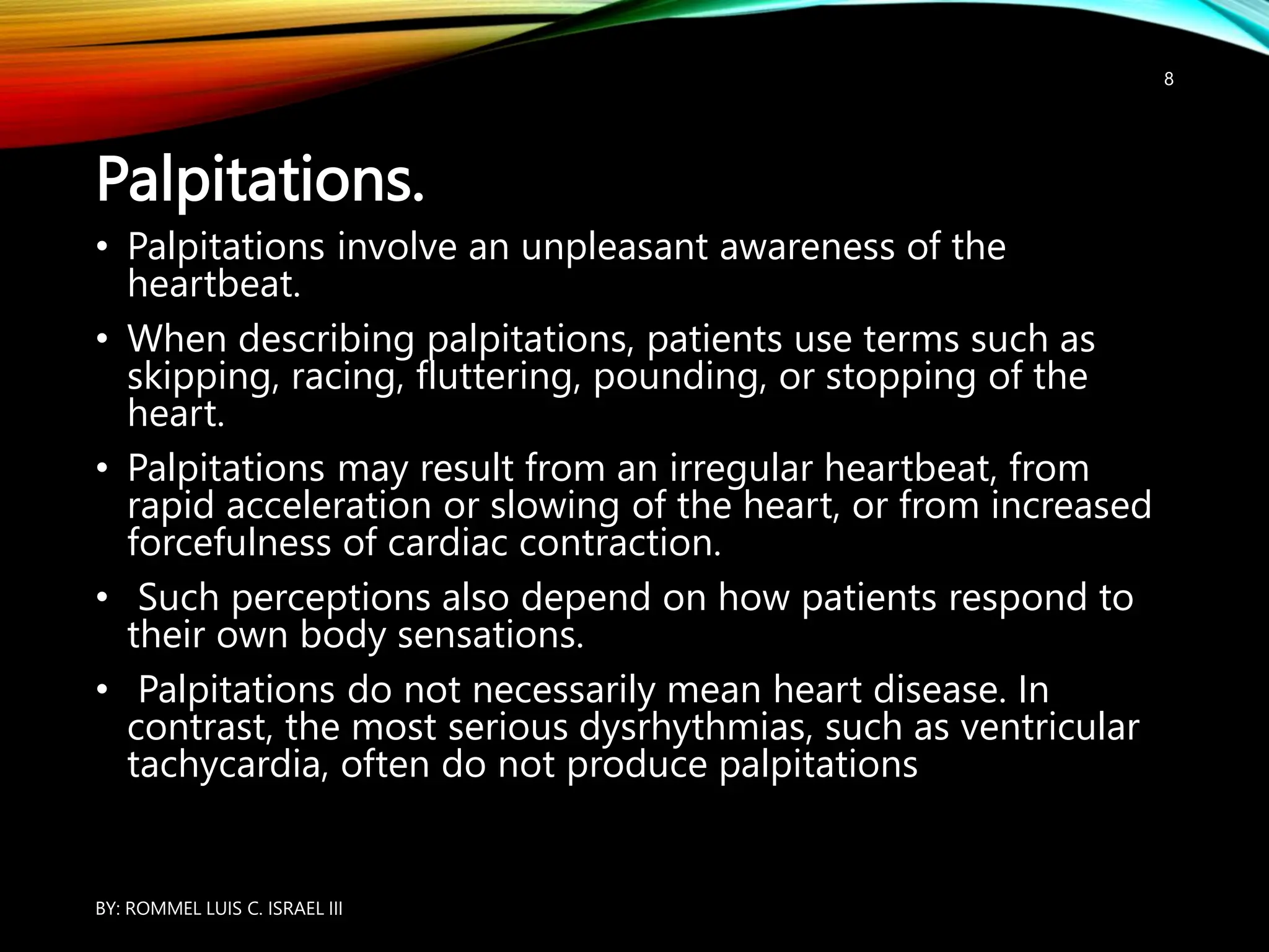
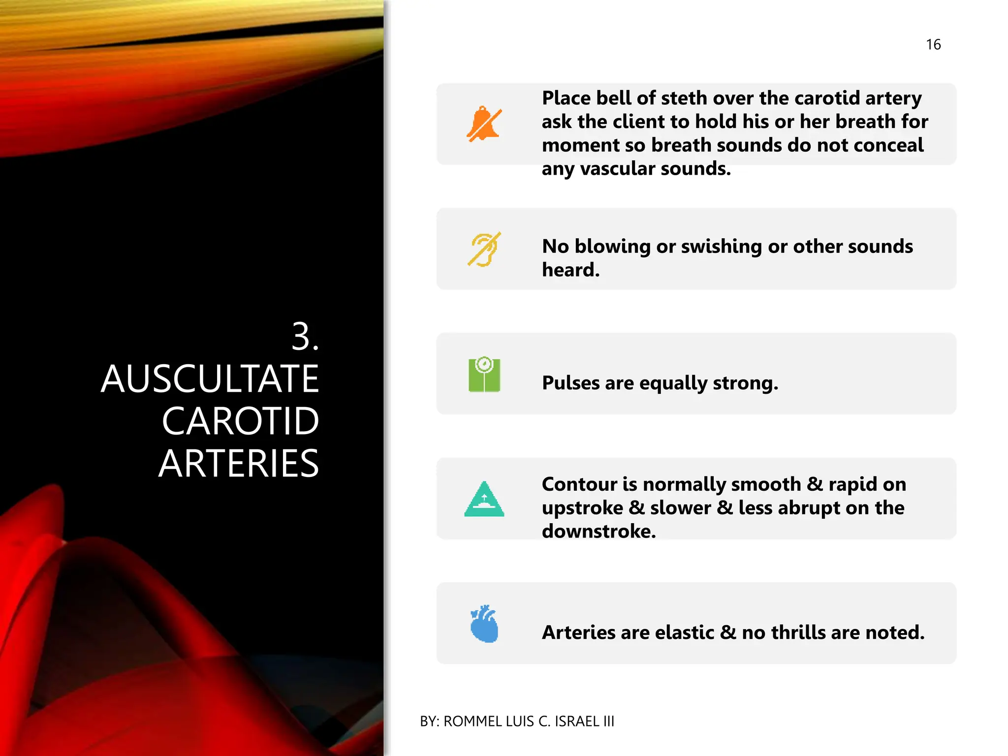
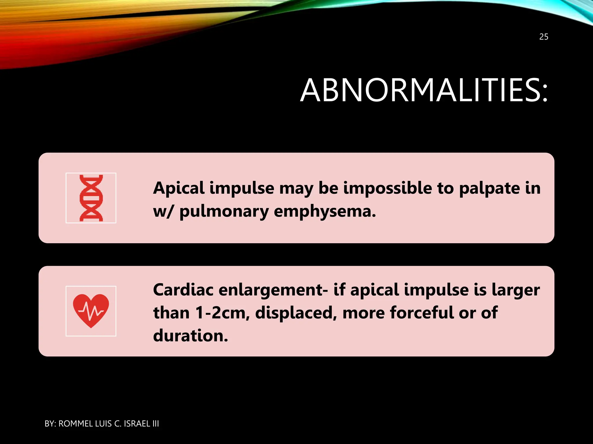
HEART AND NECK
VESSEL ASSESSMENT
BY: ROMMEL LUIS C. ISRAEL III
1
THE HEALTH HISTORY
5
Common or Concerning Symptoms
• Chest pain
• Palpitations
• Shortness of breath: dyspnea,
orthopnea, or paroxysmal nocturnal
dyspnea
• Swelling or edema
BY: ROMMEL LUIS C. ISRAEL III
6
Chest Pain.
• Chest pain is one of the most serious and important
symptoms you will assess as a clinician and is the second
leading cause of emergency room visits, after abdominal pain.
BY: ROMMEL LUIS C. ISRAEL III
EXAMPLES OF ABNORMALITIES
7
• Classic exertional pain, pressure, or
discomfort in the chest, shoulder, back,
neck, or arm in angina pectoris, seen in
50% of patients with acute myocardial
infarction;
• atypical descriptors also are common, such
as cramping, grinding, pricking; rarely,
tooth or jaw pain.
BY: ROMMEL LUIS C. ISRAEL III
Palpitations.
• Palpitations involve an unpleasant awareness of the
heartbeat.
• When describing palpitations, patients use terms such as
skipping, racing, fluttering, pounding, or stopping of the
heart.
• Palpitations may result from an irregular heartbeat, from
rapid acceleration or slowing of the heart, or from increased
forcefulness of cardiac contraction.
• Such perceptions also depend on how patients respond to
their own body sensations.
• Palpitations do not necessarily mean heart disease. In
contrast, the most serious dysrhythmias, such as ventricular
tachycardia, often do not produce palpitations
BY: ROMMEL LUIS C. ISRAEL III
8
9
Shortness of
Breath.
• Shortness of breath is a
common patient concern
and may represent
dyspnea, orthopnea, or
paroxysmal nocturnal
dyspnea. Dyspnea is an
uncomfortable awareness
of breathing that is
inappropriate
BY: ROMMEL LUIS C. ISRAEL III
10
Edema.
• refers to the accumulation of
excessive fluid in the extravascular
interstitial space. Interstitial tissue can
absorb several liters of fluid,
accommodating up to a 10% weight
gain before pitting edema appears.
Causes vary from local to systemic.
• Focus your questions on the location,
timing, and setting of the swelling, and
on associated symptoms. “Have you
had any swelling anywhere? Where?
… Anywhere else? When does it
occur? Is it worse in the morning or
at night? Do your shoes get tight?”
BY: ROMMEL LUIS C. ISRAEL III
EXAMPLES OF ABNORMALITIES
11
Dependent edema appears in the lowest body parts: the feet and
lower legs when sitting, or the sacrum when bedridden.
Causes may be cardiac (congestive heart failure), nutritional
(hypoalbuminemia), or positional
Edema occurs in renal and liver disease: periorbital puffiness, tight
rings in nephrotic syndrome; enlarged waistline from ascites and
liver failure.
BY: ROMMEL LUIS C. ISRAEL III
1. OBSERVE FOR JUGULAR
VENOUS PRESSURE
• Stand straight on the Right
side of the patient.
• Patient should be in supine
position w/ torso elevated
30-45degrees.
• Ask pt. to turn head to the
Left.
• Shine light on the neck.
• Inspect suprasternal
notch/area around clavicles
for pulsations.
• JVP is not normally visible
w/ sitting upright.
• This position distends the
vein, & pulsations may or
may not be discernible.
BY: ROMMEL LUIS C. ISRAEL III
12
ABNORMALITIES:
13
• Fully distended JV in 45
degrees indicate
increased central
pressure
( Right ventricular
pressure, pulmonary
hypertension,
pulmonary emboli)
BY: ROMMEL LUIS C. ISRAEL III
2. EVALUATE JUGULAR
VENOUS PRESSURE.
14
• Position pt. with head of bed elevated
30, 45, 60, 90 degrees.
• At each increase of the elevation, have
pt’s head turned away from the side
being evaluated.
• Using tangential light, observe for
distention.
• The JV should not be distended,
bulging or protruding @ 45 degrees
or greater.
BY: ROMMEL LUIS C. ISRAEL III
15
ABNORMALITIES:
BY: ROMMEL LUIS C. ISRAEL III
Ride-sided HF=
distention, bulging
protrusion at 45,
90 degrees.
COPD= elevated
venous pressure only
during expiration.
Severe constrictive
pericarditis=
inspiratory venous
pressure.
16
3.
AUSCULTATE
CAROTID
ARTERIES
BY: ROMMEL LUIS C. ISRAEL III
Place bell of steth over the carotid artery
ask the client to hold his or her breath for
moment so breath sounds do not conceal
any vascular sounds.
No blowing or swishing or other sounds
heard.
Pulses are equally strong.
Contour is normally smooth & rapid on
upstroke & slower & less abrupt on the
downstroke.
Arteries are elastic & no thrills are noted.
17
ABNORMALITIES:
BY: ROMMEL LUIS C. ISRAEL III
BRUIT- blowing or
sound caused by turbulent
blood flow through a
narrowed vessel indicative
occlusive arterial disease.
Pulse inequality may
arterial constriction or
occlusion in one carotid.
4. PALPATE CAROTID ARTERIES
• Palpate by placing pads of index & middle fingers medial to
the sternocleidomastoid muscle of the neck.
• Note for amplitude & contour of pulse, elasticity of artery and
any thrills.
• The strength of the pulse is evaluated on the scale from 0-4:
• Pulse amplitude scale:
0 =absent 2+ = normal 4+= bounding
1+ = weak 3+ = increased
BY: ROMMEL LUIS C. ISRAEL III
18
ABNORMALITIES:
19
• Weak pulses may indicate hypovolemia,
shock, or decreased CO.
• Bounding pulse, firm pulse may indicate
hypervolemia or increased CO.
• Variations in strength from beat to beat or with
respirations are abnormal & may indicate
variety of problems.
• Loss of elasticity may indicate arteriosclerosis.
• Thrills may indicate narrowing of artery.
BY: ROMMEL LUIS C. ISRAEL III
20
5. INSPECT
PULSATIONS.
BY: ROMMEL LUIS C. ISRAEL III
Place pt. in supine w/ HOB elevated
bet.30 & 45 degrees.
Stand on the R side & look for the
impulse & any abdominal pulsations.
The apical pulse may or may not be
visible.
If apparent, it would be in the MITRAL
AREA( L midclavicular line, 4th 0r 5th
Apical pulse is the result of L ventricle
moving outward during systole.
21
7.
PALPATE
APICAL
PULSE.
BY: ROMMEL LUIS C. ISRAEL III
Remain pt. on R side
ask to remain in
supine.
Use finger pads to
palpate apical pulse
the mitral are( 4th or
5th ICS)
After locating, use
finger pad for more
accurate palpations.
Apical pulse is palpated in the mitral area ( 4th or
5th ICS) & the size of a nickel ( 1-2cm).
Amplitude is small-like a gentle tap & duration is
brief, lasting through the 1st 2/3 of systole &
often less.
In obese pts. Or with large breasts, the apical
impulse may not b palpable.
BY: ROMMEL LUIS C. ISRAEL III
22
BY: ROMMEL LUIS C. ISRAEL III
23
BY: ROMMEL LUIS C. ISRAEL III
24
ABNORMALITIES:
Apical impulse may be impossible to palpate in
w/ pulmonary emphysema.
Cardiac enlargement- if apical impulse is larger
than 1-2cm, displaced, more forceful or of
duration.
BY: ROMMEL LUIS C. ISRAEL III
25
26
8. PALPATE
FOR
ABNORMAL
PULSATIONS.
BY: ROMMEL LUIS C. ISRAEL III
Use palmar surfaces to
palpate apex. Left
border & base.
NO pulsations/
are palpated in the
of the apex, Left sternal
border, or base.
ABNORMALITIES:
27
• A thrill ( similar to
a purring ca) or
pulsation is usually
associated w/
grade IV or higher
murmur.
BY: ROMMEL LUIS C. ISRAEL III
28
9.
AUSCULTATE
HEART RATE
& RHYTHM.
BY: ROMMEL LUIS C. ISRAEL III
Place diaphragm of
steth at the apex &
listen closely to the
& rhythm of apical
impulse.
Rate=60-100per
w/ regular rhythm.
Pulse rate in females is
5-10 bpm faster than
males.
29
ABNORMALITIES:
BY: ROMMEL LUIS C. ISRAEL III
Bradycardia( less
than 60 beats per
minute)
Tachycardia ( more
than 100 beats per
minute) may result
decreased CO.
10. IF YOU
DETECT
IRREGULAR
RHYTHM,
AUSCULTATE
FOR PULSE
RATE DEFICIT.
30
BY: ROMMEL LUIS C. ISRAEL III
Palpate radial pulse
while you
auscultate the
apical pulse
Count for full
minute.
The radial &
rates should be
identical.
31
ABNORMALITIES:
BY: ROMMEL LUIS C. ISRAEL III
Pulse deficit ( difference between
radial pulse)
Atrial fibrillation ( an abnormal heart
rhythm characterized by rapid &
beating of the atria)
Atrial flutter ( a common abnormal
rhythm that starts in the atrial
of the heart associated with a fast HR)
PVCs ( premature heartbeats
from the venticles of the heart)
NORMAL ECG TRACING
BY: ROMMEL LUIS C. ISRAEL III
32
ATRIAL FIBRILLATION AND ATRIAL
FLUTTER WITH VARYING AV
BLOCK
BY: ROMMEL LUIS C. ISRAEL III
33
PREMATURE VENTRICULAR
CONTRACTIONS
BY: ROMMEL LUIS C. ISRAEL III
34
35
11.
AUSCULTATE
TO IDENTIFY
S1 & S2.
BY: ROMMEL LUIS C. ISRAEL III
Auscultate S1( the first heart
“lub”) &
S2 ( 2nd heart sound “ dub”).
S1 corresponds with each carotid
pulsation & is the loudest at the
apex of the heart.
S2 immediately follows after S1
is the loudest at the base of the
heart.
36
ABNORMALITIES:
BY: ROMMEL LUIS C. ISRAEL III
LIFT- associated with right ventricular
hypertrophy caused by pulmonic valve
disease, pulmonic HPN & chronic lung
disease.
THRILL-palpated over the 2nd & 3rd ICS;
indicate severe aortic stenosis & systemic
HPN.
ACCENTUATED APICAL IMPULSE- sign of
pressure overload.
LATERALLY DISPLACED APICAL IMPULSE-
of volume overload.
37
12.
AUSCULTATE
FOR EXTRA
HEART
SOUNDS.
BY: ROMMEL LUIS C. ISRAEL III
Use diaphragm first
the bell to auscultate
the entire heart area.
Note characteristics (
location, timing)
Normally, no sounds are
heard.
38
KNOW YOUR
STETHOSCOPE!
BY: ROMMEL LUIS C. ISRAEL III
It is important to understand the uses of both the diaphragm and
bell.
The diaphragm.
The diaphragm is better for picking up the relatively high-pitched
of S1 and S2, the murmurs of aortic and mitral regurgitation, and
pericardial friction rubs. Listen throughout the precordium with the
diaphragm, pressing it firmly against the chest.
The bell.
The bell is more sensitive to the low-pitched sounds of S3 and S4
murmur of mitral stenosis. Apply the bell lightly, with just enough
pressure to produce an air seal with its full rim. Use the bell at the
then move medially along the lower sternal border. Resting the heel
your hand on the chest like a fulcrum may help you to maintain
pressure.
BY: ROMMEL LUIS C. ISRAEL III
39
BY: ROMMEL LUIS C. ISRAEL III
40
41
ABNORMALITIES:
BY: ROMMEL LUIS C. ISRAEL III
Ejection sounds/
clicks- associated w/
mitral valve
Heard just after S1
Friction rub- may be
heard during
pause.
42
13.
AUSCULTATE
FOR
MURMURS.
BY: ROMMEL LUIS C. ISRAEL III
Auscultate murmurs across
the entire heart. Use
diaphragm & bell of the
steth in all areas in
positions.
MURMUR-swishing sound
caused by turbulent blood
flow through the heart
valves or vessels.
No murmurs heard.
43
ABNORMALITIES:
BY: ROMMEL LUIS C. ISRAEL III
Midsystolic Murmur-
type of murmur; occur
ventricular ejection.
Pansystolic murmur-
when blood flows from a
chamber with high pressure
a chamber of low pressure
an orifice that should be
clossed.
BY: ROMMEL LUIS C. ISRAEL III
44
45
14.
AUSCULTATE
CLIENT WHILE
ASSUMING
OTHER
POSITIONS.
BY: ROMMEL LUIS C. ISRAEL III
Ask pt. to assume left
position.
Use bell of the steth &
at the apex of the heart.
Ask patient to sit up, lean
forward & exhale. Use
diaphragm of steth &
over the apex & left
border.
S1 & S2 heart sounds are
normally present.
ABNORMALITIES:
• S3 ( 3rd heart sound)
• Ventricular gallop
• Has low frequency & is heard using the bell of steth at the
apical area.
• Accentuated during inspiration & has rhythm of the word “
KEN-TUC-KY”
• Normal to young children, people with high CO.
• Associated with myocardial failure, CHF.
BY: ROMMEL LUIS C. ISRAEL III
46
ABNORMALITIES:
• S4 ( 4th heart sound)
• Atrial gallop
• Low-frequency sound occurring at the end of diastole when
atria contract.
• Has rhythm word “ TEN-NES-SEE”
• Normal in trained athletes & some older patients.
• Abnormal to coronary artery dse. , HPN, AMI.
BY: ROMMEL LUIS C. ISRAEL III
47
48
RECORDING
YOUR
FINDINGS
BY: ROMMEL LUIS C. ISRAEL III
Recording the Physical
Examination—The
Cardiovascular Examination
“The jugular venous pulse
is 3 cm above the sternal
with the head of bed elevated
30°. Carotid upstrokes are
without bruits.
The point of maximal impulse
(PMI) is tapping, 7 cm lateral
the midsternal line in the 5th
intercostal space. Crisp S1 and
49
RECORDING
YOUR
FINDINGS
BY: ROMMEL LUIS C. ISRAEL III
The JVP is 5 cm above the sternal
with the head of bed elevated to 50°.
Carotid upstrokes are brisk; a bruit is
heard over the left carotid artery.
The PMI is diffuse, 3 cm in diameter,
palpated at the anterior axillary line
the 5th and 6th intercostal spaces.
S1 and S2 are soft. S3 present at the
apex. High-pitched harsh 2/6
holosystolic murmur best heard at
apex, radiating to the axilla.”

ASSESSMENT OF THE HEART AND NECK VESSEL .pptx

  • 1.
    HEART AND NECK VESSELASSESSMENT BY: ROMMEL LUIS C. ISRAEL III BY: ROMMEL LUIS C. ISRAEL III 1
  • 5.
    THE HEALTH HISTORY 5 Commonor Concerning Symptoms • Chest pain • Palpitations • Shortness of breath: dyspnea, orthopnea, or paroxysmal nocturnal dyspnea • Swelling or edema BY: ROMMEL LUIS C. ISRAEL III
  • 6.
    6 Chest Pain. • Chestpain is one of the most serious and important symptoms you will assess as a clinician and is the second leading cause of emergency room visits, after abdominal pain. BY: ROMMEL LUIS C. ISRAEL III
  • 7.
    EXAMPLES OF ABNORMALITIES 7 •Classic exertional pain, pressure, or discomfort in the chest, shoulder, back, neck, or arm in angina pectoris, seen in 50% of patients with acute myocardial infarction; • atypical descriptors also are common, such as cramping, grinding, pricking; rarely, tooth or jaw pain. BY: ROMMEL LUIS C. ISRAEL III
  • 8.
    Palpitations. • Palpitations involvean unpleasant awareness of the heartbeat. • When describing palpitations, patients use terms such as skipping, racing, fluttering, pounding, or stopping of the heart. • Palpitations may result from an irregular heartbeat, from rapid acceleration or slowing of the heart, or from increased forcefulness of cardiac contraction. • Such perceptions also depend on how patients respond to their own body sensations. • Palpitations do not necessarily mean heart disease. In contrast, the most serious dysrhythmias, such as ventricular tachycardia, often do not produce palpitations BY: ROMMEL LUIS C. ISRAEL III 8
  • 9.
    9 Shortness of Breath. • Shortnessof breath is a common patient concern and may represent dyspnea, orthopnea, or paroxysmal nocturnal dyspnea. Dyspnea is an uncomfortable awareness of breathing that is inappropriate BY: ROMMEL LUIS C. ISRAEL III
  • 10.
    10 Edema. • refers tothe accumulation of excessive fluid in the extravascular interstitial space. Interstitial tissue can absorb several liters of fluid, accommodating up to a 10% weight gain before pitting edema appears. Causes vary from local to systemic. • Focus your questions on the location, timing, and setting of the swelling, and on associated symptoms. “Have you had any swelling anywhere? Where? … Anywhere else? When does it occur? Is it worse in the morning or at night? Do your shoes get tight?” BY: ROMMEL LUIS C. ISRAEL III
  • 11.
    EXAMPLES OF ABNORMALITIES 11 Dependentedema appears in the lowest body parts: the feet and lower legs when sitting, or the sacrum when bedridden. Causes may be cardiac (congestive heart failure), nutritional (hypoalbuminemia), or positional Edema occurs in renal and liver disease: periorbital puffiness, tight rings in nephrotic syndrome; enlarged waistline from ascites and liver failure. BY: ROMMEL LUIS C. ISRAEL III
  • 12.
    1. OBSERVE FORJUGULAR VENOUS PRESSURE • Stand straight on the Right side of the patient. • Patient should be in supine position w/ torso elevated 30-45degrees. • Ask pt. to turn head to the Left. • Shine light on the neck. • Inspect suprasternal notch/area around clavicles for pulsations. • JVP is not normally visible w/ sitting upright. • This position distends the vein, & pulsations may or may not be discernible. BY: ROMMEL LUIS C. ISRAEL III 12
  • 13.
    ABNORMALITIES: 13 • Fully distendedJV in 45 degrees indicate increased central pressure ( Right ventricular pressure, pulmonary hypertension, pulmonary emboli) BY: ROMMEL LUIS C. ISRAEL III
  • 14.
    2. EVALUATE JUGULAR VENOUSPRESSURE. 14 • Position pt. with head of bed elevated 30, 45, 60, 90 degrees. • At each increase of the elevation, have pt’s head turned away from the side being evaluated. • Using tangential light, observe for distention. • The JV should not be distended, bulging or protruding @ 45 degrees or greater. BY: ROMMEL LUIS C. ISRAEL III
  • 15.
    15 ABNORMALITIES: BY: ROMMEL LUISC. ISRAEL III Ride-sided HF= distention, bulging protrusion at 45, 90 degrees. COPD= elevated venous pressure only during expiration. Severe constrictive pericarditis= inspiratory venous pressure.
  • 16.
    16 3. AUSCULTATE CAROTID ARTERIES BY: ROMMEL LUISC. ISRAEL III Place bell of steth over the carotid artery ask the client to hold his or her breath for moment so breath sounds do not conceal any vascular sounds. No blowing or swishing or other sounds heard. Pulses are equally strong. Contour is normally smooth & rapid on upstroke & slower & less abrupt on the downstroke. Arteries are elastic & no thrills are noted.
  • 17.
    17 ABNORMALITIES: BY: ROMMEL LUISC. ISRAEL III BRUIT- blowing or sound caused by turbulent blood flow through a narrowed vessel indicative occlusive arterial disease. Pulse inequality may arterial constriction or occlusion in one carotid.
  • 18.
    4. PALPATE CAROTIDARTERIES • Palpate by placing pads of index & middle fingers medial to the sternocleidomastoid muscle of the neck. • Note for amplitude & contour of pulse, elasticity of artery and any thrills. • The strength of the pulse is evaluated on the scale from 0-4: • Pulse amplitude scale: 0 =absent 2+ = normal 4+= bounding 1+ = weak 3+ = increased BY: ROMMEL LUIS C. ISRAEL III 18
  • 19.
    ABNORMALITIES: 19 • Weak pulsesmay indicate hypovolemia, shock, or decreased CO. • Bounding pulse, firm pulse may indicate hypervolemia or increased CO. • Variations in strength from beat to beat or with respirations are abnormal & may indicate variety of problems. • Loss of elasticity may indicate arteriosclerosis. • Thrills may indicate narrowing of artery. BY: ROMMEL LUIS C. ISRAEL III
  • 20.
    20 5. INSPECT PULSATIONS. BY: ROMMELLUIS C. ISRAEL III Place pt. in supine w/ HOB elevated bet.30 & 45 degrees. Stand on the R side & look for the impulse & any abdominal pulsations. The apical pulse may or may not be visible. If apparent, it would be in the MITRAL AREA( L midclavicular line, 4th 0r 5th Apical pulse is the result of L ventricle moving outward during systole.
  • 21.
    21 7. PALPATE APICAL PULSE. BY: ROMMEL LUISC. ISRAEL III Remain pt. on R side ask to remain in supine. Use finger pads to palpate apical pulse the mitral are( 4th or 5th ICS) After locating, use finger pad for more accurate palpations.
  • 22.
    Apical pulse ispalpated in the mitral area ( 4th or 5th ICS) & the size of a nickel ( 1-2cm). Amplitude is small-like a gentle tap & duration is brief, lasting through the 1st 2/3 of systole & often less. In obese pts. Or with large breasts, the apical impulse may not b palpable. BY: ROMMEL LUIS C. ISRAEL III 22
  • 23.
    BY: ROMMEL LUISC. ISRAEL III 23
  • 24.
    BY: ROMMEL LUISC. ISRAEL III 24
  • 25.
    ABNORMALITIES: Apical impulse maybe impossible to palpate in w/ pulmonary emphysema. Cardiac enlargement- if apical impulse is larger than 1-2cm, displaced, more forceful or of duration. BY: ROMMEL LUIS C. ISRAEL III 25
  • 26.
    26 8. PALPATE FOR ABNORMAL PULSATIONS. BY: ROMMELLUIS C. ISRAEL III Use palmar surfaces to palpate apex. Left border & base. NO pulsations/ are palpated in the of the apex, Left sternal border, or base.
  • 27.
    ABNORMALITIES: 27 • A thrill( similar to a purring ca) or pulsation is usually associated w/ grade IV or higher murmur. BY: ROMMEL LUIS C. ISRAEL III
  • 28.
    28 9. AUSCULTATE HEART RATE & RHYTHM. BY:ROMMEL LUIS C. ISRAEL III Place diaphragm of steth at the apex & listen closely to the & rhythm of apical impulse. Rate=60-100per w/ regular rhythm. Pulse rate in females is 5-10 bpm faster than males.
  • 29.
    29 ABNORMALITIES: BY: ROMMEL LUISC. ISRAEL III Bradycardia( less than 60 beats per minute) Tachycardia ( more than 100 beats per minute) may result decreased CO.
  • 30.
    10. IF YOU DETECT IRREGULAR RHYTHM, AUSCULTATE FORPULSE RATE DEFICIT. 30 BY: ROMMEL LUIS C. ISRAEL III Palpate radial pulse while you auscultate the apical pulse Count for full minute. The radial & rates should be identical.
  • 31.
    31 ABNORMALITIES: BY: ROMMEL LUISC. ISRAEL III Pulse deficit ( difference between radial pulse) Atrial fibrillation ( an abnormal heart rhythm characterized by rapid & beating of the atria) Atrial flutter ( a common abnormal rhythm that starts in the atrial of the heart associated with a fast HR) PVCs ( premature heartbeats from the venticles of the heart)
  • 32.
    NORMAL ECG TRACING BY:ROMMEL LUIS C. ISRAEL III 32
  • 33.
    ATRIAL FIBRILLATION ANDATRIAL FLUTTER WITH VARYING AV BLOCK BY: ROMMEL LUIS C. ISRAEL III 33
  • 34.
  • 35.
    35 11. AUSCULTATE TO IDENTIFY S1 &S2. BY: ROMMEL LUIS C. ISRAEL III Auscultate S1( the first heart “lub”) & S2 ( 2nd heart sound “ dub”). S1 corresponds with each carotid pulsation & is the loudest at the apex of the heart. S2 immediately follows after S1 is the loudest at the base of the heart.
  • 36.
    36 ABNORMALITIES: BY: ROMMEL LUISC. ISRAEL III LIFT- associated with right ventricular hypertrophy caused by pulmonic valve disease, pulmonic HPN & chronic lung disease. THRILL-palpated over the 2nd & 3rd ICS; indicate severe aortic stenosis & systemic HPN. ACCENTUATED APICAL IMPULSE- sign of pressure overload. LATERALLY DISPLACED APICAL IMPULSE- of volume overload.
  • 37.
    37 12. AUSCULTATE FOR EXTRA HEART SOUNDS. BY: ROMMELLUIS C. ISRAEL III Use diaphragm first the bell to auscultate the entire heart area. Note characteristics ( location, timing) Normally, no sounds are heard.
  • 38.
    38 KNOW YOUR STETHOSCOPE! BY: ROMMELLUIS C. ISRAEL III It is important to understand the uses of both the diaphragm and bell. The diaphragm. The diaphragm is better for picking up the relatively high-pitched of S1 and S2, the murmurs of aortic and mitral regurgitation, and pericardial friction rubs. Listen throughout the precordium with the diaphragm, pressing it firmly against the chest. The bell. The bell is more sensitive to the low-pitched sounds of S3 and S4 murmur of mitral stenosis. Apply the bell lightly, with just enough pressure to produce an air seal with its full rim. Use the bell at the then move medially along the lower sternal border. Resting the heel your hand on the chest like a fulcrum may help you to maintain pressure.
  • 39.
    BY: ROMMEL LUISC. ISRAEL III 39
  • 40.
    BY: ROMMEL LUISC. ISRAEL III 40
  • 41.
    41 ABNORMALITIES: BY: ROMMEL LUISC. ISRAEL III Ejection sounds/ clicks- associated w/ mitral valve Heard just after S1 Friction rub- may be heard during pause.
  • 42.
    42 13. AUSCULTATE FOR MURMURS. BY: ROMMEL LUISC. ISRAEL III Auscultate murmurs across the entire heart. Use diaphragm & bell of the steth in all areas in positions. MURMUR-swishing sound caused by turbulent blood flow through the heart valves or vessels. No murmurs heard.
  • 43.
    43 ABNORMALITIES: BY: ROMMEL LUISC. ISRAEL III Midsystolic Murmur- type of murmur; occur ventricular ejection. Pansystolic murmur- when blood flows from a chamber with high pressure a chamber of low pressure an orifice that should be clossed.
  • 44.
    BY: ROMMEL LUISC. ISRAEL III 44
  • 45.
    45 14. AUSCULTATE CLIENT WHILE ASSUMING OTHER POSITIONS. BY: ROMMELLUIS C. ISRAEL III Ask pt. to assume left position. Use bell of the steth & at the apex of the heart. Ask patient to sit up, lean forward & exhale. Use diaphragm of steth & over the apex & left border. S1 & S2 heart sounds are normally present.
  • 46.
    ABNORMALITIES: • S3 (3rd heart sound) • Ventricular gallop • Has low frequency & is heard using the bell of steth at the apical area. • Accentuated during inspiration & has rhythm of the word “ KEN-TUC-KY” • Normal to young children, people with high CO. • Associated with myocardial failure, CHF. BY: ROMMEL LUIS C. ISRAEL III 46
  • 47.
    ABNORMALITIES: • S4 (4th heart sound) • Atrial gallop • Low-frequency sound occurring at the end of diastole when atria contract. • Has rhythm word “ TEN-NES-SEE” • Normal in trained athletes & some older patients. • Abnormal to coronary artery dse. , HPN, AMI. BY: ROMMEL LUIS C. ISRAEL III 47
  • 48.
    48 RECORDING YOUR FINDINGS BY: ROMMEL LUISC. ISRAEL III Recording the Physical Examination—The Cardiovascular Examination “The jugular venous pulse is 3 cm above the sternal with the head of bed elevated 30°. Carotid upstrokes are without bruits. The point of maximal impulse (PMI) is tapping, 7 cm lateral the midsternal line in the 5th intercostal space. Crisp S1 and
  • 49.
    49 RECORDING YOUR FINDINGS BY: ROMMEL LUISC. ISRAEL III The JVP is 5 cm above the sternal with the head of bed elevated to 50°. Carotid upstrokes are brisk; a bruit is heard over the left carotid artery. The PMI is diffuse, 3 cm in diameter, palpated at the anterior axillary line the 5th and 6th intercostal spaces. S1 and S2 are soft. S3 present at the apex. High-pitched harsh 2/6 holosystolic murmur best heard at apex, radiating to the axilla.”