Endocrine System
Anatomy and Physiology
Prof.Nagamani.T
Contents
Anatomy of Endocrine System
( 4 hrs)
Structure of;
•Hypothalamus,
•Pineal Gland,
•Pituitary gland,
•Thyroid,
•Parathyroid,
•Thymus,
•Pancreas and
•Adrenal glands
Physiology of the Endocrine system
( 5 hrs)
Functions and hormones of
PinealGland,
•Pituitary gland,
•Thyroid,
•Parathyroid,
•Thymus,
•Pancreas and
•Adrenal glands.
•Other hormones
•Alterations in disease
•Application and implication in
nursing
Introduction
 The endocrine system is a messenger system in
an organism comprising feedback loops of hormones that are
released by internal glands directly into the circulatory
system and that target and regulate distant organs.
 In vertebrates, the hypothalamus is the neural control center
for all endocrine systems.
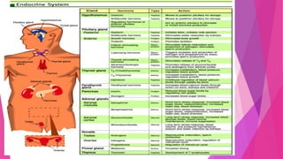
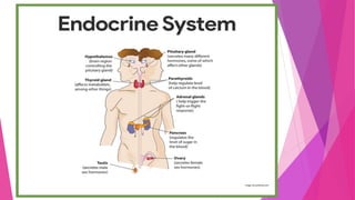
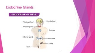
 In humans, the major endocrine glands are
the thyroid, parathyroid, pituitary, pineal, and adrenal glands,
and the (male) testicles and (female) ovaries.
The hypothalamus, pancreas, and thymus also function as
endocrine glands. (The hypothalamus and pituitary glands are
organs of the neuroendocrine system.
Functions of the Endocrine System
 Water equilibrium. The endocrine system controls water equilibrium by
regulating the solute concentration of the blood.
 Growth, metabolism, and tissue maturation. The endocrine system
controls the growth of many tissues, like the bone and muscle, and the
degree of metabolism of various tissues, which aids in the maintenance of
the normal body temperature and normal mental functions. Maturation of
tissues, which appears in the development of adult features and adult
behavior, are also determined by the endocrine system.
 Heart rate and blood pressure management. The endocrine system assists
in managing the heart rate and blood pressure and aids in preparing the
body for physical motion.
 Immune system control. The endocrine system helps
regulate the production and functions of immune cells.
 Reproductive function controls. The endocrine system
regulates the development and the functions of the
reproductive systems in males and females.
 Uterine contractions and milk release. The endocrine
system controls uterine contractions throughout the delivery
of the newborn and stimulates milk release from the breasts
in lactating females.
 Ion management. The endocrine system regulates Na+, K+,
and Ca2+ concentrations in the blood.
 Blood glucose regulator. The endocrine system
controls blood glucose levels and other nutrient levels in the
blood.
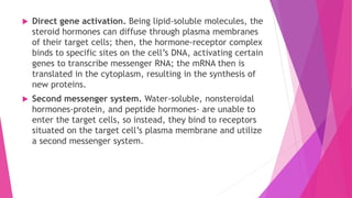
 Direct gene activation. Being lipid-soluble molecules, the
steroid hormones can diffuse through plasma membranes
of their target cells; then, the hormone-receptor complex
binds to specific sites on the cell’s DNA, activating certain
genes to transcribe messenger RNA; the mRNA then is
translated in the cytoplasm, resulting in the synthesis of
new proteins.
 Second messenger system. Water-soluble, nonsteroidal
hormones-protein, and peptide hormones- are unable to
enter the target cells, so instead, they bind to receptors
situated on the target cell’s plasma membrane and utilize
a second messenger system.
Anatomy of the Endocrine System
 Compared to other organs of the body, the organs of the endocrine
system are small and unimpressive, however, functionally the
endocrine organs are very impressive, and when their role in
maintaining body homeostasis is considered, they are true giants.
 The major endocrine organs of the body include the pituitary, thyroid,
parathyroid, adrenal, pineal, and thymus glands, the pancreas, and
the gonads.
 Endocrine glands have no ducts, are vascular, and commonly have
intracellular vacuoles or granules that store their hormones. In
contrast, exocrine glands, such as salivary glands, sweat glands, and
glands within the gastrointestinal tract, tend to be much less vascular
and have ducts or a hollow lumen. Endocrinology is a branch
of internal medicine
Types of glands
 Exocrine glands
 Endocrine Glands
Exocrine glands are the glands of external secretion. These are those that secrete
substances on the body surface through a duct. Salivary glands, mammary glands, sweat
glands, are some of the exocrine glands.
Unlike exocrine glands(sweat, salivary), endocrine glands secrete their respective
substances directly into the bloodstream rather than through a duct. These endocrine
glands belong to the body’s control system and they produce hormones which help to
regulate the functions of cells and tissues. Some glands are specific to either male (testes)
or female (ovaries).
Endocrine Glands
Hypothalamus
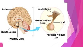
 Hypothalamus. The hypothalamus, which is part of the nervous system, is
Hypothalamus is a minute region, almost the size of an almond, present at the centre
of the human brain, near the pituitary gland.
Structure :
It consists of three main regions:
 The anterior region.
 The middle region.
 The posterior region.
 It plays a vital role in the production of hormones. Maintaining the hypothalamus
health is very important. Its improper functioning causes several disorders.
Anterior Region
 The anterior region is also known as the supraoptic region.
 It regulates body temperature and maintains the circadian rhythm.
 There are several small nuclei in the hypothalamus. The major hypothalamic nuclei
include supraoptic and paraventricular nuclei.
 The nuclei in this region are involved in the hormone secretion.
Following are the hormones secreted by the anterior region of the hypothalamus:
 Corticotropin-releasing hormone
 Thyrotropin-releasing hormone
 Gonadotropin-releasing hormone
 Oxytocin
 Vasopressin
 Somatostatin
Middle Region
 This is known as the tuberal region.
 It consists of ventromedial and arcuate nuclei.
 The ventromedial nuclei control the appetite, whereas the arcuate nuclei secrete the
growth hormone responsible for the growth and development of the body.
Posterior Region
 This region is also known as the mammillary region.
 The major nuclei include posterior hypothalamic nucleus and mammillary nuclei.
 The posterior hypothalamic nuclei cause shivering and blockage of sweat and thus
regulates the body temperature.
 The mammillary nuclei, on the contrary, are believed to be involved in the memory
function.
Functions of Hypothalamus
 The hypothalamus contains osmotic sensors that react to the concentration of sodium,
potassium, carbon dioxide, and chloride. When the blood pressure is very low these
osmotic sensors and baroreceptors intimate the kidneys to store or release water to
maintain the concentration of these substances.
Following are the important functions of the hypothalamus:
 Its main function is maintaining the body’s internal balance- homeostasis.
 It connects the endocrine and the nervous system.
 Hypothalamus stimulates or inhibits many of the body’s activities in order to maintain
homeostasis, such as regulating body temperature, appetite and body weight, heart
rate and blood pressure, etc.
 It is also involved in many essential functions of the body, including:
- Childbirth. - Emotions.
- Sleep cycles. - Balancing body fluids.
- Appetite and thirst control. - Blood pressure and heart rate.
Hormones Secreted by Hypothalamus
The anterior region of the hypothalamus is responsible for hormone secretion. The nuclei
present in this region lead the process. The important hormones secreted by
hypothalamus are:
 Corticotropin-Releasing Hormone: This hormone is responsible for the regulation of
metabolic and immune response.
 Thyrotropin Releasing Hormone: It triggers the pituitary gland to release a thyroid-
stimulating hormone which plays a major role in the functioning of organs of the body
such as heart, muscles, etc.
 Gonadotropin-Releasing Hormone: It stimulates the pituitary gland to release several
reproductive hormones.
 Oxytocin: It is involved in several processes such as lactation, childbirth, regulating
sleep cycles, maintaining body temperature.
 Somatostatin: This hormone is also known as Growth Hormone Inhibiting Hormone. It
regulates the endocrine system and affects the neurotransmission and cell
proliferation by interacting with G-protein coupled receptors.
 The middle region of hypothalamus stimulates the release of Growth Hormone
Releasing Hormone. This hormone plays a major role in the growth and development
of the body.
Oxytocin
 Oxytocin is a peptide hormone, released during childbirth and lactation. In females,
it is mainly involved in stimulating the growth of prostaglandins, increasing the
contractions of the muscles of the uterus, reduces the excess blood flow post-
childbirth, promotes milk movement into the mammary glands.
 In males, oxytocin helps in the production of testosterone and aids in the movement
of sperm. It also plays a role in social behaviour and various aspects around it.
Effects Of Oxytocin
 Consumption of oxytocin can have adverse side-effects. Such as:
 Higher levels of oxytocin have been related to Benign prostatic hyperplasia
 Lack of oxytocin can prevent breastfeeding by milk-ejection reflex
 Low oxytocin levels have also been linked to autistic disorders (Asperger’s syndrome). It
also leads to depressive symptoms
 Fetal heart rate deceleration, pulmonary oedema, and uterine hyper stimulation are
other conditions caused due to oxytocin
 Vomiting, nausea, jaundice, neonatal hyperbilirubinemia, sinus bradycardia are also
caused due to abnormal oxytocin levels
 Oxytocin imbalance in the body causes seizures in the neurons
Functions Of Oxytocin
 Affects gland growth.
 Acts as a neurotransmitter in the brain.
 Stimulates contractility of the epididymis, prostate gland, and seminiferous
tubules.
 Oxytocin plays a role in maternal and sexual behaviour, memory-related,
yawning, feeding, thermoregulation and cardiovascular regulation.
 Stimulates uterine muscle contractions. During childbirth, fetal-ejection reflex
produces oxytocin, which causes contractions of muscles of the uterus.
ADH- Anti-diuretic Hormone
 ADH, also known as arginine vasopressin, is a hormone produced by the
hypothalamus and stored in the posterior pituitary gland. It is made up of special
nerve cells found at the base of the hypothalamus.
 The hormone is transported to the pituitary gland via axons, where it is released
into the bloodstream. It regulates and balances the amount of water in the
blood.
Functions Of ADH
 It acts on the kidneys and the blood vessels and functions to control the blood
pressure. Higher concentrations of ADH constrict the blood vessels which
increases the blood pressure.
 It allows the water in the urine to be taken back in a specific area in the kidney
and thus reduces the amount of water excreted through the urine thereby
conserving the volume of the fluid in the body.
 It is mainly responsible for homeostasis.
Regulation Of ADH
 The release of ADH into the bloodstream is regulated by a number of factors:
 The decrease in the blood volume or blood pressure is detected by the large
blood vessels and the receptors present in the heart, which stimulate the release
of ADH.
 The increase in the concentration of salts in the bloodstream also controls the
secretion of ADH which is identified by special nerve cells in the hypothalamus.
 Alcohol prevents the release of ADH. This causes dehydration and an increase in
urine production.
 If the levels of ADH are higher, the water is retained by the kidney in the body. If
excess ADH is released when not required, the blood gets diluted due to excess
water retention. The salt concentration in the blood thus decreases. High levels
of ADH may be caused due to the side-effects of drugs, lung diseases, etc.
Increased ADH is associated with leukaemia, lymphoma, bladder cancer, brain
cancer, etc.
 If the levels of ADH are lower, a lot of water is excreted by the kidneys. This
increases urine volume and lowers blood pressure. Low levels of ADH indicate
primary polydipsia and damage to the hypothalamus or pituitary gland. Diabetes
insipidus is related to low levels of ADH.
Hypothalamic Disorders
Hypothalamic disorders can be due to the following problems:
 Head injuries
 Genetic disorders
 Tumours in the hypothalamus
 Disorders in eating
 Brain surgeries
 Autoimmune disorders
The symptoms that indicate a hypothalamic disorder include:
 Body temperature fluctuations
 Infertility
 Unusually high or low blood pressure
 Insomnia
 Change in appetite
 Frequent urination
 Delayed puberty
The Pituitary gland ( The hypophysis Cerebri)
 The Pituitary gland, also known as the hypophysis, is a pea-sized endocrine gland
situated at the base of our brain.
 The pituitary gland is a part of the endocrine system and controls the function of
several other endocrine glands.
 It is often referred to as the ‘Master Gland’ because it produces some of the important
hormones in the body.
 It is situated in a bony structure called the Pituitary fossa, just below the
hypothalamus, close to the optic nerve. The pituitary gland is divided into three parts,
also called lobes:
• Anterior pituitary
• Intermediate pituitary (Absent in adult human beings)
• Posterior pituitary
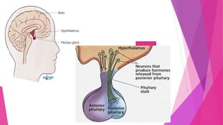
 The fully developed pituitary gland (see the image below) is pea-sized and
weighs approximately 0.5 g.
 The adenohypophysis constitutes roughly 80% of the pituitary and
manufactures an array of peptide hormones.
 It is connected to yhr hypothalamus by the pituitary or hypophysial stalk
 The release of these pituitary hormones is mediated by hypothalamic
neurohormones that are secreted from the median eminence (a site where
axon terminals emanate from the hypothalamus) and that reach the
adenohypophysis via a portal venous system.
The anterior pituitary ( Pars Anterior)
 The anterior pituitary is the front lobe of the pituitary gland, which is a
small, pea-sized gland located at the base of your brain below the
hypothalamus.
 The anterior lobe of the pituitary gland, also known as the adenohypophysis,
produces and secretes the majority of pituitary hormones. Its function is
controlled by the releasing-hormones of the hypothalamus.
 Anterior lobe consists of thick irregular cords of cells, branching by sinusoid.
The anterior pituitary (adenohypophysis) is made of three distinctive parts:
 Anterior part (distal or glandular part) is the part with the strongest secretory activity.
It is composed of follicles that vary in size, but essentially contain three types of cells.
These cells are classified according to their histological staining and
include chromophils (acidophilic and basophilic) and chromophobes.
 The acidophilic cells further divide into somatotrophs and lactotrophs. The former produce
the growth hormone, while the latter produce prolactin.
 The basophilic cells are divided into gonadotrophs (producing FSH and LH), corticotrophs
(ACTH) and thyrotropes (TSH).
 The chromophobes stain weakly, and they are the progenitor cells.
 Tuberal part extends from the anterior (distal) part of the pituitary gland. The majority
of its cells are gonadotrophs, with the small remainder being the thyrotrophs. The
tuberal part surrounds the infundibular stem, a collection of axons that project into
the pituitary from the hypothalamus. These axons are filled with the accumulated
hormones (oxytocin and vasopressin).
 Intermediate part is located between the anterior part of adenohypophysis and
neurohypophysis. It contains follicles that are made up of colloidal matrices. The
majority of the cells in this part are the corticotropic basophils, with less abundant
pituitary stem cells. These corticotropic cells of this part produce melanocyte-
stimulating hormone and endorphins.
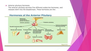
 Anterior pituitary hormones
The anterior pituitary secretes five different endocrine hormones, and
releases them into the bloodstream. These hormones are the:
The anterior pituitary gland produces and releases six main hormones:
 Adrenocorticotropic hormone (ACTH, or corticotropin): This hormone
stimulates your adrenal glands to produce cortisol and other hormones.
 Follicle-stimulating hormone (FSH): This hormone stimulates the testes to
produce sperm and stimulates the ovaries to produce eggs and estrogen.
 Luteinising hormone (LH): This hormone stimulates the ovaries and testes to
produce hormones.
 Thyroid-stimulating hormone (TSH): This hormone stimulates the thyroid
gland to produce thyroid hormones.
 Prolactin (PRL): This hormone stimulates milk production in women.
 Growth hormone (GH): This hormone stimulates growth and cell reproduction
in humans and other animals.
Regulation of the anterior pituitary function
 The hypothalamus is the main regulator of the anterior pituitary function. I
 The hypothalamus regulates the activity of the anterior pituitary by releasing
its stimulating and/or inhibitory hormones that include:
 Corticotropin-releasing hormone (CRH)
 Growth hormone-releasing hormone (GHRH)
 Gonadotropin-releasing hormone (GnRH)
 Thyrotropin-releasing hormone (TRH)
 Dopamine (DA)
 Somatostatin (SS), also known as the growth hormone inhibiting hormone
(GHIH)
Posterior pituitary
(neurohypophysis / Pars posterior)
 The posterior portion of the pituitary gland is a specialized neuroendocrine
structure.
 Unlike the anterior pituitary, it doesn’t contain the secretory cells, but rather
many unmyelinated axons that originate from the secretory hypothalamic
neurons, specifically from the magnocellular neurons of the paraventricular
and supraoptic nuclei.
 The only cellular components of the posterior pituitary are the glial cells
called the pituicytes.
The neurohypophysis is divided into the following parts:
 Infundibulum consists of the many unmyelinated axons that form the
hypothalamohypophyseal tracts. These tracts connect the pituitary gland to
the hypothalamus and serve to transport the neurohormones from the
hypothalamic nuclei to the neurohypophysis.
 Neural lobe (pars nervosa, posterior lobe) is a collection of approximate
100,000 unmyelinated axons and their endings. These axon terminals contain
the neurosecretory bodies (Herring’s bodies) that are filled with granules that
contain the hypothalamic hormones destined to be released by the
neurohypophysis. Each granule contains either antidiuretic hormone (ADH) or
oxytocin.
Posterior pituitary hormones
 Note that the posterior portion of the pituitary doesn't contain glandular
tissue, which means that it is not actively producing hormones.
 Its main function is to store and release two hormones secreted by the
hypothalamus; ADH and oxytocin.
 Oxytocin is a neuropeptide produced by the neurosecretory cells of the
supraoptic and paraventricular nuclei. It participates mainly in the processes
related to reproduction and childbirth (such as uterine contractions and
lactation), as well as in human behaviour (e.g. social bonding).
 Antidiuretic hormone (ADH), or arginine-vasopressin, is mainly produced in
the supraoptic and paraventricular regions of the hypothalamus. It is essential
in the control of the body’s electrolyte balance, blood pressure
and kidney functioning.
 Its controlled by the releasing-hormones of the hypothalamus.
Intermediate Lobe ( Pars Intermedia)
 Intermediate lobe has an irregular row of follicles containing pale staining
colloid material and clusters of cells.
Pineal Gland ( Epiphysis Cerebri)
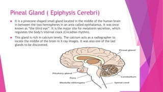
 It is a pinecone-shaped small gland located in the middle of the human brain
in between the two hemispheres in an area called epithalamus. It was once
known as “the third eye”. It is the major site for melatonin secretion, which
regulates the body’s internal clock (Circadian rhythm).
 This gland is rich in calcium levels. The calcium acts as a radiographer to
locate the middle of the brain in X-ray images. It was also one of the last
glands to be discovered.
Anatomy of Pineal Gland
 The pineal gland is about 0.3 inches long and weighs 0.1 grams. The
adrenergic nerves entering the pineal gland regulate its functions.
 The pineal gland is composed of pinealocytes and supporting cells that
resemble the astrocytes present in the brain.
 Some lower vertebrates have a well-developed eye-like structure that acts as
a light receptor.
Hormones Secreted by Pineal Gland– Melatonin
 The pineal gland synthesises melatonin and serotonin, hence they are also
called as the Pineal Gland Hormone. The pineal gland also produces
neurosteroids.
 Serotonin is the precursor of melatonin. Serotonin is acetylated and
methylated to yield melatonin within the pineal gland. The light exposure to
the eyes affects the synthesis and secretion of melatonin.
 Two melatonin receptors have been found in mammals- Mel1A and Mel1B.
These are G-protein coupled cell surface receptors.
 Melatonin affects circadian rhythm.
 Our circadian rhythm is a 24-hour biological cycle, characterised by the sleep-
wake patterns. The daylight and darkness regulate our circadian rhythms.
 The secretion of melatonin is stopped on exposure to light which in turn
controls the circadian rhythm. The secretion of melatonin is high during dark
and low during daylight. This influences our reaction to photoperiod.
Functions Of Pineal Gland
Secretion Of Melatonin
 This gland secretes the hormone melatonin which regulates the circadian
rhythm of the body and also certain reproductive hormones. The secretion of
this hormone depends upon the amount of light a person is exposed to. This
hormone is produced in large amounts when it is dark. The presence of light
inhibits the secretion of melatonin which controls our circadian rhythms.
Cardiovascular Health
 The melatonin secretion has a positive impact on the heart and blood
pressure. It may also be used for the treatment of cardiovascular diseases.
Reproduction
 Melatonin inhibits the secretion of reproductive hormones from the anterior
pituitary, which are responsible for the development and functioning of
reproductive organs.
Disorders Of Pineal Gland
 Listed below are few disorders caused by the malfunctioning of the pineal
gland.
 Depression.
 Mood swings.
 Peptic or stomach ulcers.
 Disruption in sleep patterns.
 An impaired pineal gland leads to hormonal imbalance.
 Sexual disorders are caused by the dysfunctioning of the pineal gland.
 Other disorders include:
 Low melatonin secretion also leads to anxiety, low thyroid hormone
production, menopause symptoms, etc.
 Over secretion of melatonin can lead to low blood pressure and improper
functioning of the thyroid glands and adrenal glands.
 If a tumour develops in the pineal gland, it affects several other factors in the
body:
 Nausea.
 Seizures.
 Headache.
 Memory disruption.
 Impaired vision and other senses.
Thyroid Gland
 This is the largest endocrine gland in our body
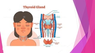
 The thyroid gland is a ductless endocrine gland situated in the anterior/front
portion of the neck.
 It is yellowish brown in color and highly vascular.
 It roughly resembles the shape of a butterfly. It is also one of the largest
endocrine glands, weighing an average of 25 – 30 g.
 This gland has two lobes on either side of the trachea, with each lobe
measuring 4 – 6 cm in length and 1.3 – 1.8 cm in width.
 The primary function of the thyroid gland is to secrete two hormones, namely,
Triiodothyronine (T3) hormone and the Thyroxine hormone (T4).
 Both T3 and T4 hormones play a very important role and affect almost every
tissue in the body.
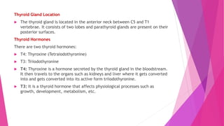
Thyroid Gland Location
 The thyroid gland is located in the anterior neck between C5 and T1
vertebrae. It consists of two lobes and parathyroid glands are present on their
posterior surfaces.
Thyroid Hormones
There are two thyroid hormones:
 T4: Thyroxine (Tetraiodothyronine)
 T3: Triiodothyronine
 T4: Thyroxine is a hormone secreted by the thyroid gland in the bloodstream.
It then travels to the organs such as kidneys and liver where it gets converted
into and gets converted into its active form triiodothyronine.
 T3: It is a thyroid hormone that affects physiological processes such as
growth, development, metabolism, etc.
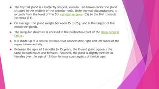
 The thyroid gland is a butterfly shaped, vascular, red-brown endocrine gland
situated in the midline of the anterior neck. Under normal circumstances, it
extends from the level of the 5th cervical vertebra (C5) to the first thoracic
vertebra (T1).
 On average, the gland weighs between 15 to 25 g, and is the largest of the
endocrine glands.
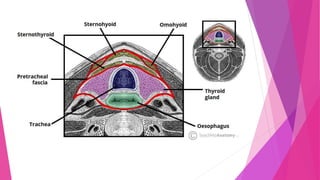
 The irregular structure is encased in the pretracheal part of the deep cervical
fascia .
 It is made up of a central isthmus that connects the right and left lobes of the
organ inferomedially.
 Between the ages of 8 months to 15 years, the thyroid gland appears the
same in both males and females. However, the gland is slightly heavier in
females over the age of 15 than in male counterparts of similar age.
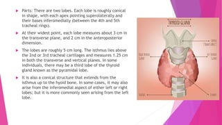
 Parts: There are two lobes. Each lobe is roughly conical
in shape, with each apex pointing superolaterally and
their bases inferomedially (between the 4th and 5th
tracheal rings).
 At their widest point, each lobe measures about 3 cm in
the transverse plane, and 2 cm in the anteroposterior
dimension.
 The lobes are roughly 5 cm long. The isthmus lies above
the 2nd or 3rd tracheal cartilages and measures 1.25 cm
in both the transverse and vertical planes. In some
individuals, there may be a third lobe of the thyroid
gland known as the pyramidal lobe.
 It is also a conical structure that extends from the
isthmus up to the hyoid bone. In some cases, it may also
arise from the inferomedial aspect of either left or right
lobes; but it is more commonly seen arising from the left
lobe.
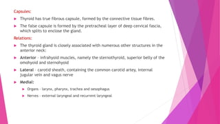
Capsules:
 Thyroid has true fibrous capsule, formed by the connective tissue fibres.
 The false capsule is formed by the pretracheal layer of deep cervical fascia,
which splits to enclose the gland.
Relations:
 The thyroid gland is closely associated with numerous other structures in the
anterior neck:
 Anterior – infrahyoid muscles, namely the sternothyroid, superior belly of the
omohyoid and sternohyoid
 Lateral – carotid sheath, containing the common carotid artey, internal
jugular vein and vagus nerve
 Medial:
 Organs – larynx, pharynx, trachea and oesophagus
 Nerves – external laryngeal and recurrent laryngeal
Vasculature
 The thyroid gland secretes hormones directly into the circulation and is highly
vascularised.
Arterial Supply
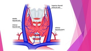
 The arterial supply to the thyroid gland is via two main arteries:
 Superior thyroid artery – arises as the first branch of the external carotid artery. It lies
in close proximity to the external branch of the superior laryngeal nerve (innervates the
larynx).
 Inferior thyroid artery – arises from the thyrocervical trunk (a branch of the subclavian
artery). It lies in close proximity to the recurrent laryngeal nerve (innervates the
larynx).
 In a small proportion of people (around 10%) there is an additional artery present –
the thyroid ima artery. It arises from the brachiocephalic trunk and supplies the
anterior surface and isthmus of the thyroid gland.
Venous Drainage
 Venous drainage is carried by the superior, middle, and inferior thyroid veins, which
form a venous plexus around the thyroid gland.
 The superior and middle veins drain into the internal jugular vein and the inferior
empties into the brachiocephalic vein.
Lymphatic Drainage
 The lymphatic drainage of the thyroid is to the paratracheal and deep cervical nodes.
Functions of Thyroid Gland
Hormones of Thyroid Gland
The thyroid gland secretes three different hormones; triiodothyronine, thyroxine, and
calcitonin.
1. Thyroid Hormone (thyroxine and triiodothyronine)
 Thyroid hormone consists of two iodine-containing hormones, thyroxine and
triiodothyronine.
 Thyroxine is also known as T4 as it contains four atoms of iodine, whereas triiodothyronine
is also called T3 as it contains three atoms of iodine.
 The follicular cells of the thyroid gland produce thyroxine as the major hormone, which is
then converted to T3.
 Both thyroxine and triiodothyronine are composed of two tyrosine amino acid units linked
together with iodine atoms. The number of iodine atoms differs between the two
hormones.
 The primary function or role of thyroid hormone is to increase the basal metabolic rate and
heat production via glucose oxidation.
 Besides, it is also necessary for tissue growth and development, especially in the case of
skeletal and nerve tissues.
 The level of thyroid hormone in the blood is regulated by the pituitary gland by the release
of thyroid-stimulating hormone. The regulation and control of the release of thyroid
hormone work by a negative feedback mechanism.
2. Calcitonin
 Calcitonin is secreted by the parafollicular cells or C cells of the thyroid gland
as a response to an increased level of calcium in the blood.
 However, the hormone is not given as much importance as the increased level
of calcium in the blood doesn’t have a physiological effect on the body.
 Calcitonin is given to patients suffering from osteoporosis as it has a bone-
sparing effect.
 Calcitonin acts on bones where it inhibits osteoclastic activity, decreasing the
release of calcium into the blood. It also stimulates the uptake of calcium
from the blood into the bone matrix.
Functions of Thyroid Gland
 The following are some of the functions of the thyroid gland:
 The most important function of the thyroid gland is to produce thyroid
hormones that are essential for metabolic activities and growth.
 The thyroid hormone regulates the basal metabolic rate in the body by
influencing glucose, fat, and protein metabolism.
 The hormones are composed of iodine atoms; thus, these also play an
essential role in iodine metabolism in the body.
 Calcitonin produced by the C cells of the thyroid gland regulates the levels of
calcium ions in the blood.
 The thyroid hormone plays a crucial role in the growth and development of
the target organs in the body like the brain and kidneys.
Clinical applications
 Diseases and Disorders of Thyroid Gland. The disorders and diseases associated
with the thyroid gland are usually due to the hypersecretion or hyposecretion of the
hormones.
The following are some of the disorders associated with the thyroid gland and its
secretions;
1. Grave’s disease
 Grave’s disease or Grave’s thyroiditis is a condition caused due to the
hypersecretion of the thyroid hormone.
 The condition is more common in women than in men and can occur at any age but
is common among individuals between 30-50 years.
 Grave’s disease is an autoimmune condition in which the abnormal antibodies of the
body are directed against the thyroid follicular cells.
 These unusual antibodies mimic the thyroid-stimulating hormone of the pituitary
gland and stimulate the thyroid gland to produce the hormone.
 Some of the common symptoms associated with this condition are nervousness,
elevated metabolism, sweating, weight loss, etc.
2. Simple goiter
 Simple goiter is a condition caused by the hyposecretion of the thyroid hormone as
a result of enlargement of the gland.
 Goiter is characterized by the formation of extra thyroid tissue, which causes the
enlargement of the gland.
 It is caused due to persistent iodine deficiency, which causes a relative lack of T3
and T4 in the body.
 In some cases, the increased size of the gland can cause damage to the adjacent
tissues like the esophagus and nerves.
3. Cretinism
 Cretinism is caused due to the hyposecretion of thyroid hormone in children.
 Clinical features of the condition are observed in the form of mental retardedness
and a disproportionately sized body.
 The effect of the condition depends on the age and physiological activity of the
individual, but it is usually more severe in children than in adults.
 Cretinism might be caused due to genetic deficiency of the thyroid gland or
maternal factors like the lack of iodine.
4. Thyroid tumor
 Malignant tumors of the thyroid gland are rare, but benign tumors, including single
adenomas, are fairly common.
 However, in the case of older adults, the tumor might become malignant.
 Even with benign tumors, some might produce a large amount of hormones,
resulting in hyperthyroidism.
5. Hyperthyroidism
 This condition is caused when thyroid glands excessively produce a hormone called
thyroxine.
 The symptoms include a change in appetite, unexpected weight loss, insomnia,
fatigue, irritability, frequent urination, increased sweating and heat intolerance.
But this condition usually resolves within a few months of relevant treatments and
proper medication.
6. Hypothyroidism
 This condition is caused by the under-secretion of the thyroid hormones. It is a
very common problem and often stays undetected for years.
 One of the main reasons that trigger hypothyroidism is an autoimmune disorder
called Hashimoto’s disease. In this condition, the antibodies produced by the body
attack the thyroid gland and this results in the glands producing fewer hormones.
The parathyroid Gland
 The parathyroid glands are small endocrine glands situated just below the
thyroid glands in the anterior neck.
 They are usually four in number, two behind each thyroid gland. They are
very small, pea-sized and weigh about 50 mg.
 They are responsible for the production of parathyroid hormone (PTH),
which acts to increase the level of serum calcium.
 The glands function to maintain the calcium and phosphorus levels in our
bodies.
 Until the 20th century, scientists failed to recognise a gland separate from the
thyroid glands, because of the small size of the parathyroid glands. Now, the
two glands can be differentiated easily as the thyroid glands have a follicular
structure, and the cells of a parathyroid gland are densely packed.
Anatomical Location
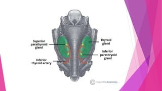
 The parathyroid glands are usually located on the posterior aspect of the thyroid
gland. They are flattened and oval in shape – situated external to the thyroid
gland itself but within the pretracheal fascia.
 Most individuals have four parathyroid glands, although variation in number
(from two to six) is common:
 Superior parathyroid glands (x2) – derived from the fourth pharyngeal pouch.
They are located at the middle of the posterior border of each thyroid lobe,
approximately 1cm superior to the entry of the inferior thyroid artery into the
thyroid gland.
 Inferior parathyroid glands (x2) – derived from the third pharyngeal pouch.
Although inconsistent in location between individuals, the inferior parathyroid
glands are usually found near the inferior poles of the thyroid gland.
 In a small number of people, the inferior parathyroid glands can be found as far
inferiorly as the superior mediastinum.
Vasculature
 The vascular supply is similar to that of the thyroid gland.
 Arterial supply is chiefly via the inferior thyroid artery (as this artery
supplies the posterior aspect of the thyroid gland – where the parathyroids
are located).
 Collateral arterial supply is from the superior thyroid artery and thyroid ima
artery.
 Venous drainage is into the superior, middle, and inferior thyroid veins.
Lymphatics
 The lymphatic drainage from the parathyroid glands is to
the paratracheal and deep cervical nodes.
Nerves
 The parathyroid glands have an extensive supply of sympathetic nerves
derived from thyroid branches of the cervical ganglia.
 Note: these nerves are vasomotor, not secretomotor – endocrine secretion of
parathyroid hormone is under hormonal control.
Functions of the Parathyroid Gland
 The parathyroid gland releases a parathyroid hormone (PTH). Also known as the
parathormone or parathyrine, it is a small peptide that maintains the homeostasis of
calcium and phosphate and also aids in bone physiology. Let us see how this hormone
functions.
 Calcium: The parathyroid hormone functions to release calcium from the bones by
stimulating the osteoblasts. It facilitates gastrointestinal calcium absorption by
activating Vitamin D. It also promotes calcium reabsorption in the kidneys. Normal
calcium levels in the body support strong nervous and muscular systems.
 Phosphorus: It inhibits the absorption of phosphorus in the kidneys, however, the
hormone increases the absorption of phosphorus in the gastrointestinal tract by
activating Vitamin D.
 Activation of Vitamin D: The PTH also helps in the activation of Vitamin D by
upregulating the enzyme 1-α-hydroxylase.
Disorders of the Parathyroid Gland
 Hyperparathyroidism: This condition is developed when an individual
produces too much of the PTH hormone. Overproduction of the hormone
releases large amounts of calcium, which causes hypercalcemia. The
symptoms include extreme thirst, more urine production, abdominal pain and
mood changes.
 Undetected high levels of calcium lead to bone thinning or osteoporosis and
can also cause stones in the kidneys.
 Hypoparathyroidism: Low levels of PTH hormone causes hypoparathyroidism
and hypocalcemia. The symptoms include tingling sensations, muscle cramps
and spasms.
The Adrenal Gland
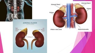
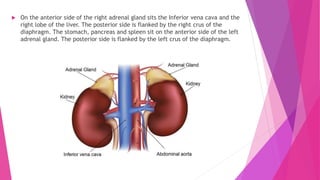
 The adrenal (or suprarenal) glands are paired endocrine glands situated over
the medial aspect of the upper poles of each kidney. ven the name “Adrenal”
directly refers to their location: (Latin: ad– “near” and renes – “kidneys.”)
 They secrete steroid and catecholamine hormones directly into the blood.
Anatomical Location and Relations
 The adrenal glands are located in the posterior abdomen, between the
superomedial kidney and the diaphragm. They are retroperitoneal, with
parietal peritoneum covering their anterior surface only.
 The right gland is pyramidal in shape, contrasting with the semi-lunar shape
of the left gland.
 Perinephric (or renal) fascia encloses the adrenal glands and the kidneys.
This fascia attaches the glands to the crura of the diaphragm. They are
separated from the kidneys by the perirenal fat.
 The adrenal glands sit in close proximity to many other structures in the
abdomen:
 On the anterior side of the right adrenal gland sits the Inferior vena cava and the
right lobe of the liver. The posterior side is flanked by the right crus of the
diaphragm. The stomach, pancreas and spleen sit on the anterior side of the left
adrenal gland. The posterior side is flanked by the left crus of the diaphragm.
Adrenal Gland Anatomy
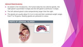
 As stated in the introduction, the human body has two adrenal glands, the
right gland is pyramidal in shape and the left gland is semilunar in shape.
 The left adrenal gland is also comparatively larger than the right.
 The glands are usually about 5×3 cm in size and their combined weight ranges
from 7 to 10 grams. Healthy glands are yellowish in colour.
There are three distinct layers of the adrenal glands.
Adrenal Capsule
 The capsule is a protective layer of fat that surrounds each adrenal glands. They are
not strictly a part of the adrenal glands, but the primary function of this capsule
layer is to enclose and protect each of the adrenal glands.
Adrenal Cortex
 The adrenal cortex is the outermost layer of the adrenal gland which is devoted to
the production of aldosterone, cortisol, and androgens hormones. There are three
layers of cortex called zones which can be examined and easily differentiated under
a microscope:
 Zona glomerulosa – It secretes mineralocorticoids like aldosterone
 Zona fasciculata – It is responsible for producing corticosteroids like cortisol. Also
secrets small quantities of androgens (such as testosterone and androstenedione)
 Zona reticularis – It produces DHES or dehydroepiandrosterone (also called
androstenolone)
Adrenal Medulla
 The last and innermost part of the adrenal gland is the medulla. It contains the
Chromaffin cells, which produce the body’s main source of catecholamines (such as
adrenaline and noradrenaline) and endorphins. These are stored and released in
response to stress.
Adrenal Gland Function
 One of the most well-known responses – the Fight or Flight Response is
triggered by the release of stress hormones from the adrenal glands.
 The adrenal glands produce a variety of hormones. These hormones are very
crucial for the normal functioning of the body. For instance, the glands
secrete cortisol, which has anti-inflammatory properties and aids the immune
system.
 The adrenal gland also helps to regulate metabolism and blood pressure
through various other hormones.
Adrenal Gland Hormones
 Epinephrine: Also called adrenaline, this hormone rapidly responds to stress
by increasing the heart rate and raising blood glucose levels in the blood.
 Norepinephrine: Also called noradrenaline, this hormone works with
epinephrine in reacting to stress. Its primary function is to mobilize the body
and brain for action.
 Hydrocortisone: It is commonly known as cortisol or a steroid hormone. It is
involved in regulating body functions like the conversion of fats, and
carbohydrates to energy and also plays a vital role in other metabolic
processes.
 Corticosterone: This hormone works with hydrocortisone to control the
immune response and prevents inflammatory reactions.
Adrenal Gland Disorders
 Adrenal Gland disorders appear when not enough hormones or inadequate
hormones are produced by the adrenal glands. Even abnormal growths or tumours
can cause certain illness.
 Cushing’s Syndrome is a condition where the cortisol levels in the body are very
high. The cause can be a tumour in the adrenal gland or the pituitary gland.
 Adrenocortical carcinoma is a cancerous tumour that usually develops in the
outer layer of the adrenal gland. This type of tumour is typically found years after
they have spread to other organs in the body.
 Congenital Adrenal Hyperplasia (CAH) is a genetic disorder characterized by very
low levels of cortisol production. The people inflicted with this condition may also
have other hormonal imbalances where their bodies may make very little
aldosterone, but too much androgen.
 Addison’s Disease is caused when adrenal glands produce insufficient cortisol or
aldosterone. It results in weakness, fatigue, low blood pressure, nausea, etc. In
70% of cases, Addisone disease is caused due to autoimmune disorder, where the
body mistakenly attacks the adrenal glands. As a result, the adrenal gland does
not produce the hormone aldosterone in sufficient quantities.
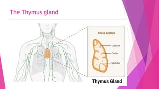
The Thymus gland
The Thymus gland
 The thymus gland is one of the key organs in the lymphatic system, which
occurs in the mediastinum below the sternum.
 The thymus extends from the lower edge of the thyroid gland to the fourth
intercostal space of the vertebrae.
 The thymus gland is distinct from other endocrine glands in that it is active
only before puberty. The gland grows the largest at puberty, after which the
gland is slowly replaced by adipose tissue.
 The thymus gland is a critical organ of the immune system as it serves as a
defense mechanism against different pathogens, tumors, and antigens.
 The thymus is particularly important for the thymus-dependent or adaptive
arm of the immune system as the thymus is essential for the development and
activation of immune cells.
 The term thymus is taken from Greek literature, where it means ‘soul’ to
indicate that the soul resides in the thymus.
 In addition to the secretory cells of the thymus, it also contains other immune
cells like macrophages, neutrophils, and dendritic cells.
 The proliferation of the secretory cells and the regulation of their secretions
is maintained by cytokines like tumor necrosis factor (TNF) and interferon.
 The structure and function of the gland can be influenced by autoimmune
diseases like myasthenia gravis.
Structure of Thymus Gland
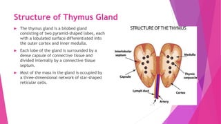
 The thymus gland is a bilobed gland
consisting of two pyramid-shaped lobes, each
with a lobulated surface differentiated into
the outer cortex and inner medulla.
 Each lobe of the gland is surrounded by a
dense capsule of connective tissue and
divided internally by a connective tissue
septum.
 Most of the mass in the gland is occupied by
a three-dimensional network of star-shaped
reticular cells.
 The epithelial cells of the thymus gland are divided into four distinct subtypes
depending on different factors like antigenic expression, ultrastructure, and
their capacity to produce thymic hormones.
 The subtypes are subcapsular cortical, inner cortical, medullary, and Hassall’s
corpuscles.
 The outer cortex of the gland contains loosely packed lymphocytes, and the
medulla contains reticulocytes rich in the cytoplasm.
 The structure also contains small bodies called Hassall’s concentric corpuscles
or Hassall’s bodies, or thymic corpuscles, which are concentric arrays of
squamous cells.
 The size of the thymus is large in infants and young children, with the largest
size at puberty. The gland then slowly begins to coalesce after puberty to be
replaced by adipose tissue.
 The blood supply to the thymus gland is provided by the inferior thyroid,
internal thyroid, and intercostals arteries.
 The gland is attached to the sternum by sternohyoid and sternothyroid
muscles, both of which are bilateral.
Hormones of Thymus Gland
 The thymus gland produces three different hormones; thymosin, thymopoietin, and the
serum thymic factor. The thymic hormones do not act on the T cells identically. Their
mode of action at the cellular level is based on the binding of the hormones to the
specific cell receptors and the interaction with adenyl cyclase.
1. Thymosin
 Thymosin is produced by the epithelial cells of the cortex and medulla, and it is the
primary hormone secreted by the gland.
 The most important function of thymosin is to induce T cell differentiation and enhance
the immunological function of different immune cells.
 Thymosin has also been associated with increasing the phenotypic markers in
lymphocytes.
 Thymosin is a protein that is heat stable up to 80°C temperature and might contain a
small amount of carbohydrates.
2. Thymopoietin
 Thymopoietin is a polypeptide hormone of the thymus that has neuromuscular functions
than immunological ones.
 However, the increased levels of the hormone are known to induce T cell activation and
differentiation as well.
Functions of Thymus Gland
The following are some of the functions of the thymus gland;
 The most important function of the thymus gland is to induce the
development, activation, and differentiation of T cells so that they can
perform their function as mediators of cellular immunity.
 The thymic hormones, thymosin, and thymopoietin stimulate the
prothymocytes to become thymocytes and T lymphocytes.
 The gland also induces the release of cytokines that are essential for
controlling the development of T cells through different stages.
 The thymus is a part of fetal immunity by the 12th week of pregnancy and
remains active through fetal life as well as childhood.
 As an endocrine gland, the thymus is known to produce human growth
hormone, which is essential for the growth and development of the body.
Diseases and Disorders of Thymus Gland
1. Thymic cysts
 Congenital thymic cysts might be present within the thymus gland, resulting in the inhibition of
proliferation of the thymocytes.
 These cysts can be removed and are usually benign. The individual with thymic cysts shows symptoms
like cough and upper respiratory tract infections as the immune system of the individual is affected.
2. Involution
 Thymic involution is common in infants that are exposed to malnutrition, neglect, and abuse.
 Besides, it can also occur after chemotherapy, radiation, and steroid therapy as the thymus is sensitive
to stress.
 Involution of the thymus is much rare in neonates, infants of early childhood.
 Premature involution can be taken as a symptom of social problems like abuse.
3. Hypoplasia
 Hypoplasia is the decreased number of cells in an organ, usually during neonatal conditions.
 Hypoplasia of the thymus occurs due to the development defect of the third and fourth pharyngeal
pouches.
 It has been assumed that alcohol or organic acids tend to injure the neural crest that disturbs the
differentiation of the pharyngeal pouch.
 The clinical manifestation of the condition depends on the extent of hypoplasia, and the amount of
genetic material defected.
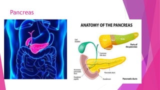
Pancreas
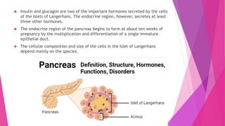
 The pancreas is a complex organ consisting of both exocrine and endocrine
areas where the endocrine part is composed of discrete islets of Langerhans
that secrete numerous hormones.
 The studies related to the Islets of Langerhans have increased over the years
due to the involvement of the cells in diabetes.
 The islets composed of a few thousand endocrine cells account for about 2%
of the total pancreatic mass.
 The primary function of the endocrine region of the pancreas is to assist
digestion by the release of hormones that help in the metabolism of different
biomolecules.
 The pancreas is located behind the stomach as a soft tadpole-shaped organ. It
has been believed that the pancreas developed as an out pocketing of the
epithelial lining of the gastrointestinal tract.
 Insulin and glucagon are two of the important hormones secreted by the cells
of the Islets of Langerhans. The endocrine region, however, secretes at least
three other hormones.
 The endocrine region of the pancreas begins to form at about ten weeks of
pregnancy by the multiplication and differentiation of a single immature
epithelial duct.
 The cellular composition and size of the cells in the Islet of Langerhans
depend mainly on the species.
Structure of Pancreas
 The proportion of Islet of Langerhans in the pancreas is usually about 2%, with the
total number of islets in humans ranging between 3.2 to 14.8 million.
 The cellular composition of the islets differs in different species, and it has been
studied that there are functional differences in islets of different species as well.
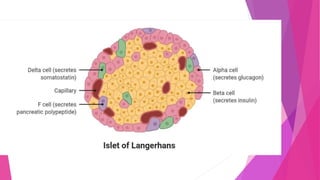
 The Islets of Langerhans in humans consist of four different types of cells; α cells, β
cells, δ cells, and γ or PP (pancreatic polypeptide-producing) cells.
 These four different types of cells of the islets are arranged randomly through the
gland.
 The size of the islets in the pancreas indicates variation as most islets follow the
mantle-core pattern.
 The β cells of the pancreas account for about 60% of the total islet mass, followed by α
cells that occupy 30% of the mass. The remaining 10% is covered by the δ and PP cells.
 The blood and nerve supply to the gland is facilitated by a neuromuscular bundle that
penetrates the central core of β cells.
 The four different cells of the islets secrete four different hormones that influence the
metabolism of the body.
Hormones of Pancreas
There are four different types of cells found in the pancreas that secrete four different
hormones (insulin, glucagon, somatostatin, and pancreatic polypeptide).
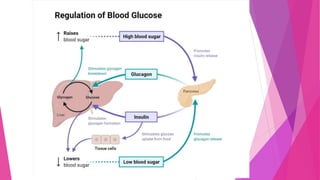
 Insulin
 Insulin is secreted by the β cells of the Islets of Langerhans as a polypeptide consisting
of about 50 amino acids.
 The primary function of insulin is to lower the blood nutrient level, mostly of glucose,
but it might include amino acids and fatty acids.
 The effect of insulin is defined as anabolic as it tends to facilitate the storage of
nutrients in the body.
 Insulin is synthesized as a part of a larger polypeptide unit called proinsulin which
then undergoes clipping to release the insulin.
 Insulin works by one of the three mechanisms; it enhances the transport of glucose
into different cells of the body and also inhibits the breakdown of glycogen into
glucose. It also inhibits the interconversion of amino acids or fats into glucose.
Glucagon
 Glucagon is secreted by the α cells of the pancreas, which works by the
opposite mechanism of insulin.
 Glucagon is a polypeptide consisting of 29 amino acids linked together to form
a longer polypeptide.
 Glucagon is an extremely potent hyperglycemic agent that causes the release
of glucose into the blood.
 The target organ of the glucagon hormone is the liver where it promotes the
breakdown of glycogen into glucose so as it increases the blood glucose level.
 The hormone also activates the synthesis of glucose from lactic acid and
noncarbohydrate molecules.
 The increase in blood glucose level results in a secondary effect of decreased
amino acid levels in the blood.
 The release of glucagon by the α cells is stimulated by falling blood glucose
levels via sympathetic nervous system stimulation as well as rising amino
acids levels
Functions of Pancreas
The following are some of the functions of the pancreatic gland;
 The endocrine part of the gland release different hormones that work to
maintain the blood nutrient level in the body.
 Insulin is secreted by the α cells of the islets of Langerhans that is responsible
for decreasing the blood glucose level in humans.
 Pancreas also secretes glucagon, which works to increase the blood glucose
levels if the levels go down beyond normal.
 The δ cells of the Islets of Langerhans release the somatostatin hormone,
which inhibits the secretion of both insulin and glucagon while inhibiting the
secretion of growth hormone by the pituitary.
Diseases and Disorders of Pancreas
 The following is one of the disorders and diseases associated with the pancreas;
Diabetes Mellitus
 Diabetes mellitus is a condition resulting from either the hyposecretion or hypoactivity
of insulin.
 Diabetes mellitus is of two types; type 1 diabetes mellitus results from the
hyposecretion of insulin, whereas type 2 diabetes mellitus results from the hypoactivity
of released insulin.
 The lack of insulin in the body results in increased blood glucose levels which then
causes excess loss of glucose via urine.
 Type 1 diabetes mellitus is common among children and is characterized by the
immediate onset of symptoms.
 Type 2 diabetes mellitus, in turn, occurs more in adults and often results in late-onset of
symptoms.
 Diabetes mellitus can be life-threatening in some conditions, resulting in kidney failure
and other serious conditions. In most cases, however, the condition is chronic and
doesn’t result in death.
 The treatment of diabetes mellitus is performed in the form of insulin shots which helps
maintain the level of insulin in the body.
Interstitial cells of testes
 The testes secrete various hormones that are involved in male reproduction and
development
 The testes secretes several male sex hormones, which are collectively called androgens, which
include testosterone, dihydro testosterone and androstenedione.
 Testosterone is much more abundent and potent than the others.
 Testosterone is formed by the interstititial cells of Leydig, which lie in the interstices between
the seminiferous tubules.
Testosterone plays multiple important roles in the body, such as:
 development of the penis and testes
 deepening of the voice during puberty
 appearance of facial and pubic hair starting at puberty; later in life, it may play a role
in balding
 muscle size and strength
 bone growth and strength
 sex drive (libido)
 sperm production.
Ovarian hormones
 The main function of the ovaries, maturation and release of oocytes, is accomplished
via the production of several steroidal and nonsteroidal hormones.
 The ovary contains multiple distinctive steroid-producing cells, including stromal cells,
theca cells, granulosa cells, and luteinized granulosa cells. Each cell type contains all
the enzymes necessary for synthesis of androgens, estrogens, and progesterone.
 Estrogens are essential in the development and maintenance ofthe female phenotype,
germ cell maturation, and pregnancy. In addition to their reproductive effects,
estrogens also have many other nonreproductive systemic effects, such as bone
metabolism/remodeling, nervous system maturation, and endothelial responsiveness.
 Progesterone plays a critical role in reproduction. It inhibits further endometrial
proliferation mediated by estrogen and converts the endometrium into a secretory
type, preparing it not only to receive the blastocyst for implantation, but also to
maintain the pregnancy. It inhibits uterine contractions and increases the viscosity of
cervical mucus. Progesterone also inhibits the action of prolactin so that lactation
occurs only after delivery.
 In women, androgens originate as 19-carbon steroids from the adrenals and ovaries.
The major androgens produced in the ovary, primarily by the thecal cells. However,
these biologically inactive androgens are converted by extraglandular metabolism to
biologically active androgens such as testosterone and dihydrotestosterone (DHT).
Placenta
 Placenta refers to the temporary vascular organ found in mammals, which
attaches the fetus to the uterus of the mother during pregnancy.
 A vascular structure that develops in the uterus of mammalian females and acts as
the organ of attachment between the embryo and uterine wall is called placenta.
 In appearance, the placenta is disk-shaped and measures up to 22 cm in length.
The placenta is also rich in blood vessels.
 Functions of the placenta:
(a) It provides nourishment to the developing foetus.
(b) It transfers oxygen, water and hormone from the mother to the developing
foetus.
(c) It removes waste products from the blood of foetus.
(d) It provides protection to the foetus from microorganisms as they cannot pass
through the placenta.
(e) It produces hormones which are necessary to maintain pregnancy, e.g.
oestrogen, progesterone and relaxin.
THANK YOU