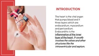
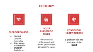
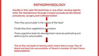
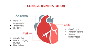
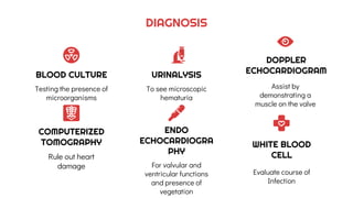
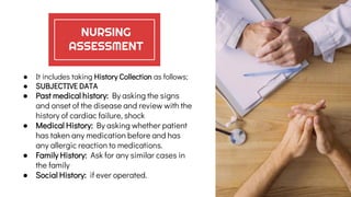
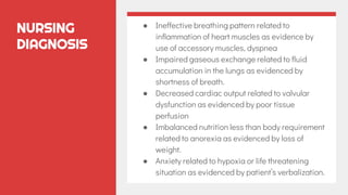
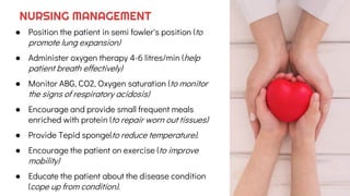
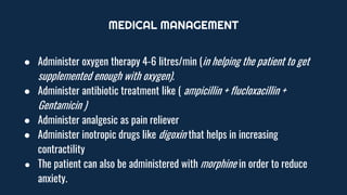
NURSING
MANAGEMENT
FOR PATIENTS ENDOCARDITIS
By
S.Vishali
CONTENTS
Definition
Of Endocarditis
01
Pathophysiology
and Clinical
Manifestation
Of Endocarditis
03
Nursing
Management
For Patients with
Endocarditis
05
Etiology and Risk
Factor
Of Endocarditis
02
Diagnostic
Assessment
Of Endocarditis
04
ENDOCARDITIS
01
Endo+Cardio+itis=
Inflammation of Inner Layer of
Heart
INTRODUCTION
The heart is the vital organ
that pumps blood and it
three layers which are
endocardium, myocardium
and pericardium.
Endocarditis is the
inflammation of the inner
layer of the heart. It usually
involves the valves and other
structures like the
intraventricular and septum.
ETIOLOGY
MICROORGANISM
● FUNGUS:
Candida,
Aspergillus
● Gram -ve org:
Pseudomonas
● BACTERIA:
Staphylococci
ACUTE
RHEUMATIC
FEVER
Which causes
enlargement and
tender lymph nodes,
damages the valves
CONGENITAL
HEART DISEASE
a problem with the
structure of the
heart
Some of main risk factors
are:
● Previous Heart Damage
● Dental procedures which lead to
infection
● Heart surgery
● Procedure like enemas, barium,
sigmoidoscopy, catheterisation, and
cystoscopy.
● Reproductive condition like delivery of
new babies, abortions and pelvic
inflammatory disease.
RISK FACTORS
Then they accumulate in the valves of the heart
Finally they form vegetations or crusters
These vegetation lead into damage heart valves by perforating and
deforming the valves leaflets
PATHOPHYSIOLOGY
Usually in this case the bacterias or any other causing agents
enter the bloodstream through invasive procedures like Dental
procedures, surgery and Catheterization
This at the end leads to tearing which means there is poor flow of
blood and lead into accumulation of blood in chamber of heart hence
ENDOCARDITIS
CLINICAL MANIFESTATION
SKIN
● Osler’s node
● Janeway lesions
● Splinter
hemorrhages
COMMON
● Elevated
temperature
● Tachycardia
● Clubbing
CVS
● Arrhythmias
● New or changing
murmur
● Heart failure
DIAGNOSIS
COMPUTERIZED
TOMOGRAPHY
Rule out heart
damage
ENDO
ECHOCARDIOGRA
PHY
For valvular and
ventricular functions
and presence of
vegetation
WHITE BLOOD
CELL
Evaluate course of
Infection
BLOOD CULTURE
Testing the presence of
microorganisms
URINALYSIS
To see microscopic
hematuria
DOPPLER
ECHOCARDIOGRAM
Assist by
demonstrating a
muscle on the valve
NURSING
ASSESSMENT
● It includes taking History Collection as follows;
● SUBJECTIVE DATA
● Past medical history: By asking the signs
and onset of the disease and review with the
history of cardiac failure, shock
● Medical History: By asking whether patient
has taken any medication before and has
any allergic reaction to medications.
● Family History: Ask for any similar cases in
the family
● Social History: if ever operated.
NURSING ASSESSMENT
● OBJECTIVE DATA
● Assess for temperature
elevation
● Heart murmur
● Evidence of cough
● Peripheral edema and
embolism
● Auscultate for heart sounds
1. Monitor: ABG
2. Rapid pulse rate
3. Dyspnea
4. Restlessness and
5. Manifestation of Heart
Failure.
NURSING
DIAGNOSIS
NURSING
DIAGNOSIS
● Ineffective breathing pattern related to
inflammation of heart muscles as evidence by
use of accessory muscles, dyspnea
● Impaired gaseous exchange related to fluid
accumulation in the lungs as evidenced by
shortness of breath.
● Decreased cardiac output related to valvular
dysfunction as evidenced by poor tissue
perfusion
● Imbalanced nutrition less than body requirement
related to anorexia as evidenced by loss of
weight.
● Anxiety related to hypoxia or life threatening
situation as evidenced by patient’s verbalization.
● Position the patient in semi fowler's position (to
promote lung expansion)
● Administer oxygen therapy 4-6 litres/min (help
patient breath effectively)
● Monitor ABG, CO2, Oxygen saturation (to monitor
the signs of respiratory acidosis)
● Encourage and provide small frequent meals
enriched with protein (to repair worn out tissues)
● Provide Tepid sponge(to reduce temperature).
● Encourage the patient on exercise (to improve
mobility)
● Educate the patient about the disease condition
(cope up from condition).
NURSING MANAGEMENT
MEDICAL MANAGEMENT
● Administer oxygen therapy 4-6 litres/min (in helping the patient to get
supplemented enough with oxygen).
● Administer antibiotic treatment like ( ampicillin + flucloxacillin +
Gentamicin )
● Administer analgesic as pain reliever
● Administer inotropic drugs like digoxin that helps in increasing
contractility
● The patient can also be administered with morphine in order to reduce
anxiety.
COMPLICATION
CHRONIC
HEART FAILURE
STROKE
SHOCK
EMBOLISM
Endocarditis as explained
above it is the inflammation
of endocardium and it bring
a lot complications.
CONCLUSIONS
CREDITS: This presentation template was
created by Slidesgo, including icons by Flaticon,
and infographics & images by Freepik.
THANKS!
Reference:
● PAUL O'NEILL (2012), MASTER MEDICINE. 2nd
Edition
● LINTON (2007). INTRO. TO MED-SURG NURSING
4th Edition
● S.Lewis, M.Heitkemper, S.Dirksen, P.O'brien (2007)
Medical Surgical Nursing; Assessment and
management of Clinical Problems 7th ed Mosby
PLEASE KEEP THIS SLIDE FOR ATTRIBUTION

Endocarditis

  • 1.
  • 2.
    CONTENTS Definition Of Endocarditis 01 Pathophysiology and Clinical Manifestation OfEndocarditis 03 Nursing Management For Patients with Endocarditis 05 Etiology and Risk Factor Of Endocarditis 02 Diagnostic Assessment Of Endocarditis 04
  • 3.
  • 4.
    INTRODUCTION The heart isthe vital organ that pumps blood and it three layers which are endocardium, myocardium and pericardium. Endocarditis is the inflammation of the inner layer of the heart. It usually involves the valves and other structures like the intraventricular and septum.
  • 5.
    ETIOLOGY MICROORGANISM ● FUNGUS: Candida, Aspergillus ● Gram-ve org: Pseudomonas ● BACTERIA: Staphylococci ACUTE RHEUMATIC FEVER Which causes enlargement and tender lymph nodes, damages the valves CONGENITAL HEART DISEASE a problem with the structure of the heart
  • 6.
    Some of mainrisk factors are: ● Previous Heart Damage ● Dental procedures which lead to infection ● Heart surgery ● Procedure like enemas, barium, sigmoidoscopy, catheterisation, and cystoscopy. ● Reproductive condition like delivery of new babies, abortions and pelvic inflammatory disease. RISK FACTORS
  • 7.
    Then they accumulatein the valves of the heart Finally they form vegetations or crusters These vegetation lead into damage heart valves by perforating and deforming the valves leaflets PATHOPHYSIOLOGY Usually in this case the bacterias or any other causing agents enter the bloodstream through invasive procedures like Dental procedures, surgery and Catheterization This at the end leads to tearing which means there is poor flow of blood and lead into accumulation of blood in chamber of heart hence ENDOCARDITIS
  • 8.
    CLINICAL MANIFESTATION SKIN ● Osler’snode ● Janeway lesions ● Splinter hemorrhages COMMON ● Elevated temperature ● Tachycardia ● Clubbing CVS ● Arrhythmias ● New or changing murmur ● Heart failure
  • 9.
    DIAGNOSIS COMPUTERIZED TOMOGRAPHY Rule out heart damage ENDO ECHOCARDIOGRA PHY Forvalvular and ventricular functions and presence of vegetation WHITE BLOOD CELL Evaluate course of Infection BLOOD CULTURE Testing the presence of microorganisms URINALYSIS To see microscopic hematuria DOPPLER ECHOCARDIOGRAM Assist by demonstrating a muscle on the valve
  • 10.
    NURSING ASSESSMENT ● It includestaking History Collection as follows; ● SUBJECTIVE DATA ● Past medical history: By asking the signs and onset of the disease and review with the history of cardiac failure, shock ● Medical History: By asking whether patient has taken any medication before and has any allergic reaction to medications. ● Family History: Ask for any similar cases in the family ● Social History: if ever operated.
  • 11.
    NURSING ASSESSMENT ● OBJECTIVEDATA ● Assess for temperature elevation ● Heart murmur ● Evidence of cough ● Peripheral edema and embolism ● Auscultate for heart sounds 1. Monitor: ABG 2. Rapid pulse rate 3. Dyspnea 4. Restlessness and 5. Manifestation of Heart Failure.
  • 12.
  • 13.
    NURSING DIAGNOSIS ● Ineffective breathingpattern related to inflammation of heart muscles as evidence by use of accessory muscles, dyspnea ● Impaired gaseous exchange related to fluid accumulation in the lungs as evidenced by shortness of breath. ● Decreased cardiac output related to valvular dysfunction as evidenced by poor tissue perfusion ● Imbalanced nutrition less than body requirement related to anorexia as evidenced by loss of weight. ● Anxiety related to hypoxia or life threatening situation as evidenced by patient’s verbalization.
  • 14.
    ● Position thepatient in semi fowler's position (to promote lung expansion) ● Administer oxygen therapy 4-6 litres/min (help patient breath effectively) ● Monitor ABG, CO2, Oxygen saturation (to monitor the signs of respiratory acidosis) ● Encourage and provide small frequent meals enriched with protein (to repair worn out tissues) ● Provide Tepid sponge(to reduce temperature). ● Encourage the patient on exercise (to improve mobility) ● Educate the patient about the disease condition (cope up from condition). NURSING MANAGEMENT
  • 15.
    MEDICAL MANAGEMENT ● Administeroxygen therapy 4-6 litres/min (in helping the patient to get supplemented enough with oxygen). ● Administer antibiotic treatment like ( ampicillin + flucloxacillin + Gentamicin ) ● Administer analgesic as pain reliever ● Administer inotropic drugs like digoxin that helps in increasing contractility ● The patient can also be administered with morphine in order to reduce anxiety.
  • 16.
  • 17.
    Endocarditis as explained aboveit is the inflammation of endocardium and it bring a lot complications. CONCLUSIONS
  • 18.
    CREDITS: This presentationtemplate was created by Slidesgo, including icons by Flaticon, and infographics & images by Freepik. THANKS! Reference: ● PAUL O'NEILL (2012), MASTER MEDICINE. 2nd Edition ● LINTON (2007). INTRO. TO MED-SURG NURSING 4th Edition ● S.Lewis, M.Heitkemper, S.Dirksen, P.O'brien (2007) Medical Surgical Nursing; Assessment and management of Clinical Problems 7th ed Mosby PLEASE KEEP THIS SLIDE FOR ATTRIBUTION