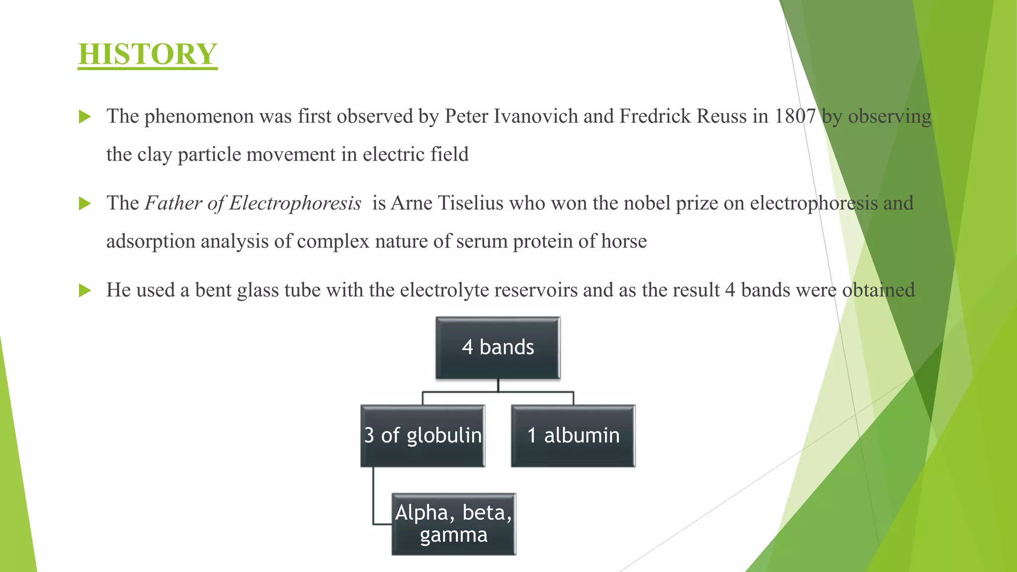
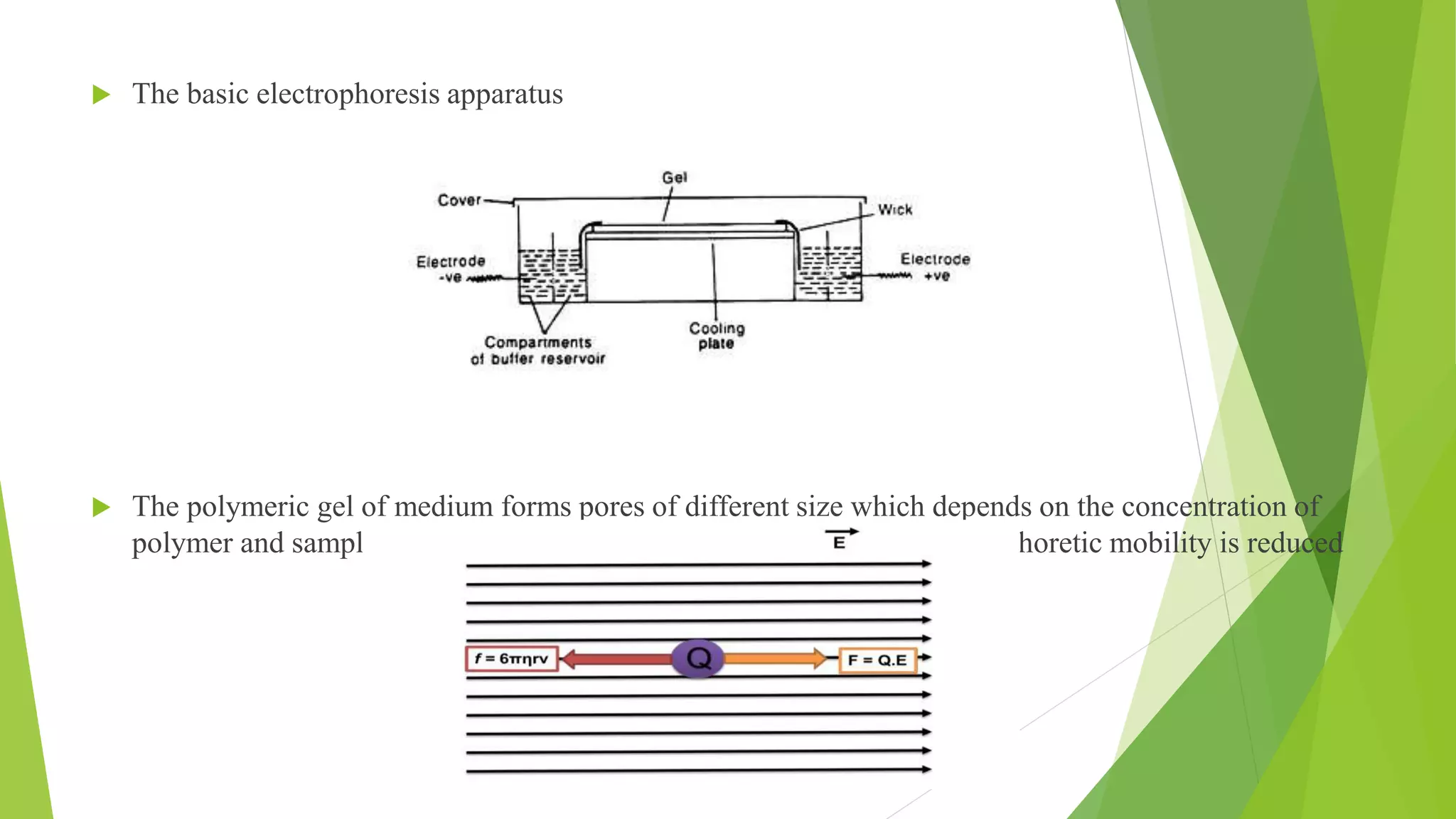
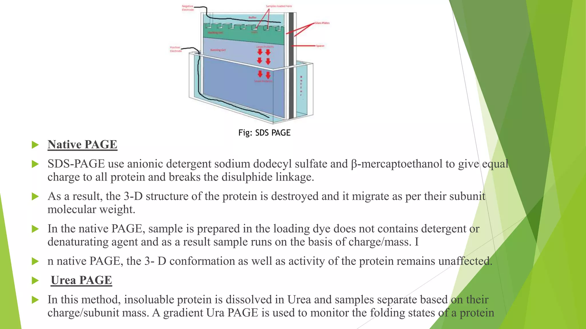
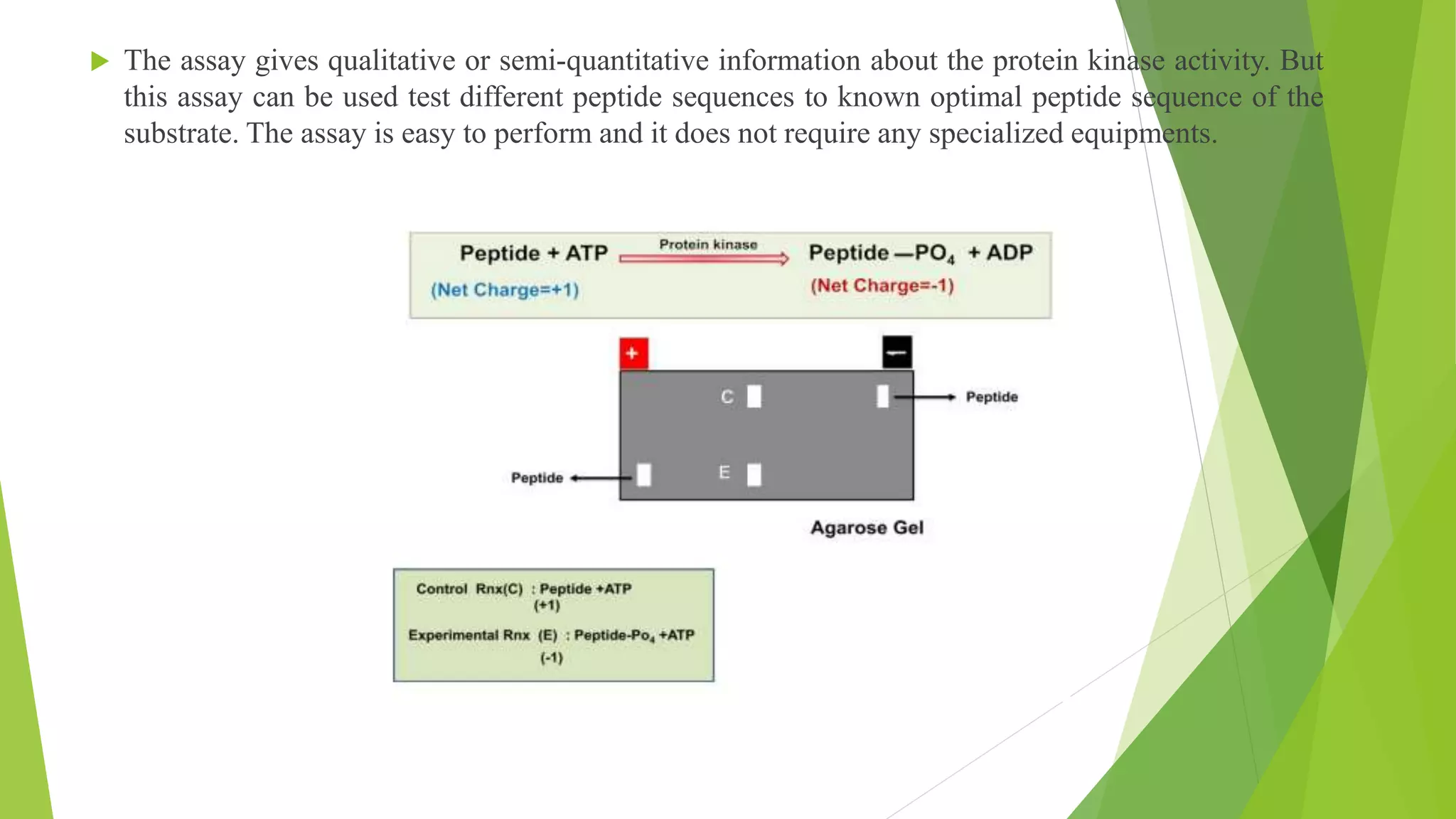
ELECTROPHORESIS
BY- MAHEK SHARAN
BY- MAHEK SHARAN
INT. MTECH BIOTECH
STUDENT
India.
ELECTROPHORESIS
 Topics discussed
 Introduction
 History
 Applications
 Principle
 Types of support medium
 Types of electrophoresis
INTRODUCTION
 The separation technique which comprise of migration of the charged particles or
molecules in a medium under the influence of uniform applied electric field
 The cataphoresis is the electrophoresis of the positively charged particles whereas
anaphoresis is the electrophoresis of the negatively charged particle
 Its is a simple, highly sensitive and rapid technique
 “Electrophoresis” literally means running in the electric field.
 The charged molecule moves to their counter charge electrodes but electric field is
removed before it reaches the electrode.
 Movement of charged species in an electric field gives differential mobility to the sample
based on the charge and consequently resolve them.
HISTORY
 The phenomenon was first observed by Peter Ivanovich and Fredrick Reuss in 1807 by observing
the clay particle movement in electric field
 The Father of Electrophoresis is Arne Tiselius who won the nobel prize on electrophoresis and
adsorption analysis of complex nature of serum protein of horse
 He used a bent glass tube with the electrolyte reservoirs and as the result 4 bands were obtained
4 bands
3 of globulin
Alpha, beta,
gamma
1 albumin
APPLICATIONS
Analysis of lipoprotein
Electrophoresis of serum protein
Diagnosis of haemoglobin A1c
Protein genotyping
Cerebrospinal fluid analysis
PRINCIPLE
 Many important biological molecules like nucleic acids, nucleotides, amino acids and proteins
possess ionisable groups at specific pH it shows positive and negative charge property
 Charged molecule moves to their counter charge electrodes but electric field is removed
before it reaches
 Movement of charged species in an electric field gives differential mobility to sample which
is based on charge and recover them
 This movement is retarted by adding polymeric gel to retain time
 The basic electrophoresis apparatus
 The polymeric gel of medium forms pores of different size which depends on the concentration of
polymer and sample pass through these pore and as a result their electrophoretic mobility is reduced
 Suppose a charged particle has net charge Q and the external electric field is E, then the force F
responsible for giving electrophoretic mobility,
F= Q.E
The friction forces F which is opposing the movement of the charged particle is as follows
F= ƒ. V
where ƒ is the friction coefficient and the v is the velocity of the electrophoretic mobility.
The movement of a spherical through a liquid medium (gel) of the viscosity η, the friction coefficient
ƒ is given by
ƒ = 6Πηrv
The place where, F=F or QE=6Πηrv The electrophoretic mobility v is given by:
𝒗 = 𝐐 𝟔𝚷𝛈𝐫
The relative mobility, v’ is as follows
𝒗′ = 𝐂𝐡𝐚𝐫𝐠𝐞/ 𝐦𝐚𝐬𝐬
 Properties of medium
inert
uncharged
Pores for movement
No binding to molecule
TYPES OF GELS FOR MEDIUM
 Agarose gel
 Linear polysaccharide
 Basic repeat unit is agarobiose
 Used in concentration 1-3%
 Mixture of polysaccharides from seaweeds
 Gelling property by inter and intramolecular hydrogen bonding
 Polyacrylamide gel
 Cross linked
 Formed by the polymerisation of acrylamide monomer in presence of small amount of N,N-
methylene bisacrylamside by free radical reaction by base TEMED
 Used for isoelectric focusing or SDS
TYPES OF ELECTROPHORESIS
 Moving boundary electrophoresis
 The electrophoresis is carried in solution, without a supporting media.
 The sample is dissolved the buffer and molecules move to their respective counter charge
electrodes and is carried out in a U shape tube with platinum electrodes attached to the end of
both arms
 At the respective ends, tube has refractometer to measure the change in refractive index of the
buffer during electrophoresis due to presence of molecule.
 Charged molecule moves to the opposite electrode as they passes through the refractometer, a
change can be measured.
 Zone electrophoresis
 In this method, an inert polymeric supporting media is used between the electrodes to separate and
analyze the sample.
 The supporting media used in zone electrophoresis are absorbent paper, gel of starch, agar and
polyacrylamide.
 The presence of supporting media minimizes mixing of the sample and that makes the analysis and
purification of the molecule from the gel much easier than the moving boundary electrophoresis.
 The gel electrophoresis is the best example of zone electrophoresis.
 Vertical Gel Electrophoresis
 The electrophoresis in this system performed in a discontinuous way with buffer in the upper and
lower tank connected by the gel slab.
 SDS-Polyacrylamide gel electrophoresis (SDS-PAGE)
 It has two buffer chamber, upper chamber and a lower chamber which are fitted with the platinum
electrodes connected to the external power supply from a power pack which supplies a direct
current or DC voltage.
 The upper and lower tank filled with the running buffer is connected by the electrophoresis gel
casted in between two glass plates (rectangular and notched).
 There are additional accessories needed for casting the polyacrylamide gel such as comb (to
prepare different well), spacer, gel caster etc.
 The acrylamide solution is mixed with the TEMED and APS and poured in between the
glass plate fitted into the gel caster as ammoinum persupfate in the presence of TEMED
forms oxygen free radicals and induces the polymerization of acryalide monomer to
form a linear polymer
 As the samples are filled vertically there is a distance drift between the molecules at
the bottom in a lane and the pH of the stacking gel is 6.8 and at this pH, glycine is
moving slowly in the front where as Tris HCl is moving fast. As a result, the sample gets
sandwiched between glycine-Tris and get stacked in the form of thin band.
 Native PAGE
 SDS-PAGE use anionic detergent sodium dodecyl sulfate and β-mercaptoethanol to give equal
charge to all protein and breaks the disulphide linkage.
 As a result, the 3-D structure of the protein is destroyed and it migrate as per their subunit
molecular weight.
 In the native PAGE, sample is prepared in the loading dye does not contains detergent or
denaturating agent and as a result sample runs on the basis of charge/mass. I
 n native PAGE, the 3- D conformation as well as activity of the protein remains unaffected.
 Urea PAGE
 In this method, insoluable protein is dissolved in Urea and samples separate based on their
charge/subunit mass. A gradient Ura PAGE is used to monitor the folding states of a protein
Fig: SDS PAGE
 Horizontal gel electrophoresis
 The electrophoresis in this gel system is performed in a continuous fashion with both electrodes and
gel cassette submersed within the buffer.
 The electrophoresis chamber has two platinum electrodes placed on the both ends are connected to
the external power supply from a power pack which supplies a direct current or DC voltage.
 The tank filled with the running buffer and the gel casted is submerged inside the buffer. There are
additional accessories needed for casting the agarose gel such as comb, spacer, gel caster etc.
 Hot agarose is poured into the gel cassette and allowed it to set. A comb can be inserted
into the hot agarose to cast the well for loading the sample.

 Electroelution
 Protein band present within the polyacrylamide gel block is removed by electroelution for further
usage
 A gel band is cut from the SDS-PAGE and placed in a dialysis bag and sealed from both ends. The
dialysis bag is chosen so that the molecular weight cut off of dialysis membrane should be lower
than the protein of interest.
 The dialysis bag is placed in the horizontal gel apparatus with buffer and electrophoresis is
performed with a constant voltage. During electrophoresis the protein band migrate and ultimately
comes out from the gel block.
 Due to dialysis bag, salt and other small molecule contaminant moves out of the dialysis bag but
protein remain trapped within the dialysis bag.
 Protein can be recovered from the dialysis bag for further use in downstream processing.
 Southern blotting
 The genomic DNA is digested with the EcoRI or BamHI and the DNA fragments are resolved on
the agarose gel.
 The gel is incubated in an alkaline solution to denature the double stranded DNA to single stranded
form.
 DNA is transferred on the nitrocellulose membrane by capillary action by applying a uniform
pressure either by suction pressure or by placing wet paper towels. The membrane is incubated with
a non-specific DNA such as sonicated calf thymus genomic DNA to block the binding sites on the
membrane.
 A single stranded radioactive probe is added to the membrane and allowed to bind. Membrane is
washed and the blot is developed by autoradiography.
 The DNA fragment complementary to the probe sequence binds the radioactive probe and give
positive signal
• Protein kinase assay
• During a kinase reaction, protein kinase transfers a phosphate group from ATP to the substrate and impart
a net negative charge on the substrate molecule.
• In the protein kinase assay, a peptide with net +1 charge is incubated in assay buffer containing enzyme,
non-radioactive ATP.
• Peptide present in the control reaction has a net +1 charge and it will migrate towards –ve electrode
where as peptide in experimental reaction has a net -1 charge and it will migrate towards +Ve electrode
 The assay gives qualitative or semi-quantitative information about the protein kinase activity. But
this assay can be used test different peptide sequences to known optimal peptide sequence of the
substrate. The assay is easy to perform and it does not require any specialized equipments.
Electrophoresis

Electrophoresis

  • 1.
    ELECTROPHORESIS BY- MAHEK SHARAN BY-MAHEK SHARAN INT. MTECH BIOTECH STUDENT India.
  • 2.
    ELECTROPHORESIS  Topics discussed Introduction  History  Applications  Principle  Types of support medium  Types of electrophoresis
  • 3.
    INTRODUCTION  The separationtechnique which comprise of migration of the charged particles or molecules in a medium under the influence of uniform applied electric field  The cataphoresis is the electrophoresis of the positively charged particles whereas anaphoresis is the electrophoresis of the negatively charged particle  Its is a simple, highly sensitive and rapid technique  “Electrophoresis” literally means running in the electric field.  The charged molecule moves to their counter charge electrodes but electric field is removed before it reaches the electrode.  Movement of charged species in an electric field gives differential mobility to the sample based on the charge and consequently resolve them.
  • 4.
    HISTORY  The phenomenonwas first observed by Peter Ivanovich and Fredrick Reuss in 1807 by observing the clay particle movement in electric field  The Father of Electrophoresis is Arne Tiselius who won the nobel prize on electrophoresis and adsorption analysis of complex nature of serum protein of horse  He used a bent glass tube with the electrolyte reservoirs and as the result 4 bands were obtained 4 bands 3 of globulin Alpha, beta, gamma 1 albumin
  • 5.
    APPLICATIONS Analysis of lipoprotein Electrophoresisof serum protein Diagnosis of haemoglobin A1c Protein genotyping Cerebrospinal fluid analysis
  • 6.
    PRINCIPLE  Many importantbiological molecules like nucleic acids, nucleotides, amino acids and proteins possess ionisable groups at specific pH it shows positive and negative charge property  Charged molecule moves to their counter charge electrodes but electric field is removed before it reaches  Movement of charged species in an electric field gives differential mobility to sample which is based on charge and recover them  This movement is retarted by adding polymeric gel to retain time
  • 7.
     The basicelectrophoresis apparatus  The polymeric gel of medium forms pores of different size which depends on the concentration of polymer and sample pass through these pore and as a result their electrophoretic mobility is reduced
  • 8.
     Suppose acharged particle has net charge Q and the external electric field is E, then the force F responsible for giving electrophoretic mobility, F= Q.E The friction forces F which is opposing the movement of the charged particle is as follows F= ƒ. V where ƒ is the friction coefficient and the v is the velocity of the electrophoretic mobility. The movement of a spherical through a liquid medium (gel) of the viscosity η, the friction coefficient ƒ is given by ƒ = 6Πηrv The place where, F=F or QE=6Πηrv The electrophoretic mobility v is given by: 𝒗 = 𝐐 𝟔𝚷𝛈𝐫 The relative mobility, v’ is as follows 𝒗′ = 𝐂𝐡𝐚𝐫𝐠𝐞/ 𝐦𝐚𝐬𝐬
  • 9.
     Properties ofmedium inert uncharged Pores for movement No binding to molecule
  • 10.
    TYPES OF GELSFOR MEDIUM  Agarose gel  Linear polysaccharide  Basic repeat unit is agarobiose  Used in concentration 1-3%  Mixture of polysaccharides from seaweeds  Gelling property by inter and intramolecular hydrogen bonding  Polyacrylamide gel  Cross linked  Formed by the polymerisation of acrylamide monomer in presence of small amount of N,N- methylene bisacrylamside by free radical reaction by base TEMED  Used for isoelectric focusing or SDS
  • 11.
    TYPES OF ELECTROPHORESIS Moving boundary electrophoresis  The electrophoresis is carried in solution, without a supporting media.  The sample is dissolved the buffer and molecules move to their respective counter charge electrodes and is carried out in a U shape tube with platinum electrodes attached to the end of both arms  At the respective ends, tube has refractometer to measure the change in refractive index of the buffer during electrophoresis due to presence of molecule.  Charged molecule moves to the opposite electrode as they passes through the refractometer, a change can be measured.
  • 12.
     Zone electrophoresis In this method, an inert polymeric supporting media is used between the electrodes to separate and analyze the sample.  The supporting media used in zone electrophoresis are absorbent paper, gel of starch, agar and polyacrylamide.  The presence of supporting media minimizes mixing of the sample and that makes the analysis and purification of the molecule from the gel much easier than the moving boundary electrophoresis.  The gel electrophoresis is the best example of zone electrophoresis.
  • 13.
     Vertical GelElectrophoresis  The electrophoresis in this system performed in a discontinuous way with buffer in the upper and lower tank connected by the gel slab.  SDS-Polyacrylamide gel electrophoresis (SDS-PAGE)  It has two buffer chamber, upper chamber and a lower chamber which are fitted with the platinum electrodes connected to the external power supply from a power pack which supplies a direct current or DC voltage.  The upper and lower tank filled with the running buffer is connected by the electrophoresis gel casted in between two glass plates (rectangular and notched).  There are additional accessories needed for casting the polyacrylamide gel such as comb (to prepare different well), spacer, gel caster etc.  The acrylamide solution is mixed with the TEMED and APS and poured in between the glass plate fitted into the gel caster as ammoinum persupfate in the presence of TEMED forms oxygen free radicals and induces the polymerization of acryalide monomer to form a linear polymer  As the samples are filled vertically there is a distance drift between the molecules at the bottom in a lane and the pH of the stacking gel is 6.8 and at this pH, glycine is moving slowly in the front where as Tris HCl is moving fast. As a result, the sample gets sandwiched between glycine-Tris and get stacked in the form of thin band.
  • 14.
     Native PAGE SDS-PAGE use anionic detergent sodium dodecyl sulfate and β-mercaptoethanol to give equal charge to all protein and breaks the disulphide linkage.  As a result, the 3-D structure of the protein is destroyed and it migrate as per their subunit molecular weight.  In the native PAGE, sample is prepared in the loading dye does not contains detergent or denaturating agent and as a result sample runs on the basis of charge/mass. I  n native PAGE, the 3- D conformation as well as activity of the protein remains unaffected.  Urea PAGE  In this method, insoluable protein is dissolved in Urea and samples separate based on their charge/subunit mass. A gradient Ura PAGE is used to monitor the folding states of a protein Fig: SDS PAGE
  • 15.
     Horizontal gelelectrophoresis  The electrophoresis in this gel system is performed in a continuous fashion with both electrodes and gel cassette submersed within the buffer.  The electrophoresis chamber has two platinum electrodes placed on the both ends are connected to the external power supply from a power pack which supplies a direct current or DC voltage.  The tank filled with the running buffer and the gel casted is submerged inside the buffer. There are additional accessories needed for casting the agarose gel such as comb, spacer, gel caster etc.  Hot agarose is poured into the gel cassette and allowed it to set. A comb can be inserted into the hot agarose to cast the well for loading the sample. 
  • 16.
     Electroelution  Proteinband present within the polyacrylamide gel block is removed by electroelution for further usage  A gel band is cut from the SDS-PAGE and placed in a dialysis bag and sealed from both ends. The dialysis bag is chosen so that the molecular weight cut off of dialysis membrane should be lower than the protein of interest.  The dialysis bag is placed in the horizontal gel apparatus with buffer and electrophoresis is performed with a constant voltage. During electrophoresis the protein band migrate and ultimately comes out from the gel block.  Due to dialysis bag, salt and other small molecule contaminant moves out of the dialysis bag but protein remain trapped within the dialysis bag.  Protein can be recovered from the dialysis bag for further use in downstream processing.
  • 17.
     Southern blotting The genomic DNA is digested with the EcoRI or BamHI and the DNA fragments are resolved on the agarose gel.  The gel is incubated in an alkaline solution to denature the double stranded DNA to single stranded form.  DNA is transferred on the nitrocellulose membrane by capillary action by applying a uniform pressure either by suction pressure or by placing wet paper towels. The membrane is incubated with a non-specific DNA such as sonicated calf thymus genomic DNA to block the binding sites on the membrane.  A single stranded radioactive probe is added to the membrane and allowed to bind. Membrane is washed and the blot is developed by autoradiography.  The DNA fragment complementary to the probe sequence binds the radioactive probe and give positive signal
  • 18.
    • Protein kinaseassay • During a kinase reaction, protein kinase transfers a phosphate group from ATP to the substrate and impart a net negative charge on the substrate molecule. • In the protein kinase assay, a peptide with net +1 charge is incubated in assay buffer containing enzyme, non-radioactive ATP. • Peptide present in the control reaction has a net +1 charge and it will migrate towards –ve electrode where as peptide in experimental reaction has a net -1 charge and it will migrate towards +Ve electrode
  • 19.
     The assaygives qualitative or semi-quantitative information about the protein kinase activity. But this assay can be used test different peptide sequences to known optimal peptide sequence of the substrate. The assay is easy to perform and it does not require any specialized equipments.