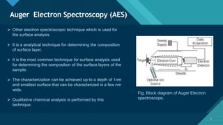
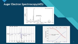
Click to edit Master title style
1
ELECTRO ION
SPECTROSOCPY
C r e a t e d B y
N i s h a n t C h a u d h a r i ( P R N : 1 8 2 2 2 0 7 )
H r u s h i k e s h P a t i l ( P R N : 1 7 2 1 0 1 4 )
T Y I n s t r u m e n t a t i o n ( B T E C H )
G o v e r n m e n t C o l l e g e o f E n g i n e e r i n g , J a l g a o n
Click to edit Master title style
2
1 - S u r f a c e S p e c t r o s c o p y Te c h n i q u e s
2 - P r i n c i p l e
3 - E l e c t r o n S p e c t r o s c o p y f o r C h e m i c a l A n a l y s i s
4 - A u g e r S p e c t r o s c o p y
5 - S e c o n d a r y i o n m a s s s p e c t r o s c o p y
6 - I o n s c a t t e r i n g s p e c t r o s c o p y
2
Content
Click to edit Master title style
3
Surface Spectroscopic
Techniques
T h e s e a r e c l a s s i f i e d a s f o l l o w s
1 - E l e c t r o n S p e c t r o s c o p y
a ) E l e c t r o n I o n s p e c t r o s c o p y f o r c h e m i c a l a n a l y s i s
( E S C A )
b ) A u g e r s p e c t r o s c o p y ( A E S )
2 - I o n S p e c t r o s c o p y
a ) S e c o n d a r y - i o n m a s s s p e c t r o s c o p y ( S I M S )
b ) I o n s c a t t e r i n g s p e c t r o s c o p y ( I S S )
3
Click to edit Master title style
4
Principle of Electron and Ion spectroscopy
4
Electron and ion spectroscopy techniques find
application for surface analysis.
They can provide chemical information, which
the classical methods like microscopy,
reflectivity and adsorption isotherms cannot.
With electron and ion spectroscopy, one can
obtain elemental analyses either as elemental
ratio or oxidation sate ratio.
Click to edit Master title style
5
When a beam is incident on the surface, it
penetrates to some depth within the surface
layer.
A second beam exits from the surface, which
can be analyzed by a spectrometer.
The beam may be photons, electrons and
ions.
5
Click to edit Master title style
6
Electron Ion spectroscopy for chemical analysis
(ESCA)
6
 Electron Ion spectroscopy for chemical analysis (ESCA), also known as x-
ray photoelectron spectroscopy (XPS) is an effective technique for detecting
the element and their bonding states on the surface of solids.
 The methods use soft X-ray to eject electrons from inner-shell orbitals.
 The kinetic energy of these photoelectron energies are dependent upon the
chemical environment of the atom, it makes XPS useful to obtain the oxide
state and ligands of an atom.
• In XPS, we are concerned with the ejection of an electron from one of the
tightly bound core or weakly bound valence shells of an atom or molecule
(usually a core level for analytical purposes, as we will see later) under the
influence of a monochromatic source of X-rays.
Click to edit Master title style
7 7
Figure 1: Schematic diagram of a typical electron spectrometer
showing the necessary components. A hemispherical electrostatic electron energy analyzer is depicted.
Click to edit Master title style
8 8
Click to edit Master title style
9
Auger Electron Spectroscopy (AES)
9
 Other electron spectroscopic technique which is used for
the surface analysis.
 It is a analytical technique for determining the composition
of surface layer.
 It is the most common technique for surface analysis used
for determining the composition of the surface layers of the
sample.
 The characterization can be achieved up to a depth of 1nm
and smallest surface that can be characterized is a few nm
wide.
 Qualitative chemical analysis is performed by this
technique.
Fig. Block diagram of Auger Electron
spectroscope.
Click to edit Master title style
10
Auger Electron Spectroscopy(AES)
10
 The technique include three main steps:
i. Emission of photoelectron or atomic ionization.
ii. Transition of valence electron or electron emission.
iii. Emission of auger electron.
 The process has an involvement of three electrons.
 Kinetic energy of auger electron is given by,
K.E. = hv – B.E. – Work function.
 Kinetic energy can be estimated from binding energies of various levels involved.
 Auger transition is characterized primarily by,
i. Location of initial hole.
ii. Location of final hole.
 Auger electron fail to emerge with their characteristic energies if they start from deeper than 0.5 to
5nm into the surface.
 Highly sensitive for all elements for all atoms but H and He.
Click to edit Master title style
11
Auger Electron Spectroscopy(AES)
11
Click to edit Master title style
12
Auger Electron Spectroscopy(AES)
12
Features Limitations
• High Resolution, spatially resolved chemical
analysis.
• Quantitative compositional analysis of surface
region specimen by comparison with standard
samples
• Highly versatile sensitive (0.1% atom) : standard
analytical tools in lab
• Stronger KLL transition in lower atomic number and
stronger MNN transition in higher atomic number
elements.
• It can’t be used to detect hydrogen and helium.
• It does not include non-destructive depth profiles.
• It requires sample to be compatible with high
vacuum.
• Non conducting samples gets charged under
electron beam bombardment and simply can’t be
analyzed.
Click to edit Master title style
13
Auger Electron Spectroscopy(AES)
13
• Instrumentation:
i. Ultra high vacuum : 10e-7 pascal or
10e-9 torr
ii. Mean free path of the electron should
be approximately 40km.
iii. Ion source: Ar+ Ions (Energy 1-5 keV)
iv. Primary Beam: focused electron beam
v. Detected: Auger electrons, Secondary
electrons
vi. Elements Detected: Li-U; Chemical
bonding information
vii. Resolution: 2 – 3 nm (Profiling mode)
viii. Detection Limits: 0.1-1% at sub-
monolayer
• Applications :
i. Surface analysis.
ii. Particle analysis.
iii. Depth profiling.
iv. Thin film composition analysis.
Click to edit Master title style
14
Ion mass spectroscopy.
14
• Technique used to analyze the composition of solid surfaces and thin film by sputtering the surface of the
specimen with a focused primary ion beam and collecting analyzing ejected secondary ions.
• This technique is used to measure up to a depth of 1 to 2nm.
• Most sensitive surface analyzing technique with elemental detection limits ranging from parts per million to
parts per billion.
• Ion mass spectroscopy is divided into two parts:
i. Secondary ion mass spectroscopy.
ii. Ion scattering spectroscopy.
Click to edit Master title style
15
Ion mass spectroscopy.
15
• An inert gas ion having kinetic energy 0.3 to 5 keV is incident on the
surface of sample.
• The ion is penetrated into the surface of the solid and cause
considerable disruption by transfer of momentum to lattice atoms or
molecules this is called sputtering.
• This results in expelling atomic and molecular fragments, these
fragments can be neutral or ions.
• These ions are referred to as secondary ions and thus termed as
secondary ion mass spectroscopy.
Click to edit Master title style
16
Thank You

Electron and ion spectroscopy.

  • 1.
    Click to editMaster title style 1 ELECTRO ION SPECTROSOCPY C r e a t e d B y N i s h a n t C h a u d h a r i ( P R N : 1 8 2 2 2 0 7 ) H r u s h i k e s h P a t i l ( P R N : 1 7 2 1 0 1 4 ) T Y I n s t r u m e n t a t i o n ( B T E C H ) G o v e r n m e n t C o l l e g e o f E n g i n e e r i n g , J a l g a o n
  • 2.
    Click to editMaster title style 2 1 - S u r f a c e S p e c t r o s c o p y Te c h n i q u e s 2 - P r i n c i p l e 3 - E l e c t r o n S p e c t r o s c o p y f o r C h e m i c a l A n a l y s i s 4 - A u g e r S p e c t r o s c o p y 5 - S e c o n d a r y i o n m a s s s p e c t r o s c o p y 6 - I o n s c a t t e r i n g s p e c t r o s c o p y 2 Content
  • 3.
    Click to editMaster title style 3 Surface Spectroscopic Techniques T h e s e a r e c l a s s i f i e d a s f o l l o w s 1 - E l e c t r o n S p e c t r o s c o p y a ) E l e c t r o n I o n s p e c t r o s c o p y f o r c h e m i c a l a n a l y s i s ( E S C A ) b ) A u g e r s p e c t r o s c o p y ( A E S ) 2 - I o n S p e c t r o s c o p y a ) S e c o n d a r y - i o n m a s s s p e c t r o s c o p y ( S I M S ) b ) I o n s c a t t e r i n g s p e c t r o s c o p y ( I S S ) 3
  • 4.
    Click to editMaster title style 4 Principle of Electron and Ion spectroscopy 4 Electron and ion spectroscopy techniques find application for surface analysis. They can provide chemical information, which the classical methods like microscopy, reflectivity and adsorption isotherms cannot. With electron and ion spectroscopy, one can obtain elemental analyses either as elemental ratio or oxidation sate ratio.
  • 5.
    Click to editMaster title style 5 When a beam is incident on the surface, it penetrates to some depth within the surface layer. A second beam exits from the surface, which can be analyzed by a spectrometer. The beam may be photons, electrons and ions. 5
  • 6.
    Click to editMaster title style 6 Electron Ion spectroscopy for chemical analysis (ESCA) 6  Electron Ion spectroscopy for chemical analysis (ESCA), also known as x- ray photoelectron spectroscopy (XPS) is an effective technique for detecting the element and their bonding states on the surface of solids.  The methods use soft X-ray to eject electrons from inner-shell orbitals.  The kinetic energy of these photoelectron energies are dependent upon the chemical environment of the atom, it makes XPS useful to obtain the oxide state and ligands of an atom. • In XPS, we are concerned with the ejection of an electron from one of the tightly bound core or weakly bound valence shells of an atom or molecule (usually a core level for analytical purposes, as we will see later) under the influence of a monochromatic source of X-rays.
  • 7.
    Click to editMaster title style 7 7 Figure 1: Schematic diagram of a typical electron spectrometer showing the necessary components. A hemispherical electrostatic electron energy analyzer is depicted.
  • 8.
    Click to editMaster title style 8 8
  • 9.
    Click to editMaster title style 9 Auger Electron Spectroscopy (AES) 9  Other electron spectroscopic technique which is used for the surface analysis.  It is a analytical technique for determining the composition of surface layer.  It is the most common technique for surface analysis used for determining the composition of the surface layers of the sample.  The characterization can be achieved up to a depth of 1nm and smallest surface that can be characterized is a few nm wide.  Qualitative chemical analysis is performed by this technique. Fig. Block diagram of Auger Electron spectroscope.
  • 10.
    Click to editMaster title style 10 Auger Electron Spectroscopy(AES) 10  The technique include three main steps: i. Emission of photoelectron or atomic ionization. ii. Transition of valence electron or electron emission. iii. Emission of auger electron.  The process has an involvement of three electrons.  Kinetic energy of auger electron is given by, K.E. = hv – B.E. – Work function.  Kinetic energy can be estimated from binding energies of various levels involved.  Auger transition is characterized primarily by, i. Location of initial hole. ii. Location of final hole.  Auger electron fail to emerge with their characteristic energies if they start from deeper than 0.5 to 5nm into the surface.  Highly sensitive for all elements for all atoms but H and He.
  • 11.
    Click to editMaster title style 11 Auger Electron Spectroscopy(AES) 11
  • 12.
    Click to editMaster title style 12 Auger Electron Spectroscopy(AES) 12 Features Limitations • High Resolution, spatially resolved chemical analysis. • Quantitative compositional analysis of surface region specimen by comparison with standard samples • Highly versatile sensitive (0.1% atom) : standard analytical tools in lab • Stronger KLL transition in lower atomic number and stronger MNN transition in higher atomic number elements. • It can’t be used to detect hydrogen and helium. • It does not include non-destructive depth profiles. • It requires sample to be compatible with high vacuum. • Non conducting samples gets charged under electron beam bombardment and simply can’t be analyzed.
  • 13.
    Click to editMaster title style 13 Auger Electron Spectroscopy(AES) 13 • Instrumentation: i. Ultra high vacuum : 10e-7 pascal or 10e-9 torr ii. Mean free path of the electron should be approximately 40km. iii. Ion source: Ar+ Ions (Energy 1-5 keV) iv. Primary Beam: focused electron beam v. Detected: Auger electrons, Secondary electrons vi. Elements Detected: Li-U; Chemical bonding information vii. Resolution: 2 – 3 nm (Profiling mode) viii. Detection Limits: 0.1-1% at sub- monolayer • Applications : i. Surface analysis. ii. Particle analysis. iii. Depth profiling. iv. Thin film composition analysis.
  • 14.
    Click to editMaster title style 14 Ion mass spectroscopy. 14 • Technique used to analyze the composition of solid surfaces and thin film by sputtering the surface of the specimen with a focused primary ion beam and collecting analyzing ejected secondary ions. • This technique is used to measure up to a depth of 1 to 2nm. • Most sensitive surface analyzing technique with elemental detection limits ranging from parts per million to parts per billion. • Ion mass spectroscopy is divided into two parts: i. Secondary ion mass spectroscopy. ii. Ion scattering spectroscopy.
  • 15.
    Click to editMaster title style 15 Ion mass spectroscopy. 15 • An inert gas ion having kinetic energy 0.3 to 5 keV is incident on the surface of sample. • The ion is penetrated into the surface of the solid and cause considerable disruption by transfer of momentum to lattice atoms or molecules this is called sputtering. • This results in expelling atomic and molecular fragments, these fragments can be neutral or ions. • These ions are referred to as secondary ions and thus termed as secondary ion mass spectroscopy.
  • 16.
    Click to editMaster title style 16 Thank You