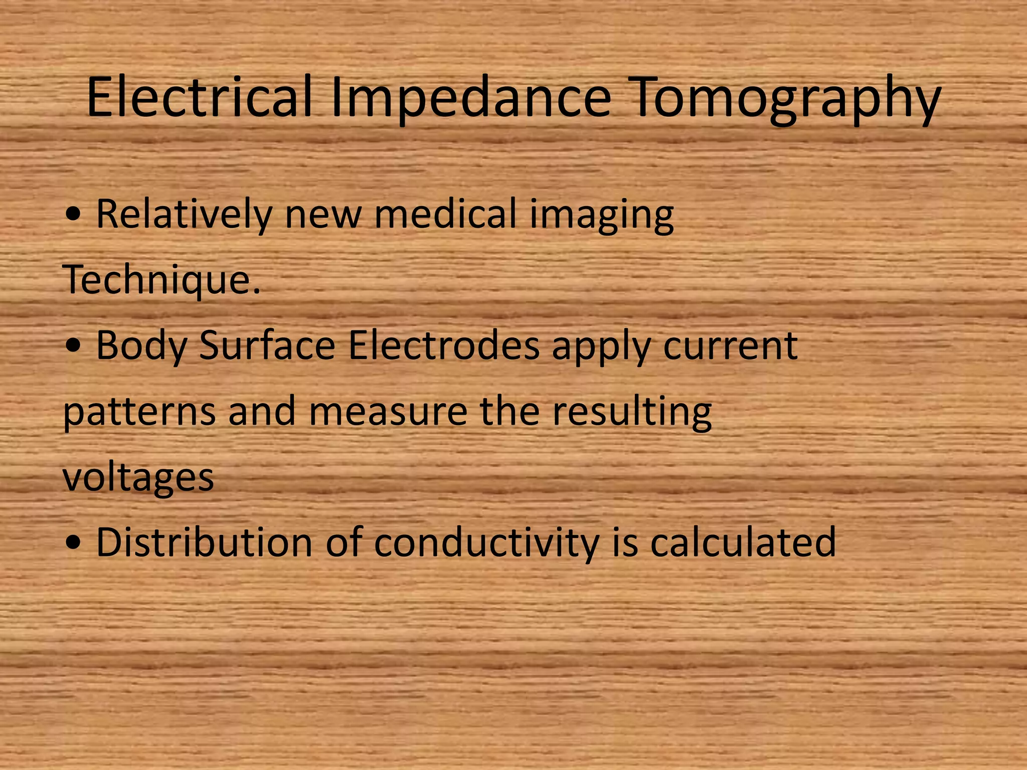
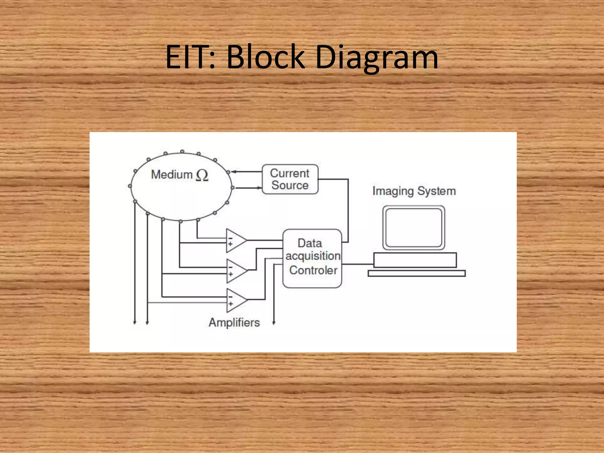
Electrical Impedance
Tomography
and
Applications
Outline
• Electrical Impedance Tomography
• Physics
• Applications
•Advantages
• Disadvantages
Electrical Impedance Tomography
• Relatively new medical imaging
Technique.
• Body Surface Electrodes apply current
patterns and measure the resulting
voltages
• Distribution of conductivity is calculated
EIT: Block Diagram
EIT: Physics
Figure 2: Electrode configuration around the
Thorax: Small alternating currents are being
applied through drive electrodes (red), the
resulting equal potentials being recorded by
the remaining (green) electrodes.
Figure 1: A cross section of a human thorax
from an X-ray CT showing current stream lines
and equi potentials from drive electrodes.
Note how lines are bent by the change in
conductivity between different organs.
In this prototype, the electrodes are attached
around a grapefruit which represents a child's
head. Liquid is injected into the grapefruit to
mimic brain haemorrhage.
•After positioning surface electrodes through adhesive electrodes, an
electrode belt or a conductive electrode vest around the body part of
interest, alternating currents of typically a few milli-Amperes at a
frequency of 10–100 kHz will be applied across two or more drive
electrodes.
The remaining electrodes will be used to measure the resulting voltage.
•The procedure will then be repeated for numerous "stimulation
patterns",
•E.x. successive pairs of adjacent electrodes until an entire circle has
been completed and image reconstruction can be carried out and
displayed by a digital workstation that incorporates complex
mathematical algorithms and a priori data
THEORY
Applications
Applications
• EIT can image physiological processes
• involving movement of conductive fluids
• and gasses
• Lungs
• Heart
• Brain
• Chest
Lung (a-EIT, f-EIT)
• EIT is particularly useful for monitoring lung
function because lung tissue resistivity is five
times higher when compared to most other soft
tissues within the thorax.
• This results in high absolute contrast of the lungs
in general. In addition, lung resistivity increases
and decreases several-fold between inspiration
and expiration
• which explains why monitoring the respiratory
cycle is currently the most promising clinical
application of functional EIT.
Resulting image (earlier
attempt)
Adhesive electrodes on chest
Brain (a-EIT, f-EIT, MF-EIT)
• EIT has been suggested as a basis for brain
imaging to enable the detection and
• Monitoring of cerebral
ischemia, haemorrhage,
• Other morphological pathologies associated
with impedance changes due to neuronal cell
swelling
ADVANTAGES
• EIT is a relatively low resolution imaging
• modality, but
• • Non-invasive
• • Non-cumbersome
• • Suitable for monitoring
• • Underlying technology is low cost
Disadvantages
• Absolute EIT approaches are targeted at
digital reconstruction of static images, i.e.
two-dimensional representations of the
anatomy within the body part of interest. As
mentioned above and unlike linear x-rays
in Computed Tomography, electrical currents
travel three-dimensionally along the path of
least resistivity, which results in partial loss of
the electrical current applied
Disadvantages
• This is one of the reasons why image reconstruction in
absolute EIT is so complex, since there is usually more
than just one solution for image reconstruction of a
three-dimensional area projected onto a two-
dimensional plane.
• Another difficulty is that given the number of
electrodes and the measurement precision at each
electrode, only objects bigger than a given size can be
distinguished. This explains the necessity of highly
sophisticated mathematical algorithms that will
account for the inverse problem and its ill-posedness.
Thank You

Electrical impedance tomography

  • 1.
  • 2.
    Outline • Electrical ImpedanceTomography • Physics • Applications •Advantages • Disadvantages
  • 3.
    Electrical Impedance Tomography •Relatively new medical imaging Technique. • Body Surface Electrodes apply current patterns and measure the resulting voltages • Distribution of conductivity is calculated
  • 4.
  • 5.
  • 7.
    Figure 2: Electrodeconfiguration around the Thorax: Small alternating currents are being applied through drive electrodes (red), the resulting equal potentials being recorded by the remaining (green) electrodes.
  • 8.
    Figure 1: Across section of a human thorax from an X-ray CT showing current stream lines and equi potentials from drive electrodes. Note how lines are bent by the change in conductivity between different organs.
  • 9.
    In this prototype,the electrodes are attached around a grapefruit which represents a child's head. Liquid is injected into the grapefruit to mimic brain haemorrhage.
  • 10.
    •After positioning surfaceelectrodes through adhesive electrodes, an electrode belt or a conductive electrode vest around the body part of interest, alternating currents of typically a few milli-Amperes at a frequency of 10–100 kHz will be applied across two or more drive electrodes. The remaining electrodes will be used to measure the resulting voltage. •The procedure will then be repeated for numerous "stimulation patterns", •E.x. successive pairs of adjacent electrodes until an entire circle has been completed and image reconstruction can be carried out and displayed by a digital workstation that incorporates complex mathematical algorithms and a priori data THEORY
  • 11.
  • 12.
    Applications • EIT canimage physiological processes • involving movement of conductive fluids • and gasses • Lungs • Heart • Brain • Chest
  • 13.
    Lung (a-EIT, f-EIT) •EIT is particularly useful for monitoring lung function because lung tissue resistivity is five times higher when compared to most other soft tissues within the thorax. • This results in high absolute contrast of the lungs in general. In addition, lung resistivity increases and decreases several-fold between inspiration and expiration • which explains why monitoring the respiratory cycle is currently the most promising clinical application of functional EIT.
  • 14.
  • 15.
    Brain (a-EIT, f-EIT,MF-EIT) • EIT has been suggested as a basis for brain imaging to enable the detection and • Monitoring of cerebral ischemia, haemorrhage, • Other morphological pathologies associated with impedance changes due to neuronal cell swelling
  • 16.
    ADVANTAGES • EIT isa relatively low resolution imaging • modality, but • • Non-invasive • • Non-cumbersome • • Suitable for monitoring • • Underlying technology is low cost
  • 17.
    Disadvantages • Absolute EITapproaches are targeted at digital reconstruction of static images, i.e. two-dimensional representations of the anatomy within the body part of interest. As mentioned above and unlike linear x-rays in Computed Tomography, electrical currents travel three-dimensionally along the path of least resistivity, which results in partial loss of the electrical current applied
  • 18.
    Disadvantages • This isone of the reasons why image reconstruction in absolute EIT is so complex, since there is usually more than just one solution for image reconstruction of a three-dimensional area projected onto a two- dimensional plane. • Another difficulty is that given the number of electrodes and the measurement precision at each electrode, only objects bigger than a given size can be distinguished. This explains the necessity of highly sophisticated mathematical algorithms that will account for the inverse problem and its ill-posedness.
  • 19.