
Recommended
More Related Content
Similar to lymphoid_tissue.pdf
Similar to lymphoid_tissue.pdf (20)
More from nandhinivenkat4
More from nandhinivenkat4 (15)
Recently uploaded
Recently uploaded (20)
lymphoid_tissue.pdf
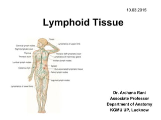
- 1. Lymphoid Tissue Dr. Archana Rani Associate Professor Department of Anatomy KGMU UP, Lucknow 10.03.2015
- 2. What is lymphoid tissue? • Specialized form of connective tissue • Supporting framework: reticular cells & reticular fibres • Large number of lymphocytes • Other cells: Plasma cells & macrophages
- 3. Consists of……. • Lymphatic vessels • Specific lymphoid organs (lymph node, spleen, thymus) • Lymphatic tissue found within the tissues of other organs (in bone marrow, GI tract, urinary tract, respiratory tract)
- 4. Functions • Defense of body • Phagocytosis of foreign cells • Involved in production of lymphocytes and plasma cells
- 5. Lymphatic Vessels –Originate as lymph capillaries –Capillaries unite to form larger lymph vessels • Resemble veins in structure • Connect to lymph nodes at various intervals
- 7. Lymphatic capillary & Vessel
- 9. Channels of Lymphatics – Lymphatics ultimately deliver lymph into 2 main channels • Right lymphatic duct –Drains right side of head & neck, right arm, right thorax –Empties into the right subclavian vein • Thoracic duct –Drains the rest of the body –Empties into the left subclavian vein
- 11. Major Lymphatic Vessel of the Trunk
- 12. Lymphatic Tissue – 3 types • Diffuse lymphatic tissue – No capsule present – Found in connective tissue of almost all organs • Lymphatic nodules – No capsule present – Oval-shaped masses – Found singly or in clusters • Lymphatic organs – Capsule present – Lymph nodes, spleen, thymus
- 13. Diffuse lymphatic tissue • Called as mucosa associated lymphatic tissue (MALT). • Accumulation of lymphatic tissue in the mucous membrane of gastrointestinal, respiratory, urinary and reproductive tracts. • Located where they come in direct contact with antigens.
- 14. Lymphatic Nodule • Circumscribed concentration of lymphatic tissue (lymphocytes and related cells). • Not surrounded by capsule.
- 15. Lymphatic Nodule
- 16. Lymphatic Organs
- 17. Lymph Node – Consists of connective tissue framework & numerous lymphocytes. – Bean shaped structures placed in pathway of lymphatic vessels. – Enclosed by a fibrous capsule. – Cortex = outer portion • Germinal centers produce lymphocytes – Medulla = inner portion • Medullary cords
- 18. Lymph Node • Lymph enters nodes through afferent lymph vessels, flows through sinuses, exits through efferent lymph vessel.
- 19. Lymph Node
- 20. 1.Capsule 2. lymphoid nodule with germinal center 3. subcapsular sinus 4. intermediate sinus 5. medullary cords 6. medullary sinuses 7. trabecula
- 21. Cortex of Lymph Node
- 22. Medulla of Lymph Node Medullary sinuses Medullary cords
- 23. Medullary sinus of a lymph node containing reticular cells with long processes and elongated nuclei, macrophages, and many lymphocytes. (1) Macrophage; (2) reticular cell; (3) trabecula. H&E stain. High magnification. (Courtesy of PA Abrahamsohn.)
- 24. Cells of Lymph Node • Lymphocytes • Plasma cells • Reticular cells • Macrophages and other phagocytic antigen processing cells • Lymphatic and vascular endothelial cells
- 25. Functions of Lymph Node • Filtration of particles and microorganisms to keep them out of general circulation. • Interaction of circulating antigens in lymph with lymphocytes to initiate immune response. • Activation, proliferation of B lymphocytes and antibody production. • Activation, proliferation of T lymphocytes.
- 26. Spleen • Largest lymphoid organ • Encapsulated • Structure is similar to a node • Capsule present • But no afferent vessels or sinuses
- 27. Spleen • Substance is arranged in form of: White pulp (basophilic) Red pulp (reddish) • Supporting Elements: Capsule Trabeculae Trabecular network Lymphocytes, macrophages, blood cells Red pulp: splenic cords
- 28. Spleen
- 29. White pulp: Lymphoid Nodule (Malpighian corpuscle) 1. Germinal center 2. Central artery
- 30. Red Pulp of Spleen (Splenic cords: Cords of Billroth)
- 31. Functions of Spleen • Filtration of blood. • Immune response against antigens circulating in blood. • Site for production of B & T lymphocytes. • Formation of blood cells during fetal life. • Storage of blood. • Site of destruction of aged erythrocytes.
- 32. Thymus – Location – behind the sternum in the mediastinum – Development: • Infant – conspicuous • Puberty – maximum size • Maturity – decreases in size – Function • Differentiation and maturation of T cells
- 33. Thymus • Consists of 2 lobes (rt. & lt.) covered by connective tissue capsule. • Septa passing inwards from the capsule subdivide the lobe into a large number of lobules. • Lobule: outer cortex, inner medulla • Supporting stroma: epithelioreticular cells
- 34. Thymic Lobule • Cortex: densely packed small lymphocytes. • Medulla: Lymphocytes are less densely packed. Presence of Hassall’s corpuscles.
- 35. Functions of thymus • Provides the environment for stem cells where they can divide and mature into T lymphocytes. • Thymopoietin induces T cell production & maturation. • Thymosin supports T cell activities.
- 36. Tonsils – Multiple groups of large lymphatic nodules – Location – mucous membrane of the oral and pharyngeal cavities. – Palatine tonsils • Posterior-lateral wall of the oropharynx – Pharyngeal tonsil • Posterior wall of nasopharynx – Lingual tonsils • Base of tongue
- 37. Palatine Tonsil • Aggregation of lymphatic nodules within diffuse lymphoid tissue. • Covered by stratified squamous epithelium. • Tonsillar crypts (opening of numerous mucous glands)
- 38. Functions of tonsil • Production of lymphocytes. • Immunological response against antigens & organisms coming in contact with epithelium.
- 39. References 1. diFiore’s Atlas of Histology with functional Correlations, 12th Edition. 2. Textbook of Human Histology. Inderbir Singh, 1st Edition. 3. Textbook of Histology. GP Pal, 3rd Edition.
- 40. MCQ • The supporting framework of lymphatic tissue is formed by all except: 1. Reticular cells 2. Plasma cells 3. Macrophages 4. Fibrocyte
- 41. MCQ • Afferent lymphatics in lymph node pour their lymph into: 1. Trabeculae 2. Subcapsular sinus 3. Medullary sinus 4. Lymph nodule
- 42. MCQ • Cords of Billroth is a feature of: 1. White pulp of spleen 2. Red pulp of spleen 3. Thymus 4. Tonsil
- 43. MCQ • The tonsil is covered by: 1. Simple squamous epithelium 2. Stratified squamous epithelium 3. Stratified cuboidal epithelium 4. Stratified columnar epithelium
- 44. MCQ • Hassall’s corpuscles is a feature of: 1. Lymph node 2. Spleen 3. Thymus 4. Tonsil