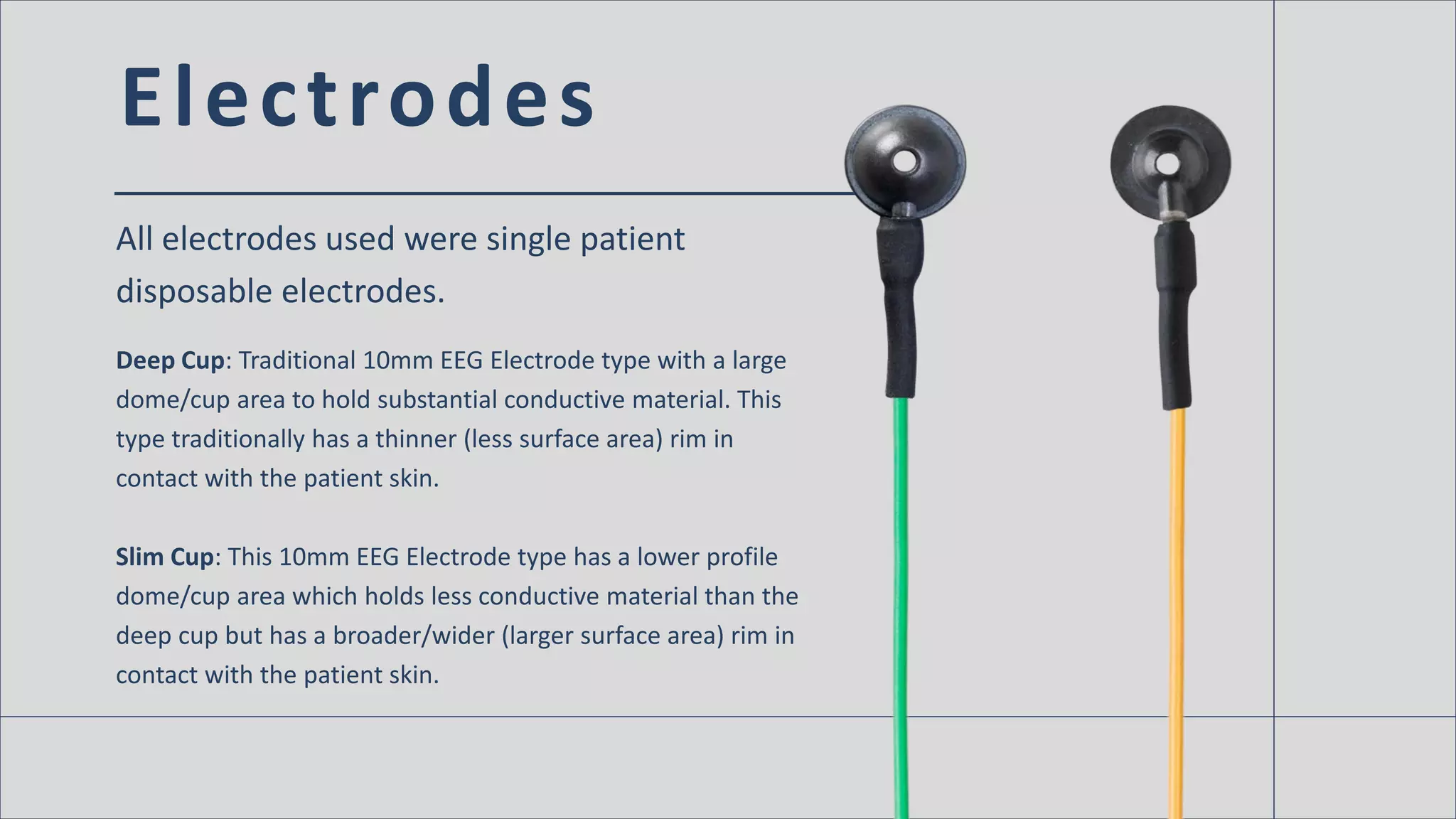
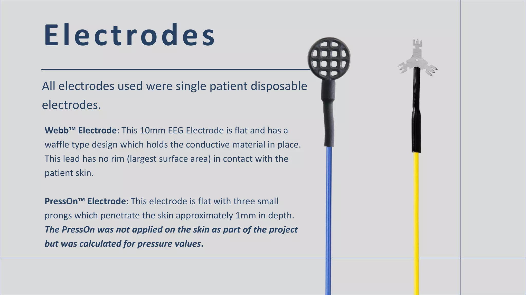
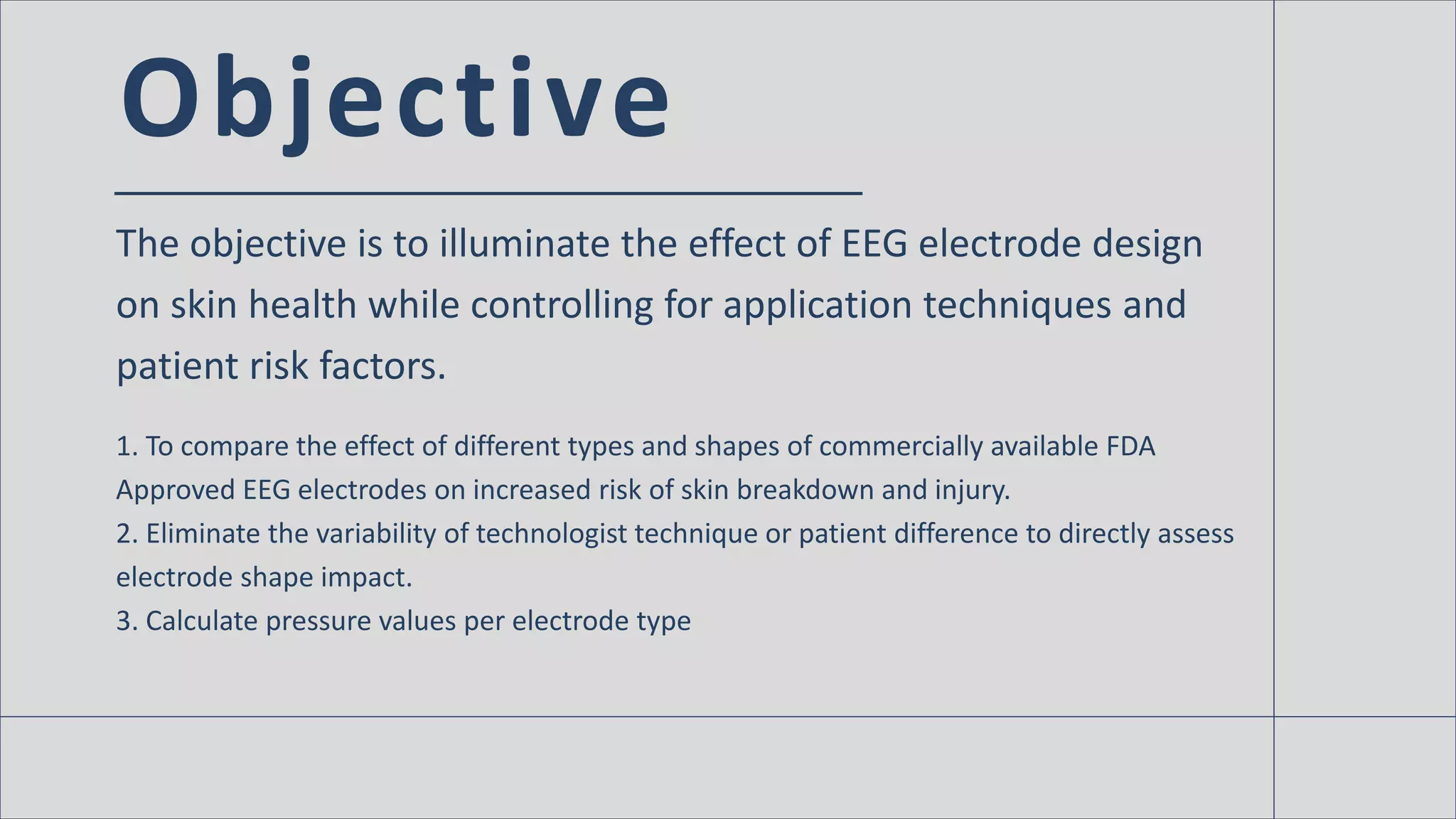
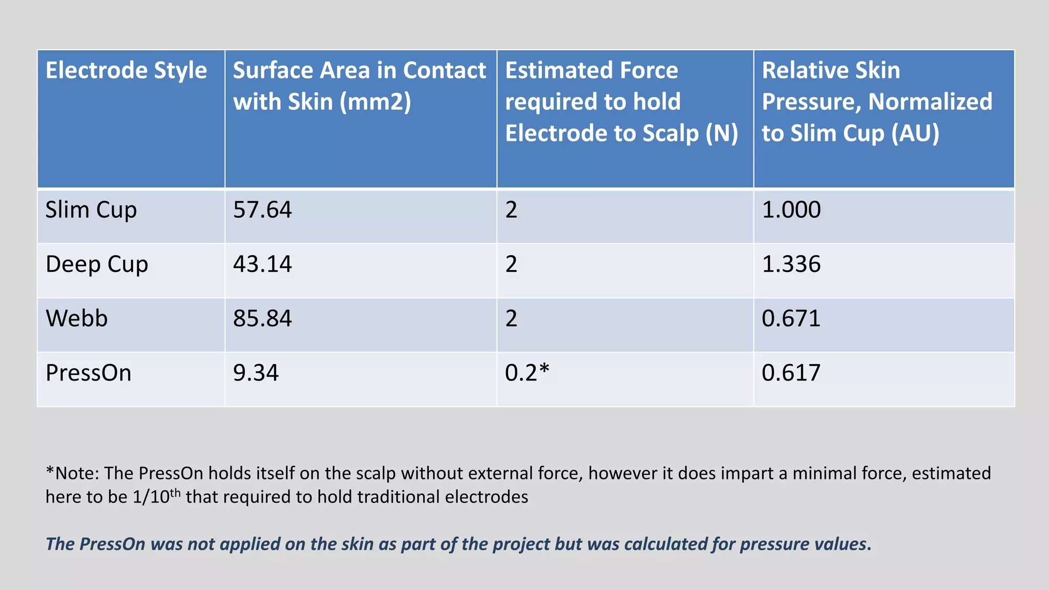
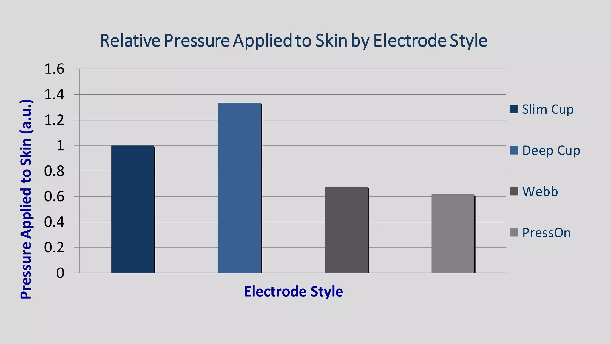
E E G E l e c t r o d e S h a p e
A f f e c t s S k i n S a f e t y
a n d B r e a kd o w n f o r
L o n g e r S t u d i e s
L e a h H a n s o n , R . E E G / E P T
V P o f G l o b a l S a l e s
G a b r i e l O rs i n g e r, P h D, P M P
M a n a g e r, P r o d u c t D e v e l o p m e n t
“EEG Electrodes have been used for decades with very little
attention being paid to the design or shape of the electrode and its
impact on skin safety and health. As a matter of fact, even in the
past few years when more attention than ever is being paid to skin
health, breakdown and injury, the electrode design continues to be
relatively dismissed and reported to have less impact on skin
breakdown than the technique in which the electrode is used.”
Introduction
The objective is to illuminate the effect of EEG electrode design
on skin health while controlling for application techniques and
patient risk factors.
Electrodes
All electrodes used were single patient
disposable electrodes.
Deep Cup: Traditional 10mm EEG Electrode type with a large
dome/cup area to hold substantial conductive material. This
type traditionally has a thinner (less surface area) rim in
contact with the patient skin.
Slim Cup: This 10mm EEG Electrode type has a lower profile
dome/cup area which holds less conductive material than the
deep cup but has a broader/wider (larger surface area) rim in
contact with the patient skin.
Electrodes
All electrodes used were single patient disposable
electrodes.
Webb™ Electrode: This 10mm EEG Electrode is flat and has a
waffle type design which holds the conductive material in place.
This lead has no rim (largest surface area) in contact with the
patient skin.
PressOn™ Electrode: This electrode is flat with three small
prongs which penetrate the skin approximately 1mm in depth.
The PressOn was not applied on the skin as part of the project
but was calculated for pressure values.
Objective
The objective is to illuminate the effect of EEG electrode design
on skin health while controlling for application techniques and
patient risk factors.
1. To compare the effect of different types and shapes of commercially available FDA
Approved EEG electrodes on increased risk of skin breakdown and injury.
2. Eliminate the variability of technologist technique or patient difference to directly assess
electrode shape impact.
3. Calculate pressure values per electrode type
Results
The Deep Cup style with the thinner (smaller surface area) rim
showed visible skin changes versus the slim (larger surface area)
and the flat Webb™ ( largest surface area) which had little or no
visible identification after removal.
Electrode Style Surface Area in Contact
with Skin (mm2)
Estimated Force
required to hold
Electrode to Scalp (N)
Relative Skin
Pressure, Normalized
to Slim Cup (AU)
Slim Cup 57.64 2 1.000
Deep Cup 43.14 2 1.336
Webb 85.84 2 0.671
PressOn 9.34 0.2* 0.617
*Note: The PressOn holds itself on the scalp without external force, however it does impart a minimal force, estimated
here to be 1/10th that required to hold traditional electrodes
The PressOn was not applied on the skin as part of the project but was calculated for pressure values.
0
0.2
0.4
0.6
0.8
1
1.2
1.4
1.6
PressureAppliedtoSkin(a.u.)
Electrode Style
RelativePressureAppliedto Skinby ElectrodeStyle
Slim Cup
Deep Cup
Webb
PressOn
Figure 1. Skin Conditions
following 74 hours of wearing
Deep Cup, Slim Cup, and
Webb Electrodes.
Images were taken
immediately following
removal (T = 0 hrs), 6 hours
after removal
(T = 6 hrs) and 24 hours after
removal (T = 24 hrs).
Methods
Same application technique for all electrodes:
• Weaver NuPrep™ used to prep the skin
• Weaver Ten20™ used as the conductive material
• Mavidon™ Collodion used as the adhesive with gauze
• Impedances recorded only at initial application-all were between 2K-5K Ohms.
• All electrodes were Rhythmlink disposable EEG Electrodes
• Electrodes (Deep Cup, Slim Cup, Webb™) applied for cont. 74 hours on left forearm with
each electrodes 1.5 inches apart
Conclusions
The data indicates the shape of a traditional style Deep Cup may
increase risks for skin challenges/issues due to the small surface
area and high relative pressure when applied.
Surface area and relative pressure were related with the smallest surface area reporting
the highest relative pressure and the largest surfaced area reporting the lowest relative
pressure when applied to the skin.
MORE INFO
This information was originally presented at the 2016
Annual Meeting for The Neurodiagnostic Society
(ASET). View the poster at the link below!
Click here to learn more!
References
• Braden Scale for Preventing Sore Risk. Prevention Plus. 1988.Accessed July 13,
2015https://en.wikipedia.org/wiki/Braden_Scale_for_Predicting_Pressure_Ulcer_Risk
• Guideline Twelve: Guidelines for Long-Term Monitoring for Epilepsy.American Clinical
Neurophysiology Society. Am JElectroneurodiagnosticTechnol. 2008 Dec:48(4):265–86
• MagnanMA, MaklebustJ. The nursing process and pressure ulcer prevention: making the
connection. AdvSkin Wound Care. 2009Feb;22(2):83–92.
• ASET The Neurodiagnostic Society Position Statement, Skin Safety During EEG Procedures –a
Guideline to Improving Outcome, 2015.
Acknowledgements
Special thanks to
The engineering team at Rhythmlink International
The clinicians surveyed and offered valuable input

EEG Electrode Shape Affects Skin Safety and Breakdown for Longer Studies

  • 1.
    E E GE l e c t r o d e S h a p e A f f e c t s S k i n S a f e t y a n d B r e a kd o w n f o r L o n g e r S t u d i e s L e a h H a n s o n , R . E E G / E P T V P o f G l o b a l S a l e s G a b r i e l O rs i n g e r, P h D, P M P M a n a g e r, P r o d u c t D e v e l o p m e n t
  • 2.
    “EEG Electrodes havebeen used for decades with very little attention being paid to the design or shape of the electrode and its impact on skin safety and health. As a matter of fact, even in the past few years when more attention than ever is being paid to skin health, breakdown and injury, the electrode design continues to be relatively dismissed and reported to have less impact on skin breakdown than the technique in which the electrode is used.”
  • 3.
    Introduction The objective isto illuminate the effect of EEG electrode design on skin health while controlling for application techniques and patient risk factors.
  • 4.
    Electrodes All electrodes usedwere single patient disposable electrodes. Deep Cup: Traditional 10mm EEG Electrode type with a large dome/cup area to hold substantial conductive material. This type traditionally has a thinner (less surface area) rim in contact with the patient skin. Slim Cup: This 10mm EEG Electrode type has a lower profile dome/cup area which holds less conductive material than the deep cup but has a broader/wider (larger surface area) rim in contact with the patient skin.
  • 5.
    Electrodes All electrodes usedwere single patient disposable electrodes. Webb™ Electrode: This 10mm EEG Electrode is flat and has a waffle type design which holds the conductive material in place. This lead has no rim (largest surface area) in contact with the patient skin. PressOn™ Electrode: This electrode is flat with three small prongs which penetrate the skin approximately 1mm in depth. The PressOn was not applied on the skin as part of the project but was calculated for pressure values.
  • 6.
    Objective The objective isto illuminate the effect of EEG electrode design on skin health while controlling for application techniques and patient risk factors. 1. To compare the effect of different types and shapes of commercially available FDA Approved EEG electrodes on increased risk of skin breakdown and injury. 2. Eliminate the variability of technologist technique or patient difference to directly assess electrode shape impact. 3. Calculate pressure values per electrode type
  • 7.
    Results The Deep Cupstyle with the thinner (smaller surface area) rim showed visible skin changes versus the slim (larger surface area) and the flat Webb™ ( largest surface area) which had little or no visible identification after removal.
  • 8.
    Electrode Style SurfaceArea in Contact with Skin (mm2) Estimated Force required to hold Electrode to Scalp (N) Relative Skin Pressure, Normalized to Slim Cup (AU) Slim Cup 57.64 2 1.000 Deep Cup 43.14 2 1.336 Webb 85.84 2 0.671 PressOn 9.34 0.2* 0.617 *Note: The PressOn holds itself on the scalp without external force, however it does impart a minimal force, estimated here to be 1/10th that required to hold traditional electrodes The PressOn was not applied on the skin as part of the project but was calculated for pressure values.
  • 9.
  • 10.
    Figure 1. SkinConditions following 74 hours of wearing Deep Cup, Slim Cup, and Webb Electrodes. Images were taken immediately following removal (T = 0 hrs), 6 hours after removal (T = 6 hrs) and 24 hours after removal (T = 24 hrs).
  • 11.
    Methods Same application techniquefor all electrodes: • Weaver NuPrep™ used to prep the skin • Weaver Ten20™ used as the conductive material • Mavidon™ Collodion used as the adhesive with gauze • Impedances recorded only at initial application-all were between 2K-5K Ohms. • All electrodes were Rhythmlink disposable EEG Electrodes • Electrodes (Deep Cup, Slim Cup, Webb™) applied for cont. 74 hours on left forearm with each electrodes 1.5 inches apart
  • 12.
    Conclusions The data indicatesthe shape of a traditional style Deep Cup may increase risks for skin challenges/issues due to the small surface area and high relative pressure when applied. Surface area and relative pressure were related with the smallest surface area reporting the highest relative pressure and the largest surfaced area reporting the lowest relative pressure when applied to the skin.
  • 13.
    MORE INFO This informationwas originally presented at the 2016 Annual Meeting for The Neurodiagnostic Society (ASET). View the poster at the link below! Click here to learn more!
  • 14.
    References • Braden Scalefor Preventing Sore Risk. Prevention Plus. 1988.Accessed July 13, 2015https://en.wikipedia.org/wiki/Braden_Scale_for_Predicting_Pressure_Ulcer_Risk • Guideline Twelve: Guidelines for Long-Term Monitoring for Epilepsy.American Clinical Neurophysiology Society. Am JElectroneurodiagnosticTechnol. 2008 Dec:48(4):265–86 • MagnanMA, MaklebustJ. The nursing process and pressure ulcer prevention: making the connection. AdvSkin Wound Care. 2009Feb;22(2):83–92. • ASET The Neurodiagnostic Society Position Statement, Skin Safety During EEG Procedures –a Guideline to Improving Outcome, 2015.
  • 15.
    Acknowledgements Special thanks to Theengineering team at Rhythmlink International The clinicians surveyed and offered valuable input