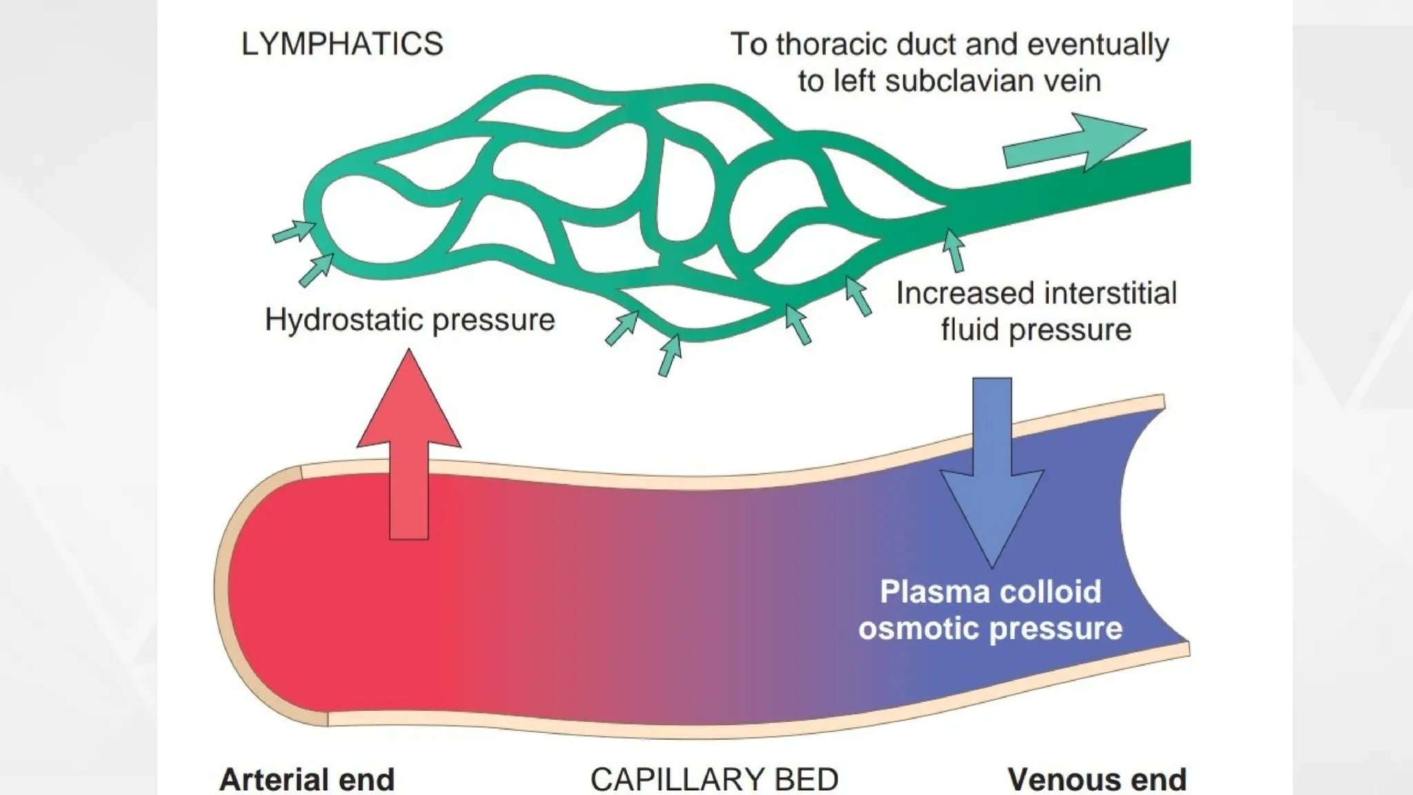
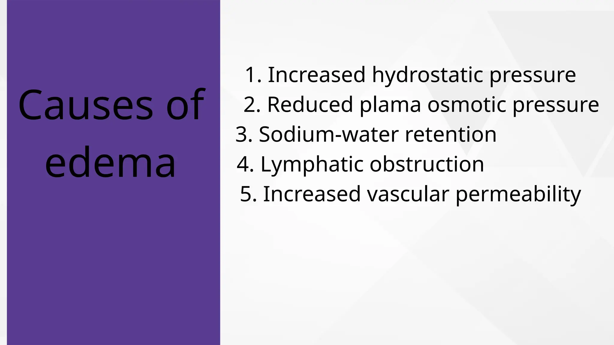
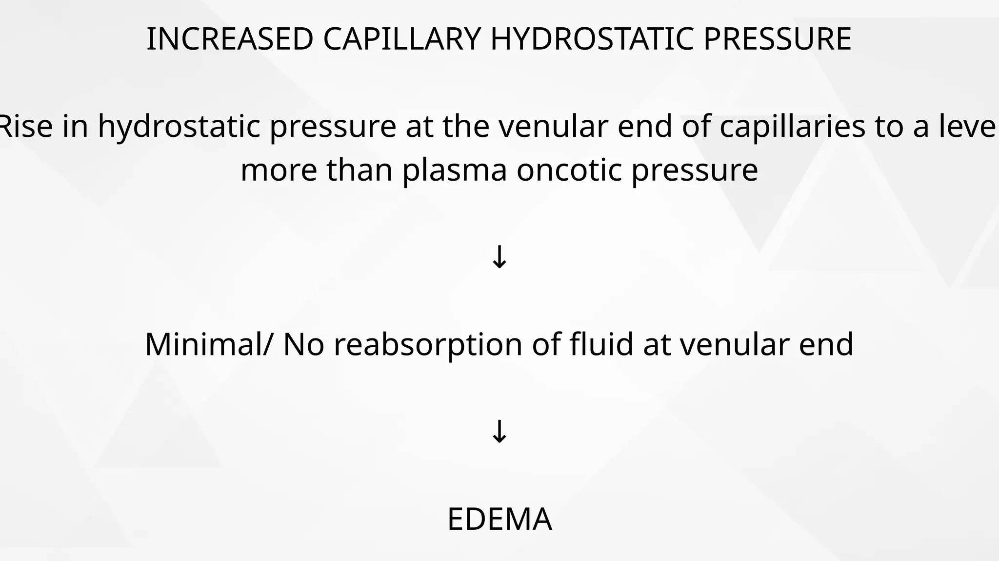
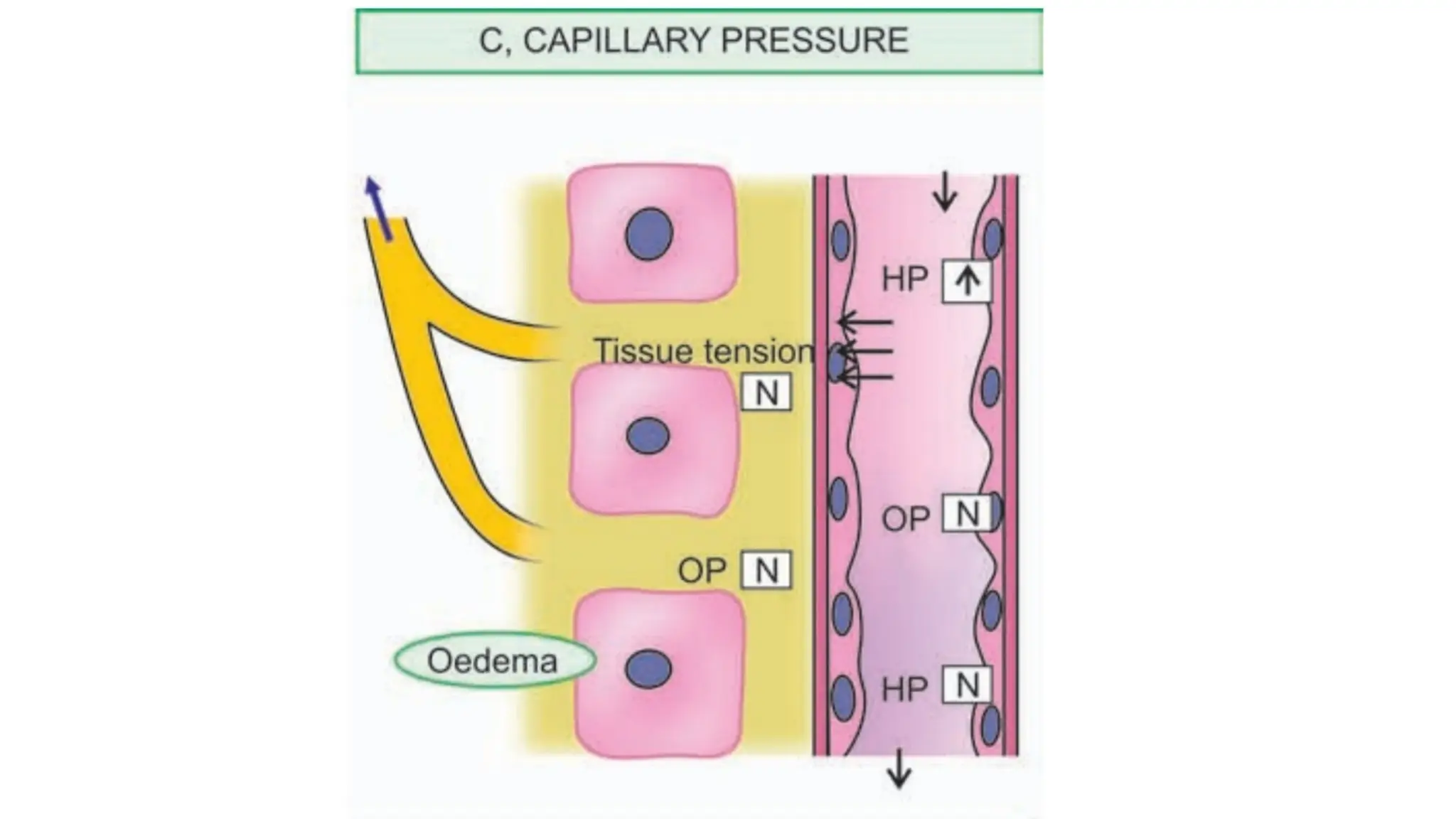
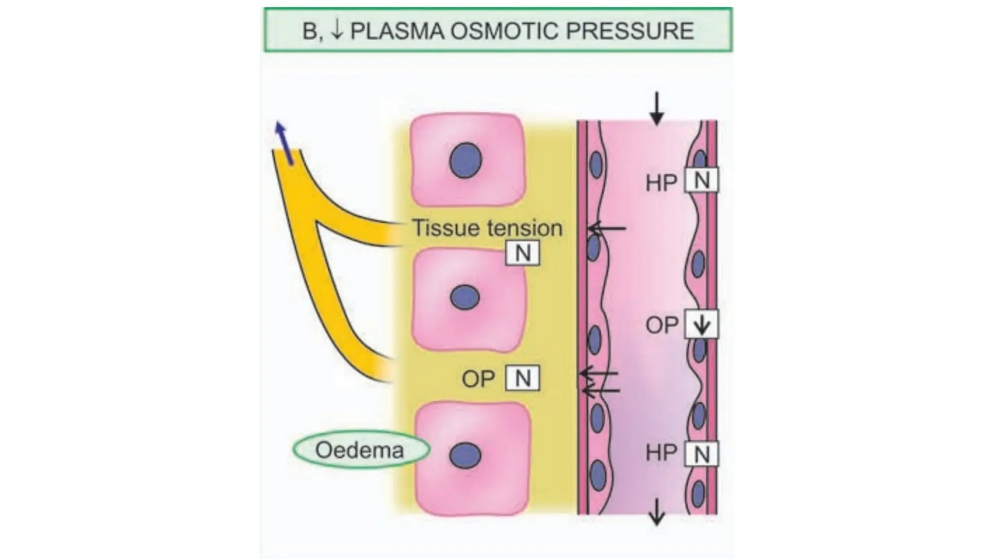
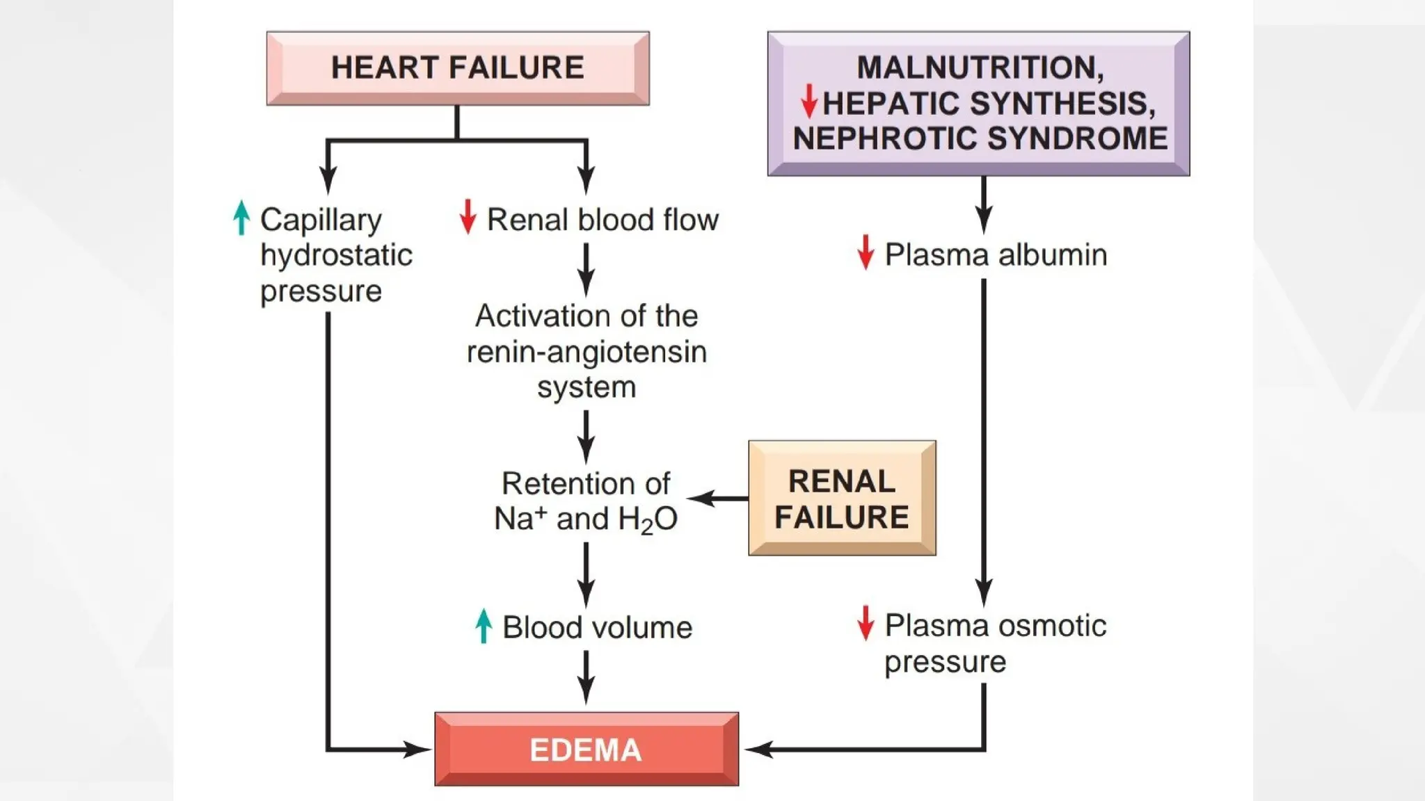
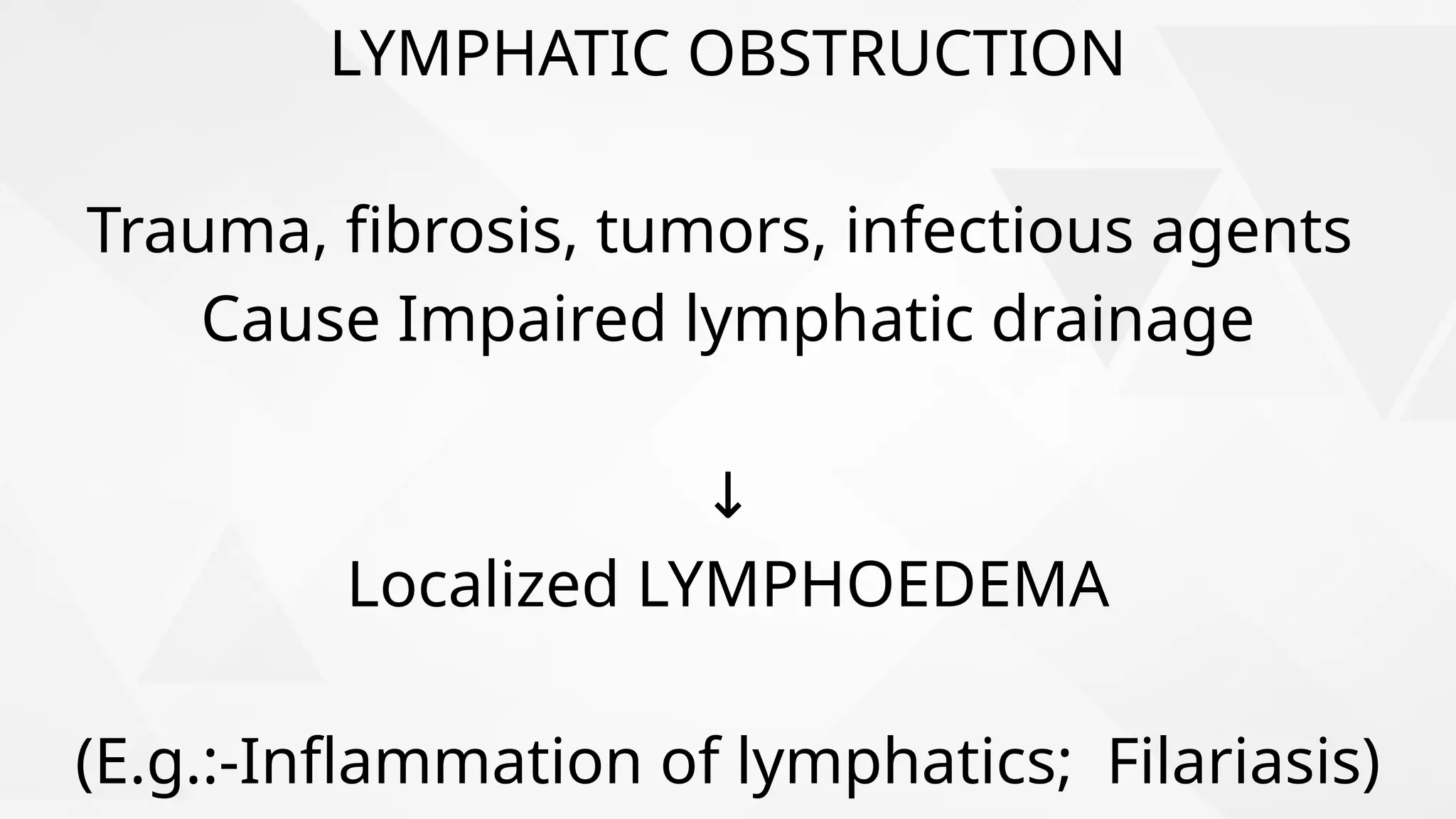
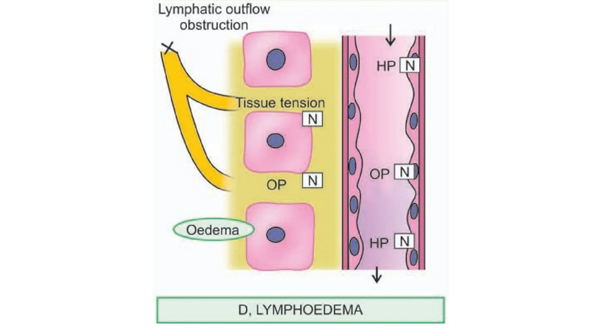
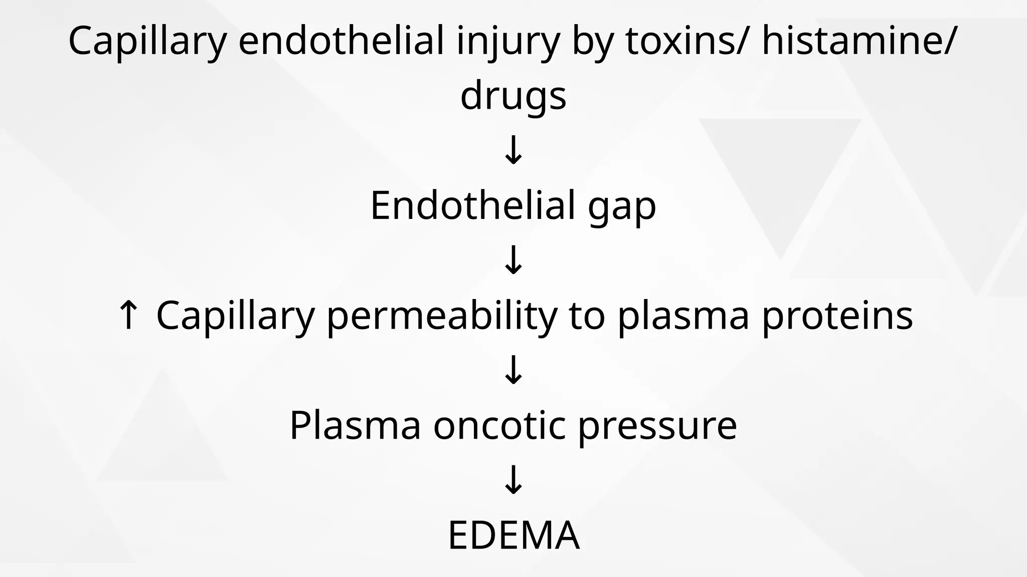
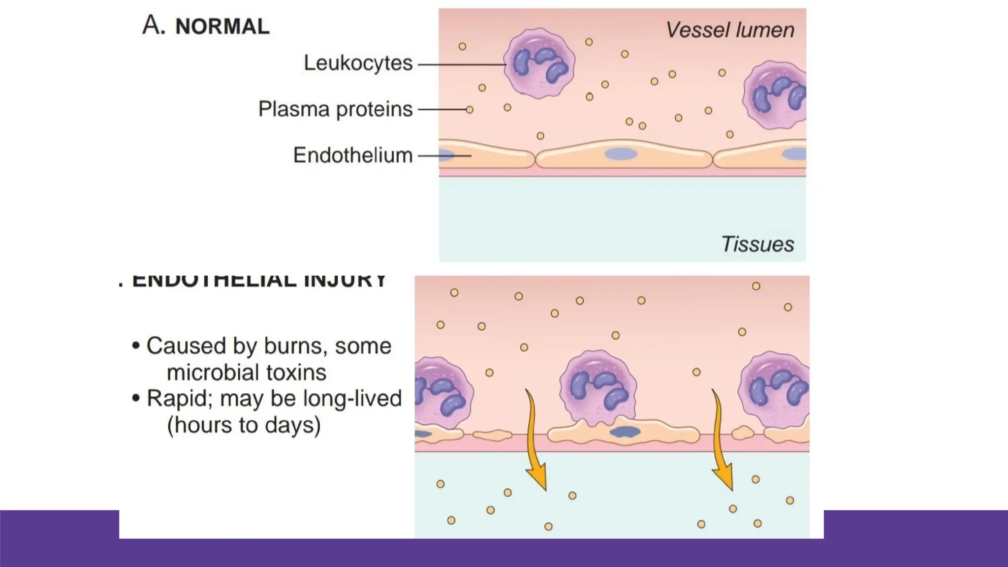
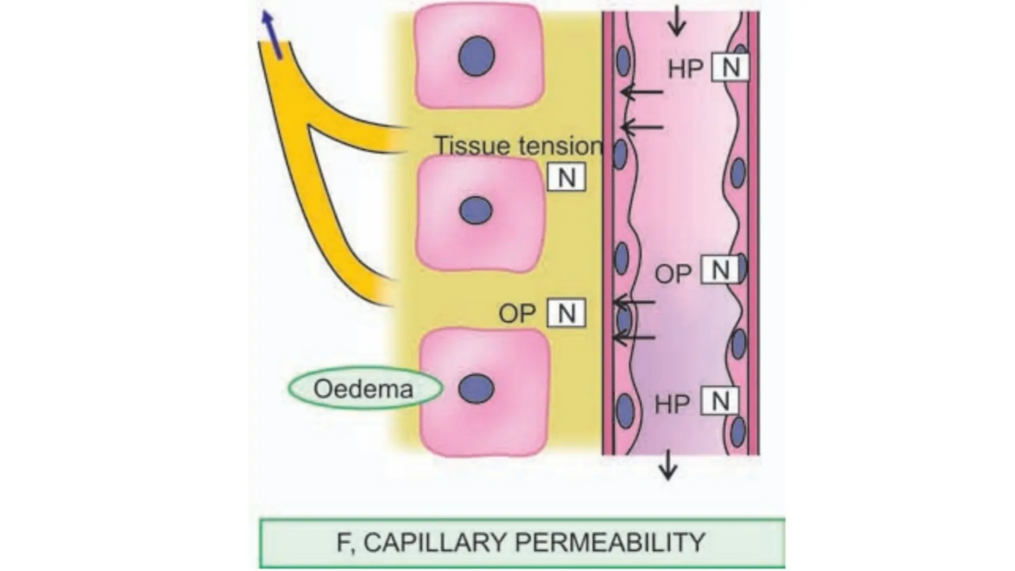
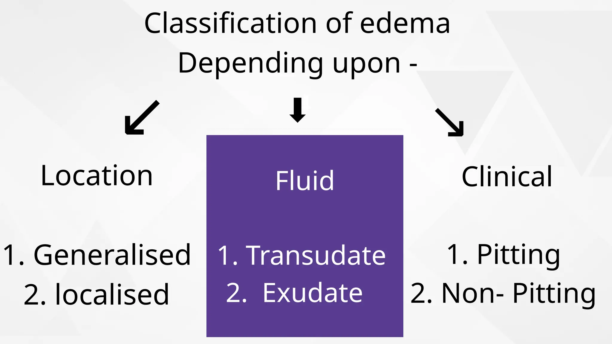
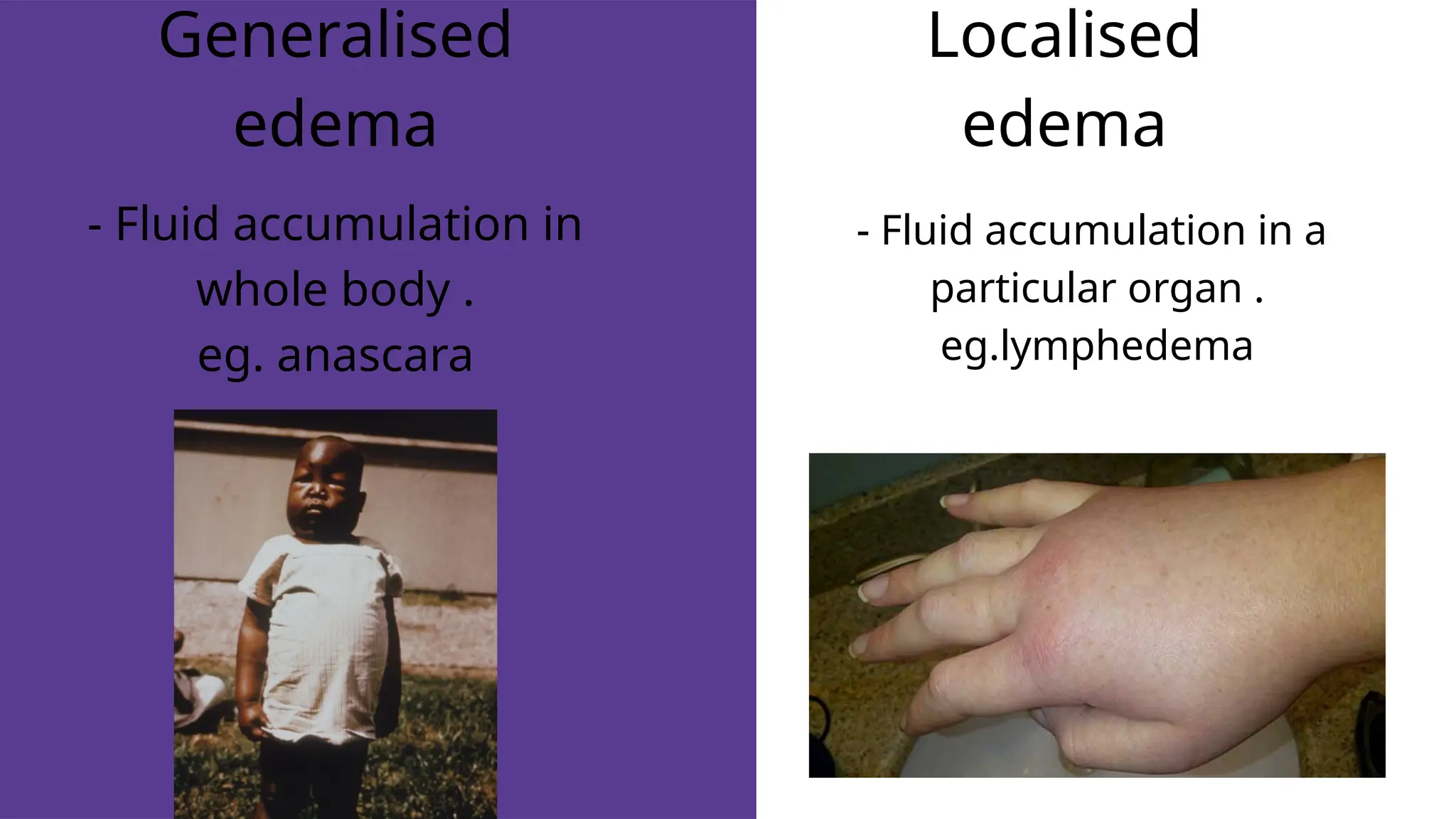
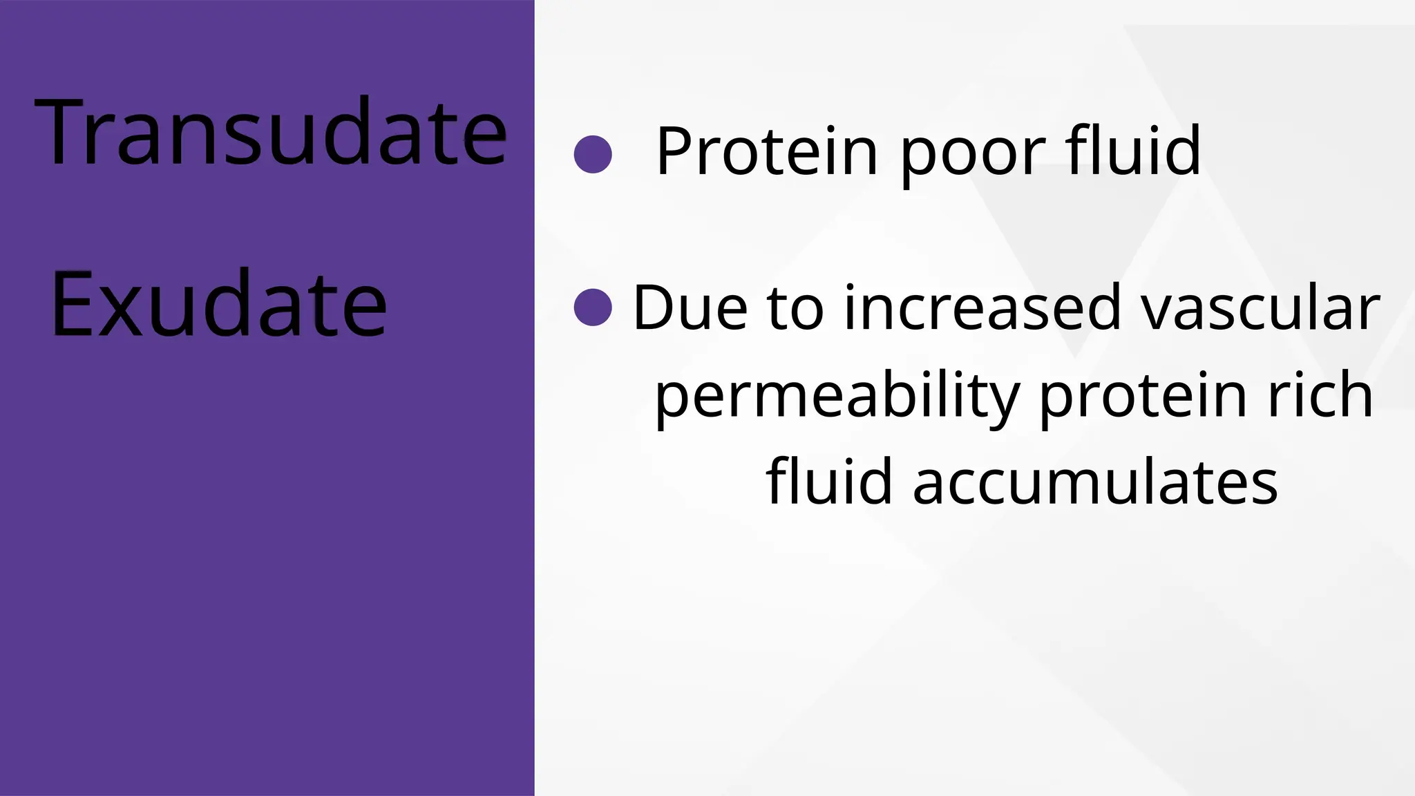
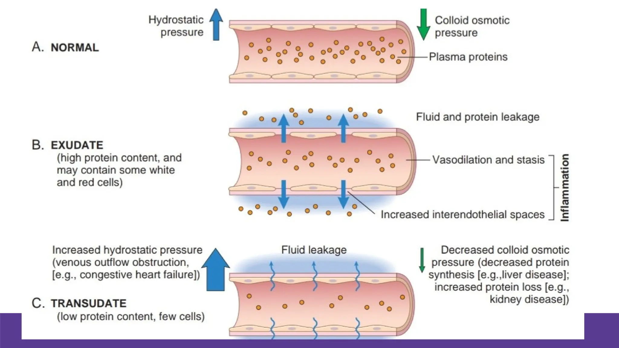
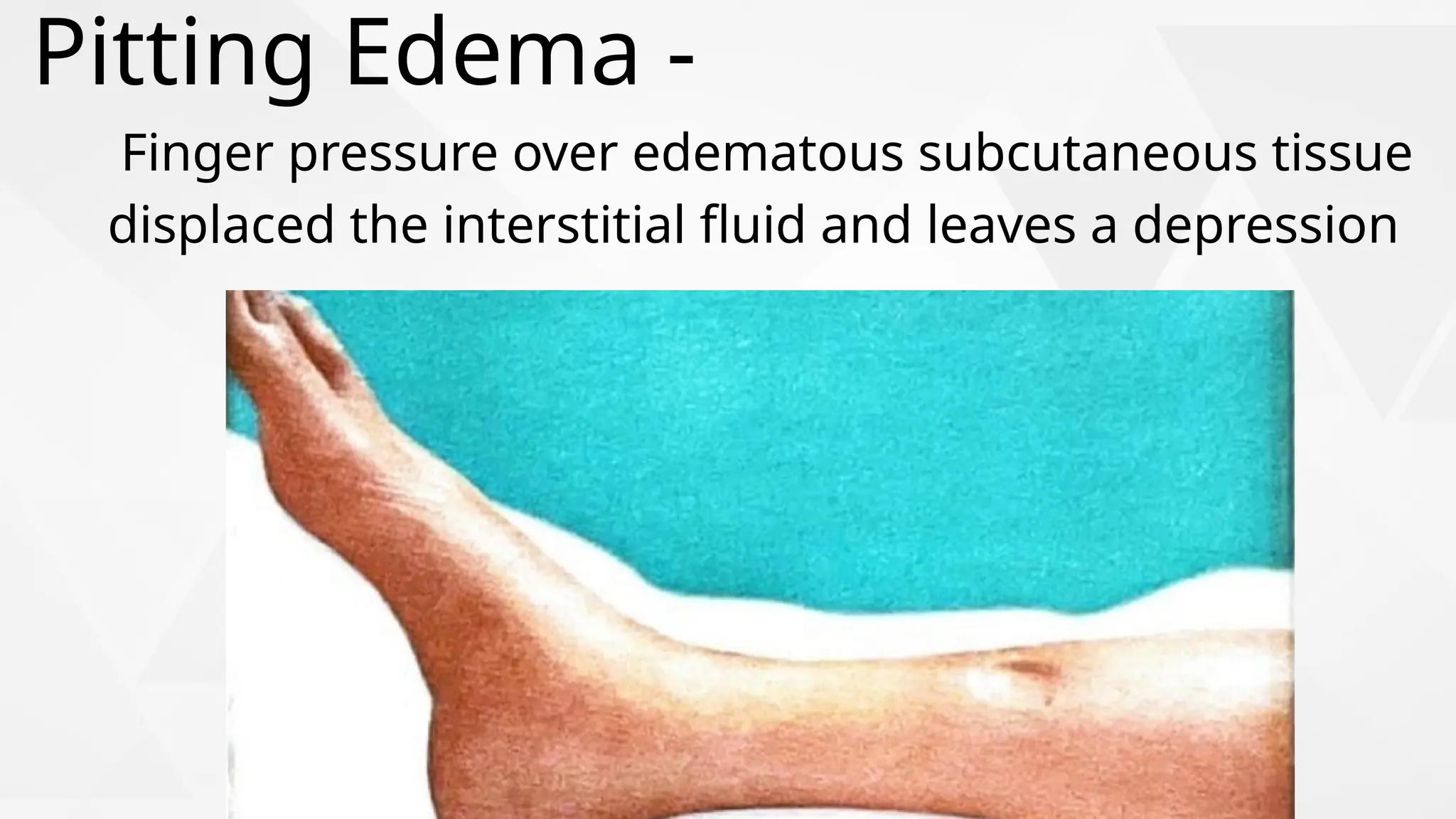
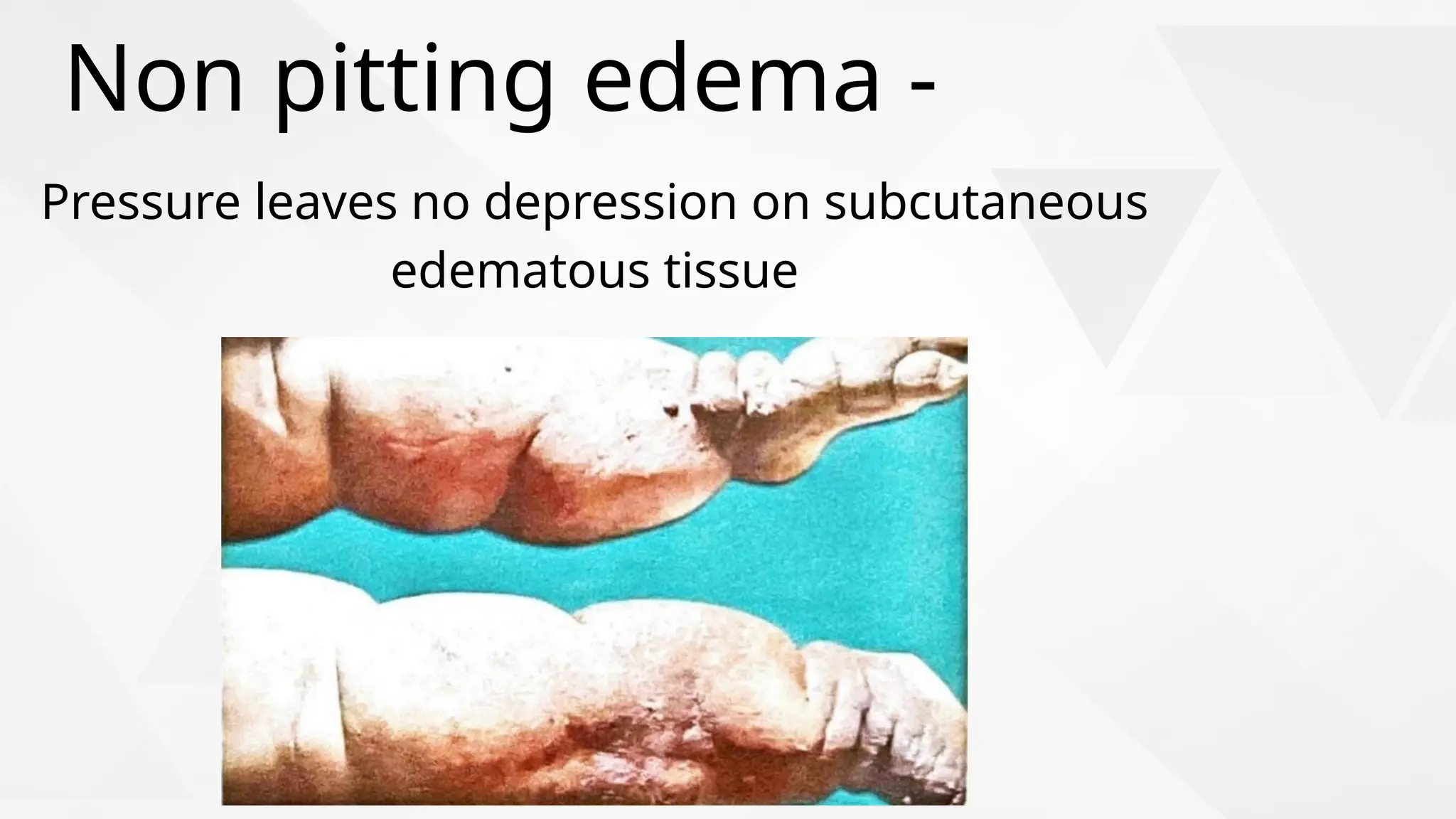
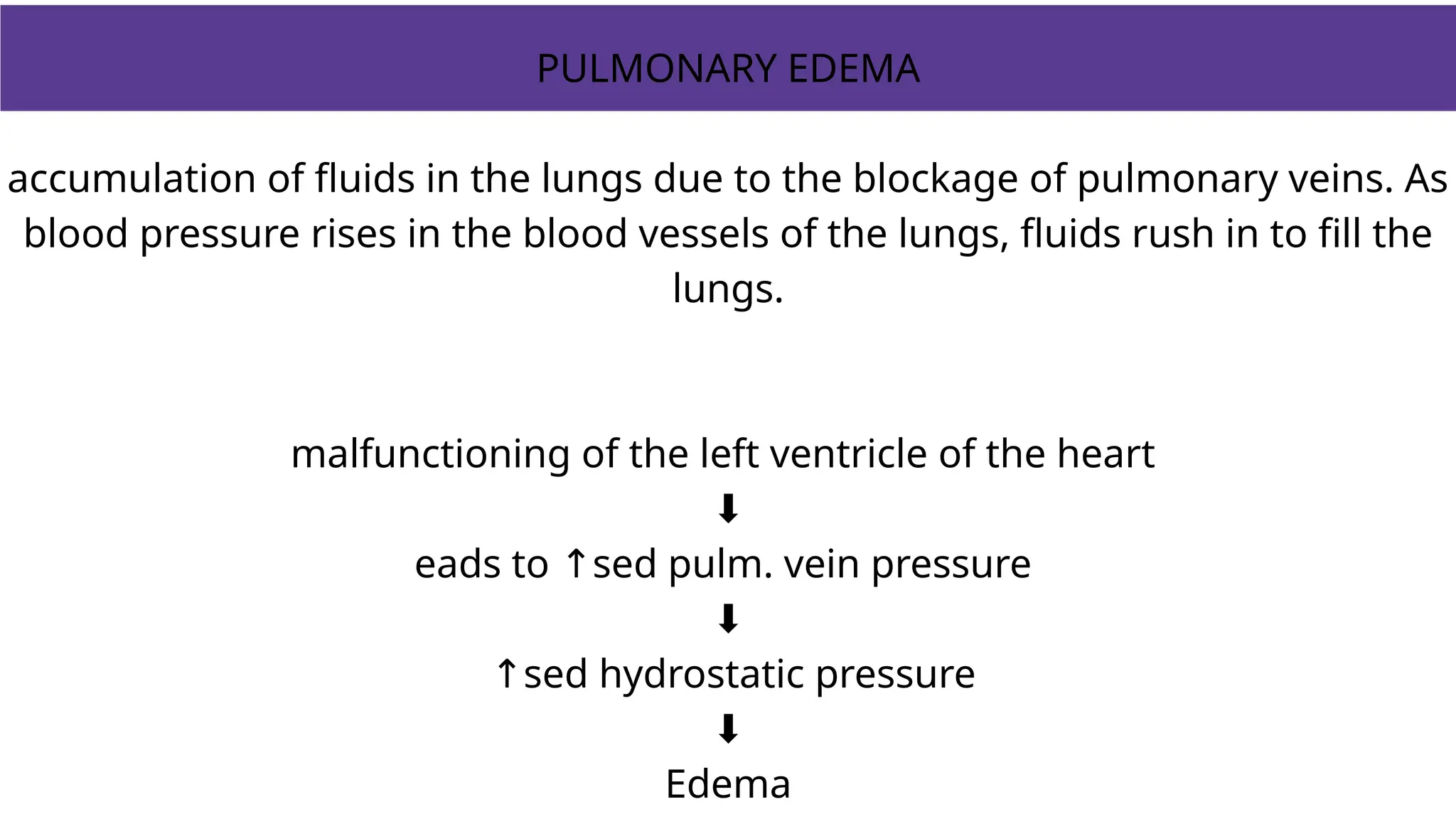
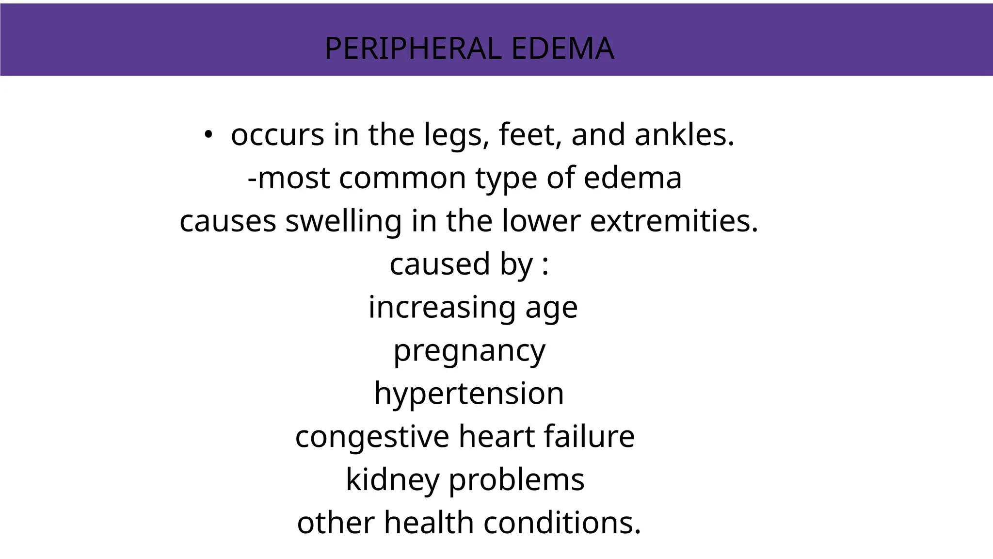
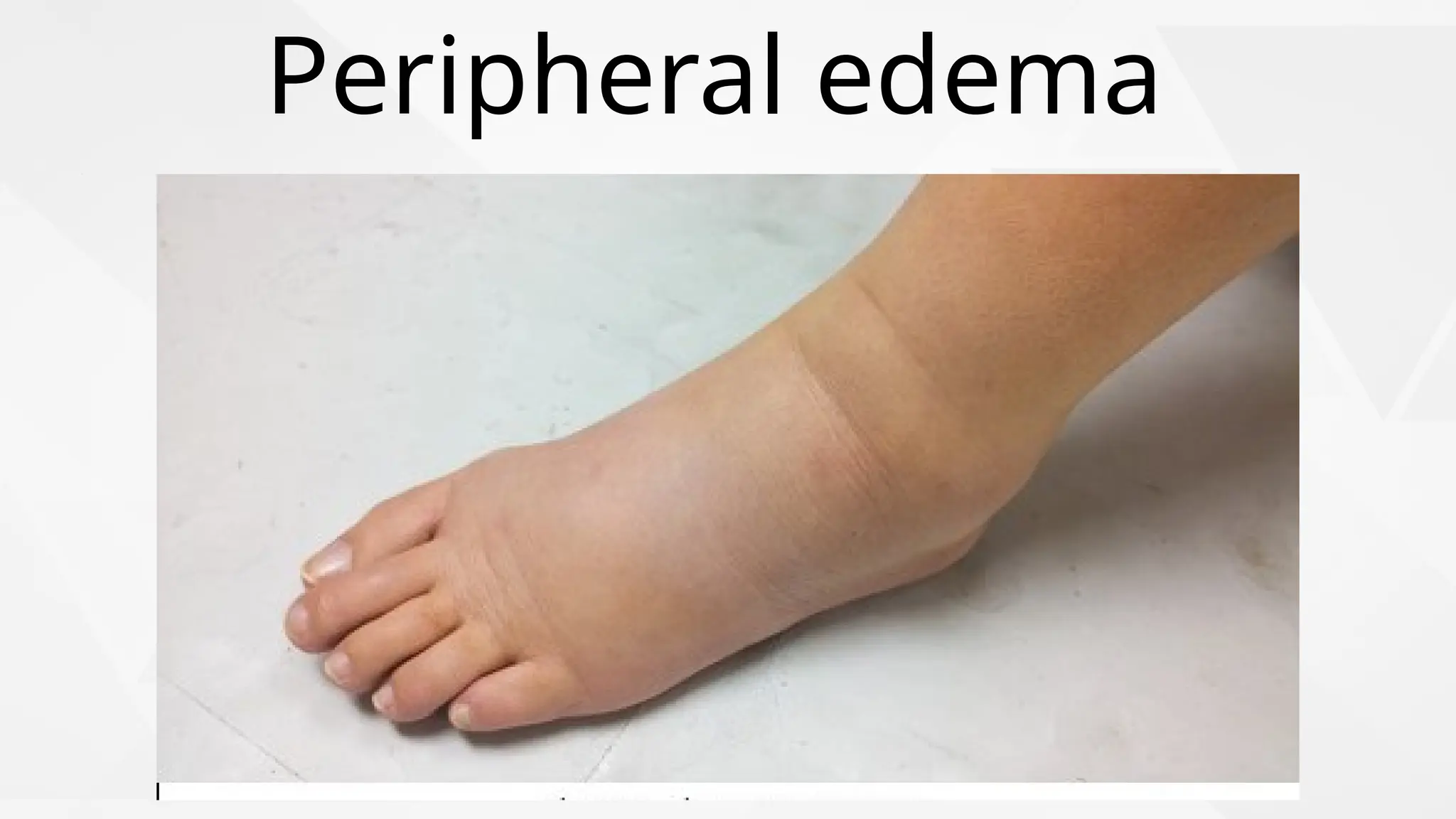
Edema
Presented by
aayudrishti.com
Definition
abnormal and excessive
accumulation of "free fluid" in the
interstitial tissue spaces and
serous cavities.
Free fluid in body cavities:
-ascites (if in the peritoneal cavity),
-hydrothorax or pleural effusion ( pleural
cavity),
-hydropericardium or pericardial effusion
( pericardial cavity).
Free fluid in interstitial space: fluid can be
displaced from one place to another.
. Normal amount of fluid in body
- 60% of body weight
2/3 -intracellular
1/3- extracellular
Extracellular is further divided into --:interstitial
fluid
-: intravascular fluid
Normal fluid circulation
-The movement of vascular fluid is maintained by vascular
hydrostatic pressure and plama collid osmotic pressure.
-Hydrostatic pressure is more at arterial end and plasma
oncontic pressure is more at venous end of capillaries.
-Small net movement of fluid into interstitium drains into
lymphatic vessel.
Causes of
edema
1. Increased hydrostatic pressure
2. Reduced plama osmotic pressure
3. Sodium-water retention
4. Lymphatic obstruction
5. Increased vascular permeability
INCREASED CAPILLARY HYDROSTATIC PRESSURE
Rise in hydrostatic pressure at the venular end of capillaries to a level
more than plasma oncotic pressure
↓
Minimal/ No reabsorption of fluid at venular end
↓
EDEMA
Reduced Plasma Oncotic Pressure
Reduced albumin synthesis in liver / protein malnutrition
↓
Fall in plasma oncotic pressure
↓
Net movement of fluid into interstitial tissues
↓
EDEMA
Sodium water retention
In some cases of nephrotic syndrome blood
volume decrease
⬇️
Activate renin angiotensin aldosterone mechanism
⬇️
Sodium and water retention by the kidney
⬇️
blood volume increase
⬇️
Increase hydrostatic pressure
⬇️
Generalized edema.
LYMPHATIC OBSTRUCTION
Trauma, fibrosis, tumors, infectious agents
Cause Impaired lymphatic drainage
↓
Localized LYMPHOEDEMA
(E.g.:-Inflammation of lymphatics; Filariasis)
Capillary endothelial injury by toxins/ histamine/
drugs
↓
Endothelial gap
↓
↑ Capillary permeability to plasma proteins
↓
Plasma oncotic pressure
↓
EDEMA
Classification of edema
Depending upon -
Location
1. Generalised
2. localised
Clinical
1. Pitting
2. Non- Pitting
Fluid
1. Transudate
2. Exudate
⬇️
⬇️
↙️ ↘️
Generalised
edema
Localised
edema
- Fluid accumulation in
whole body .
eg. anascara
- Fluid accumulation in a
particular organ .
eg.lymphedema
Transudate Protein poor fluid
Exudate Due to increased vascular
permeability protein rich
fluid accumulates
Finger pressure over edematous subcutaneous tissue
displaced the interstitial fluid and leaves a depression
Pitting Edema -
1+ 2-mm depression, immediate rebound
2+ 4-mm deep pit, a few seconds to rebound
3+ 6-mm deep pit, 10-12 seconds to rebound
4+ 8-mm deep pit, >20 seconds to rebound
Grading of Pitting Edema
Non pitting edema -
Pressure leaves no depression on subcutaneous
edematous tissue
Major types
- Pulmonary
- Cerebral
- Peripheral
PULMONARY EDEMA
accumulation of fluids in the lungs due to the blockage of pulmonary veins. As
blood pressure rises in the blood vessels of the lungs, fluids rush in to fill the
lungs.
malfunctioning of the left ventricle of the heart
⬇️
eads to sed pulm. vein pressure
↑
⬇️
↑sed hydrostatic pressure
⬇️
Edema
Pulmonary edema
PERIPHERAL EDEMA
• occurs in the legs, feet, and ankles.
-most common type of edema
causes swelling in the lower extremities.
caused by :
increasing age
pregnancy
hypertension
congestive heart failure
kidney problems
other health conditions.
Peripheral edema
CEREBRAL EDEMA
-Fluids accumulates in the intracellular and
extracellular spaces of the brain.
-caused by metabolic abnormalities due to an
underlying disease or as a response to oxygen
deprivation at high altitudes.
- serious form of edema.
- can lead to loss of consciousness and brain
damage.
Cerebral edema
Thank
you!

Edema complete details ppt (definition, types).pptx

  • 1.
  • 2.
    Definition abnormal and excessive accumulationof "free fluid" in the interstitial tissue spaces and serous cavities.
  • 3.
    Free fluid inbody cavities: -ascites (if in the peritoneal cavity), -hydrothorax or pleural effusion ( pleural cavity), -hydropericardium or pericardial effusion ( pericardial cavity). Free fluid in interstitial space: fluid can be displaced from one place to another.
  • 4.
    . Normal amountof fluid in body - 60% of body weight 2/3 -intracellular 1/3- extracellular Extracellular is further divided into --:interstitial fluid -: intravascular fluid
  • 5.
    Normal fluid circulation -Themovement of vascular fluid is maintained by vascular hydrostatic pressure and plama collid osmotic pressure. -Hydrostatic pressure is more at arterial end and plasma oncontic pressure is more at venous end of capillaries. -Small net movement of fluid into interstitium drains into lymphatic vessel.
  • 8.
    Causes of edema 1. Increasedhydrostatic pressure 2. Reduced plama osmotic pressure 3. Sodium-water retention 4. Lymphatic obstruction 5. Increased vascular permeability
  • 9.
    INCREASED CAPILLARY HYDROSTATICPRESSURE Rise in hydrostatic pressure at the venular end of capillaries to a level more than plasma oncotic pressure ↓ Minimal/ No reabsorption of fluid at venular end ↓ EDEMA
  • 11.
    Reduced Plasma OncoticPressure Reduced albumin synthesis in liver / protein malnutrition ↓ Fall in plasma oncotic pressure ↓ Net movement of fluid into interstitial tissues ↓ EDEMA
  • 13.
    Sodium water retention Insome cases of nephrotic syndrome blood volume decrease ⬇️ Activate renin angiotensin aldosterone mechanism ⬇️ Sodium and water retention by the kidney ⬇️ blood volume increase ⬇️ Increase hydrostatic pressure ⬇️ Generalized edema.
  • 15.
    LYMPHATIC OBSTRUCTION Trauma, fibrosis,tumors, infectious agents Cause Impaired lymphatic drainage ↓ Localized LYMPHOEDEMA (E.g.:-Inflammation of lymphatics; Filariasis)
  • 17.
    Capillary endothelial injuryby toxins/ histamine/ drugs ↓ Endothelial gap ↓ ↑ Capillary permeability to plasma proteins ↓ Plasma oncotic pressure ↓ EDEMA
  • 20.
    Classification of edema Dependingupon - Location 1. Generalised 2. localised Clinical 1. Pitting 2. Non- Pitting Fluid 1. Transudate 2. Exudate ⬇️ ⬇️ ↙️ ↘️
  • 21.
    Generalised edema Localised edema - Fluid accumulationin whole body . eg. anascara - Fluid accumulation in a particular organ . eg.lymphedema
  • 22.
    Transudate Protein poorfluid Exudate Due to increased vascular permeability protein rich fluid accumulates
  • 24.
    Finger pressure overedematous subcutaneous tissue displaced the interstitial fluid and leaves a depression Pitting Edema -
  • 25.
    1+ 2-mm depression,immediate rebound 2+ 4-mm deep pit, a few seconds to rebound 3+ 6-mm deep pit, 10-12 seconds to rebound 4+ 8-mm deep pit, >20 seconds to rebound Grading of Pitting Edema
  • 26.
    Non pitting edema- Pressure leaves no depression on subcutaneous edematous tissue
  • 27.
    Major types - Pulmonary -Cerebral - Peripheral
  • 28.
    PULMONARY EDEMA accumulation offluids in the lungs due to the blockage of pulmonary veins. As blood pressure rises in the blood vessels of the lungs, fluids rush in to fill the lungs. malfunctioning of the left ventricle of the heart ⬇️ eads to sed pulm. vein pressure ↑ ⬇️ ↑sed hydrostatic pressure ⬇️ Edema
  • 29.
  • 30.
    PERIPHERAL EDEMA • occursin the legs, feet, and ankles. -most common type of edema causes swelling in the lower extremities. caused by : increasing age pregnancy hypertension congestive heart failure kidney problems other health conditions.
  • 31.
  • 32.
    CEREBRAL EDEMA -Fluids accumulatesin the intracellular and extracellular spaces of the brain. -caused by metabolic abnormalities due to an underlying disease or as a response to oxygen deprivation at high altitudes. - serious form of edema. - can lead to loss of consciousness and brain damage.
  • 33.
  • 34.