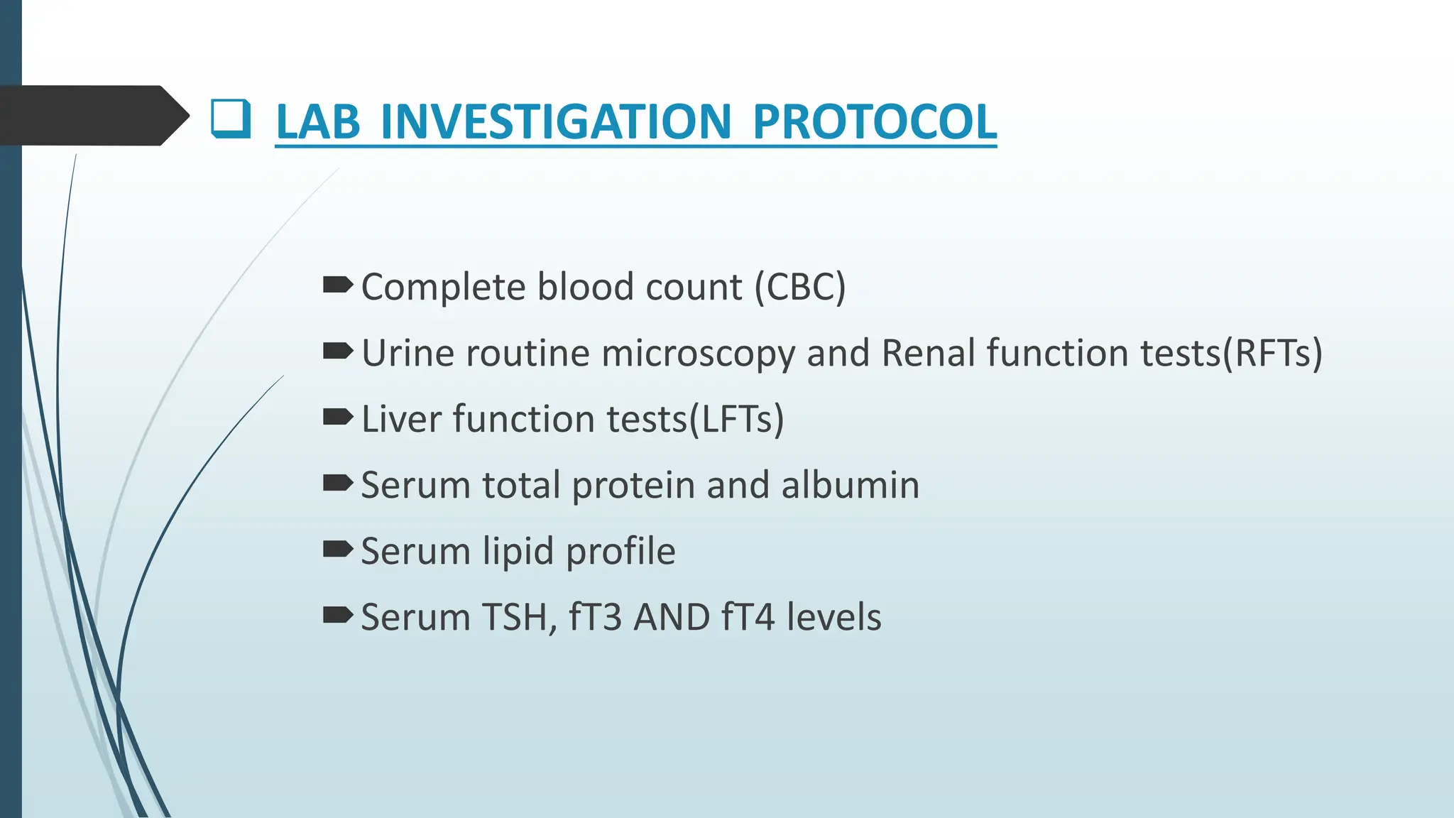
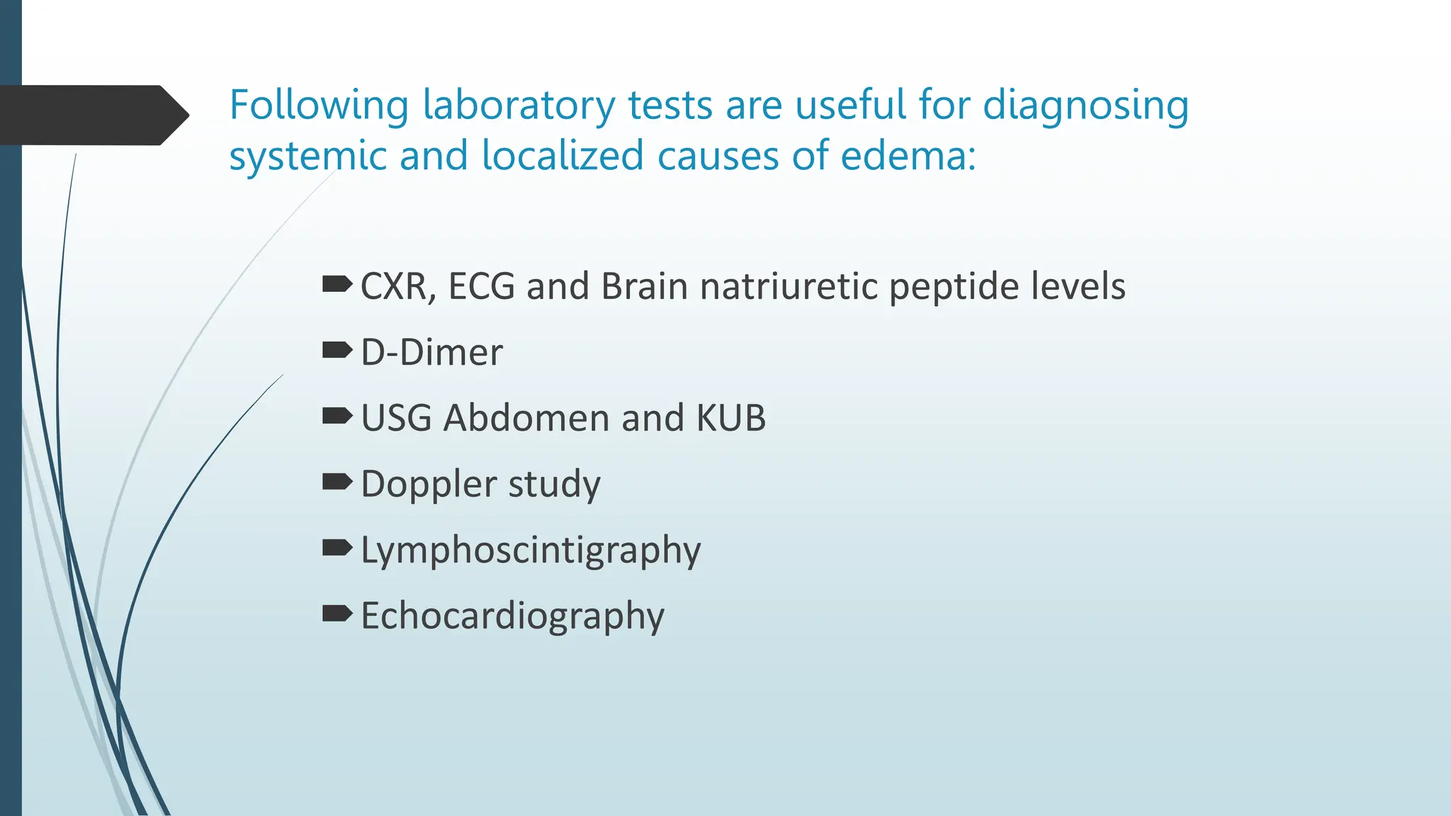
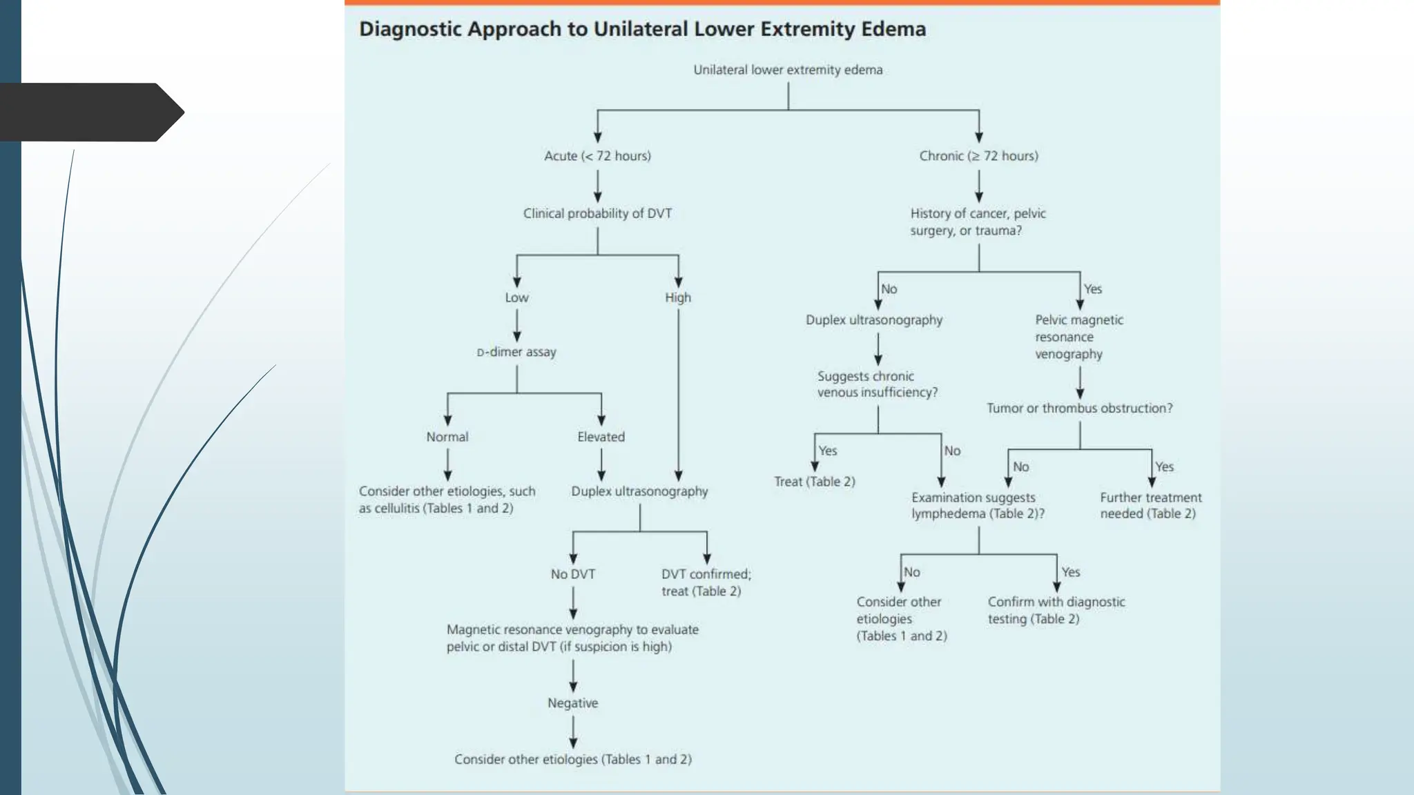
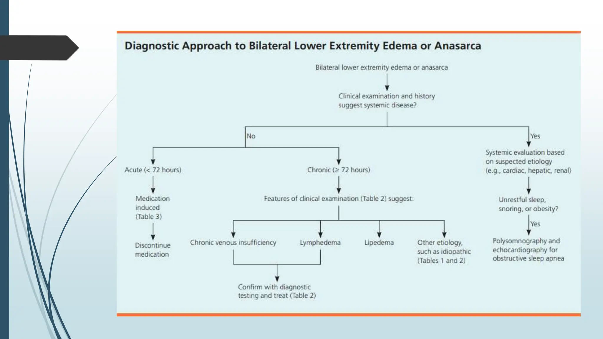
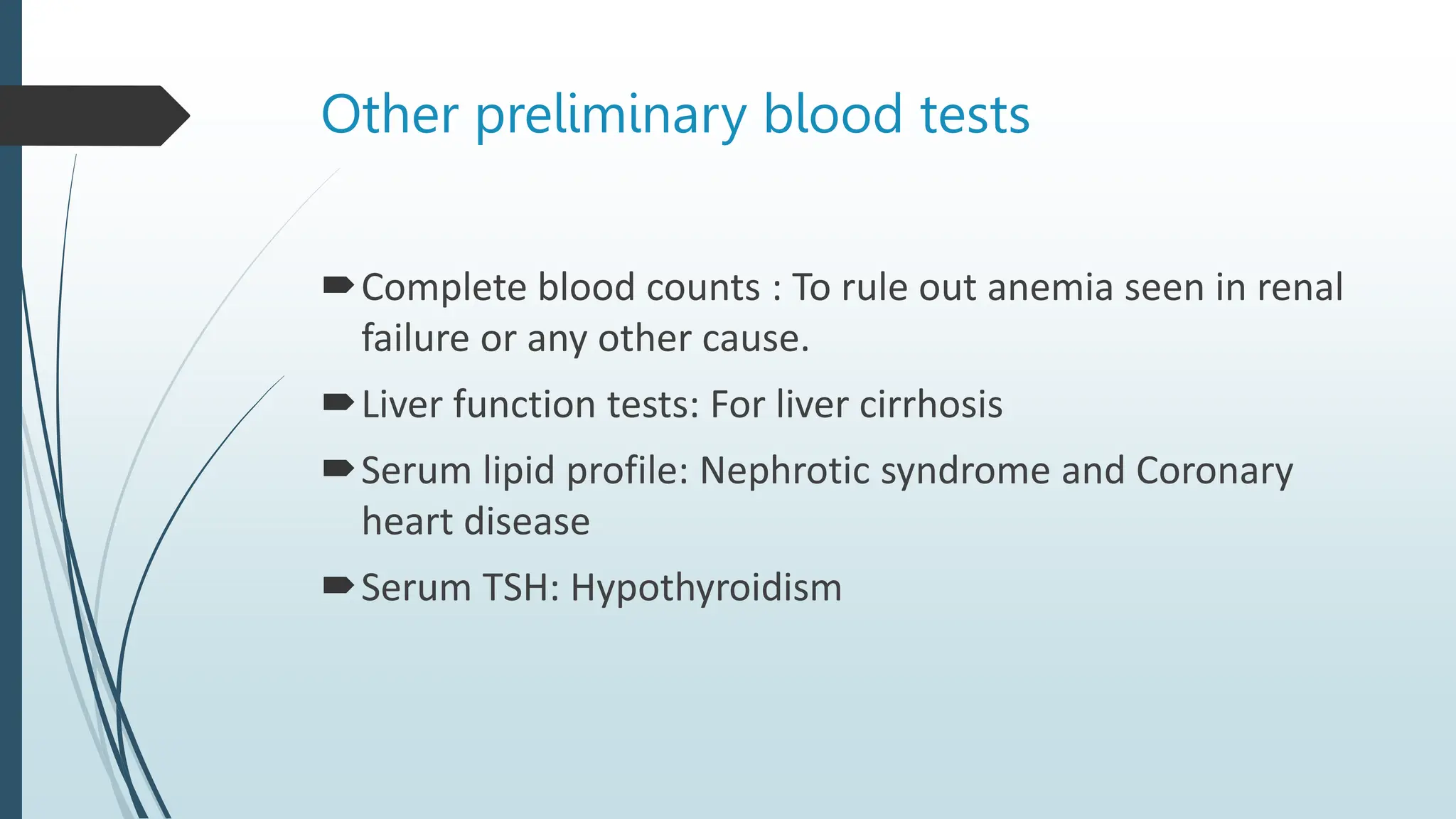
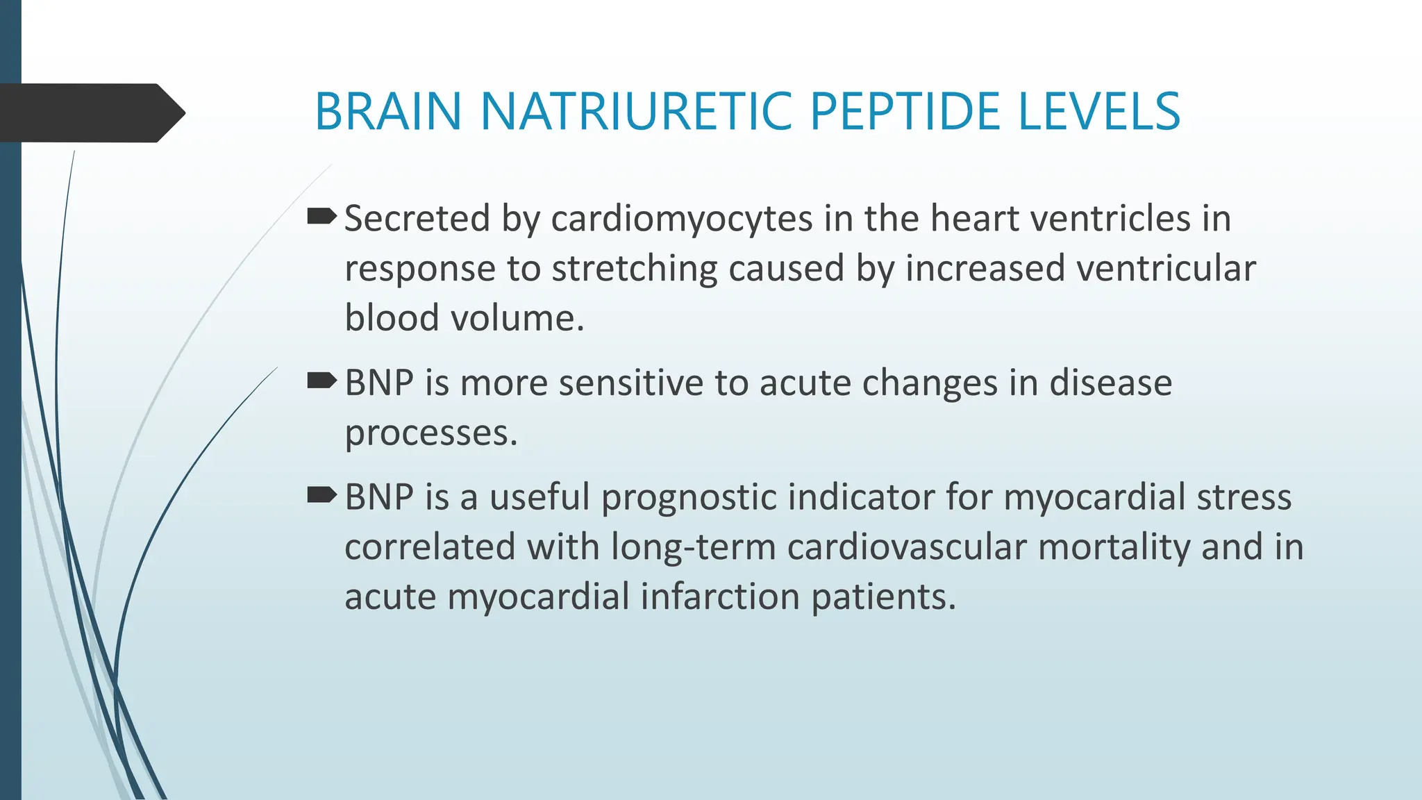
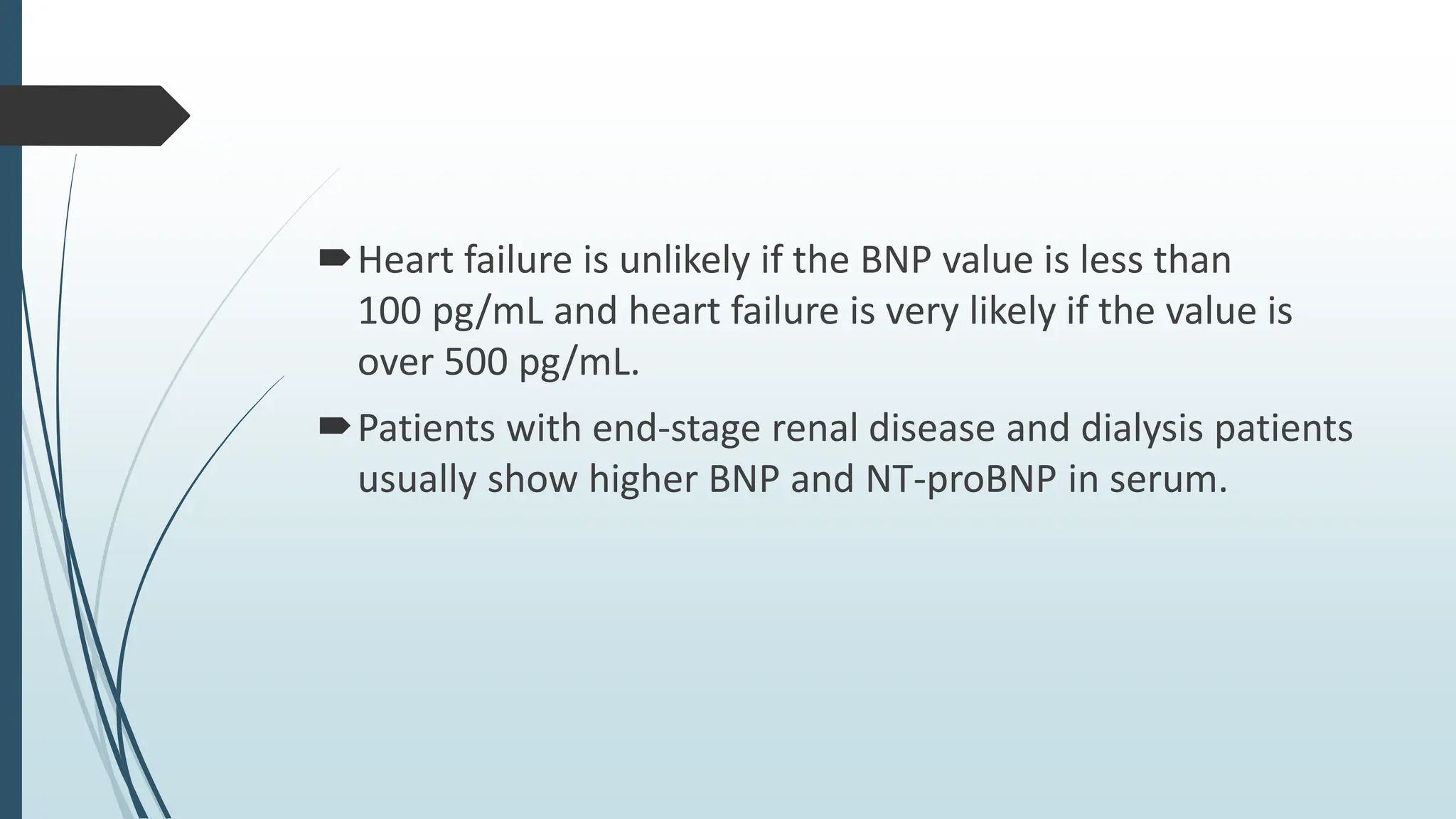
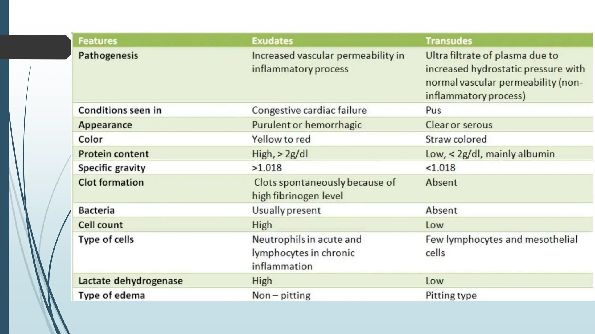
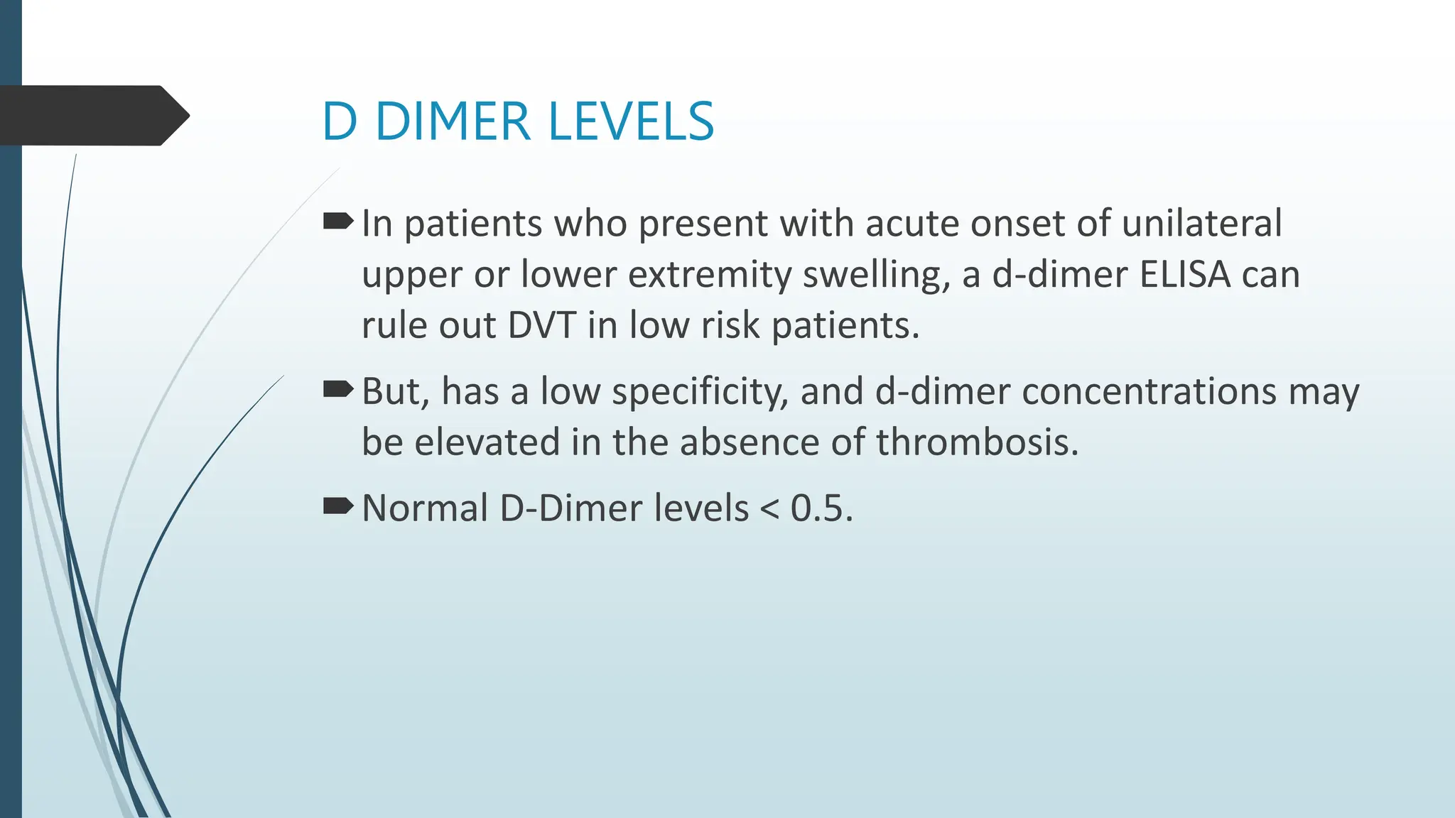
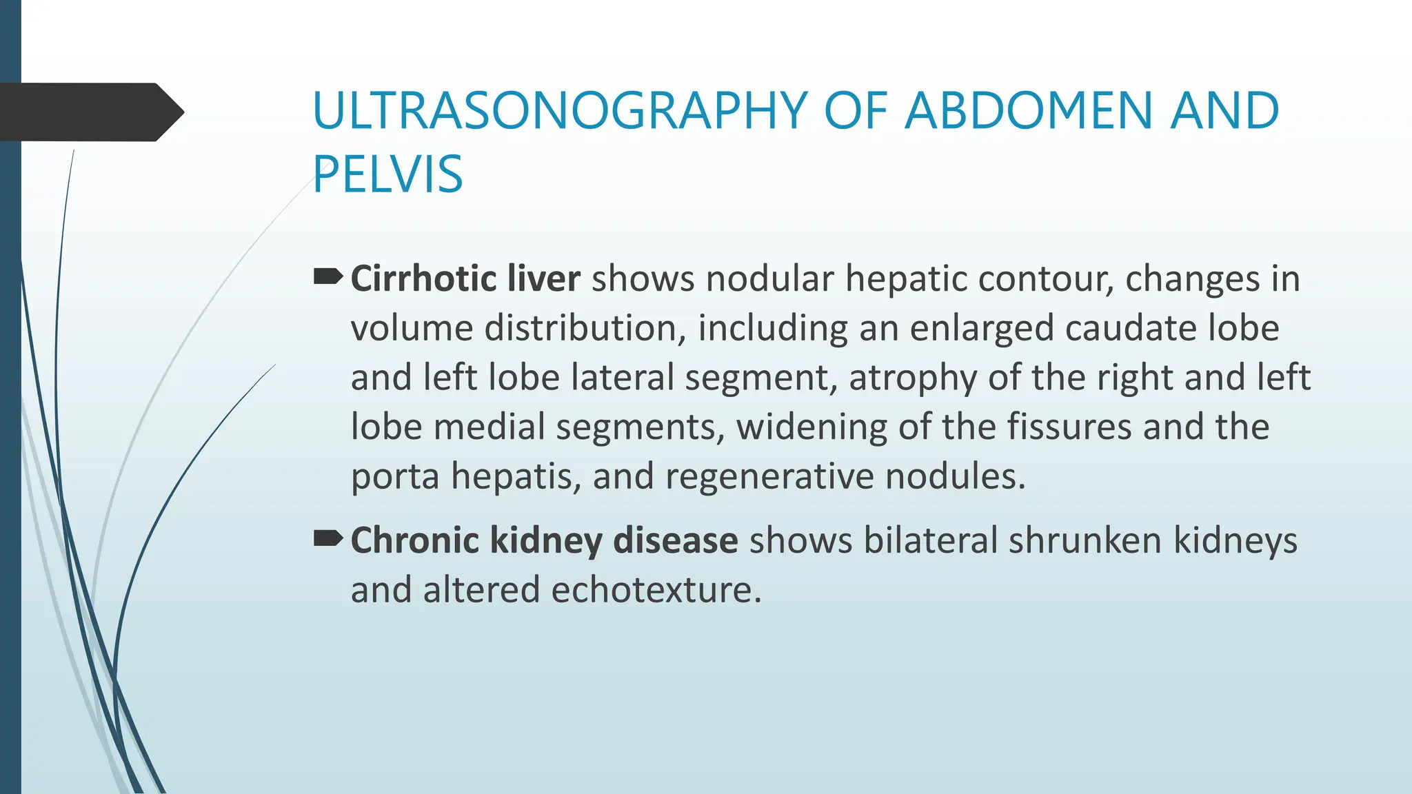
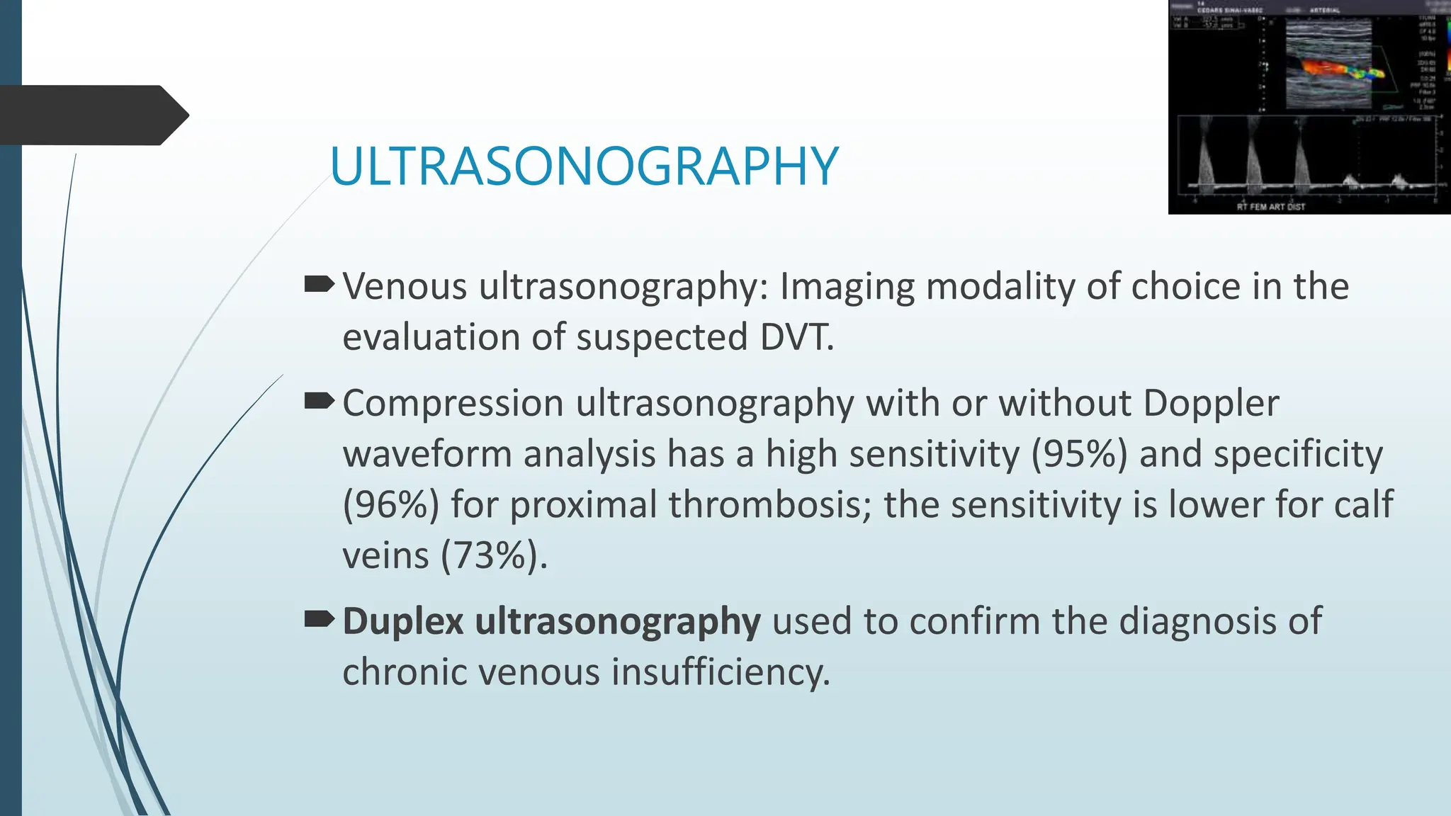
LAB INVESTIGATION
PROTOCOL OF A PATIENT
PRESENTING WITH
EDEMA
Speaker: Dr. Pratiksha Mishra
Moderator: Dr Goutami Dasnayak
EDEMA : OUTLINES
 What is edema
 Pathophysiology of edema
 Causes of edema
 Morphology and types of edema
 Laboratory investigation protocol
 Summary
 Definition
 Edema is the accumulation of fluid in interstitial tissues due to a net
movement of water into extravascular spaces.
 It is marked by swelling of tissues and accumulation of fluid in body
cavities, commonly in subcutaneous tissues, lungs and brain.
• 60% of body weight is water;
• Fluid (water) movement between the vascular and interstitial spaces is by
two opposing forces:
1) The hydrostatic pressure and
2) The osmotic pressure.
2/3rd : Intracellular
1/3rd : Extracellular
3/4th: Interstitial
1/4th : Intravascular
• Normally, outflow of fluid
produced by hydrostatic pressure
at the arteriolar end is balanced
by inflow at the venular end.
• This balance when disturbed by
pathologic conditions promote
edema.
 PATHOPHYSIOLOGY OF EDEMA
 CAUSES OF EDEMA
 MORPHOLOGY
• Edema is easily recognized on gross inspection.
• HPE shows clearing and separation of the extracellular
matrix elements.
• Edema most commonly found in subcutaneous tissues,
lungs and brain.
EDEMA » MORPHOLOGY » SUBCUTANEOUS EDEMA
• Finger pressure over affected
tissue displaces the interstitial
fluid, leaving a finger-shaped
depression; this appearance is
called pitting edema.
Fig: PITTING EDEMA; showing depression
 TYPES OF EDEMA
PITTING EDEMA NON PITTING EDEMA
• Chronic lymphedema
• Myxedema
• Angioneurotic edema
• Radiation injury
EDEMA » MORPHOLOGY » PULMONARY EDEMA
Fig: PULMONARY EDEMA
• Grossly, 2-3 times the normal
weight
• C/S: Frothy, blood tinted fluid
consisting of mixture f air, fluid
and extravasated red cells
EDEMA » MORPHOLOGY » BRAIN EDEMA
• Brain edema can be localized or
generalized
• With generalized edema, the sulci are
narrowed as the gyri swell and become
flattened against the skull.
Fig: BRAIN EDEMA
 LAB INVESTIGATION PROTOCOL
Complete blood count (CBC)
Urine routine microscopy and Renal function tests(RFTs)
Liver function tests(LFTs)
Serum total protein and albumin
Serum lipid profile
Serum TSH, fT3 AND fT4 levels
Following laboratory tests are useful for diagnosing
systemic and localized causes of edema:
CXR, ECG and Brain natriuretic peptide levels
D-Dimer
USG Abdomen and KUB
Doppler study
Lymphoscintigraphy
Echocardiography
SERUM TOTAL PROTEIN and ALBUMIN
The normal range of albumin is 3.5 to 5.5 g/dL or 35-55
g/liter.
Normal serum protein: 60-80g/L
• Increased loss – Nephrotic syndrome
• Decreased synthesis – Chronic liver disease
• Malabsorption – Protein-losing enteropathy, e.g. Crohn’s
disease and coeliac disease
• Malnutrition – Kwashiorkar
Composition of normal urine (24 hrs) in adults
19
Sl
No
Parameters Values Sl
No
Parameters Values
1 Volume 600-2000 ml 10 Ureanitrogen 12-20 gm
2 Specific gravity 1.003-1.030 11 Uric Acid 250-750 mg
3 Osmolality 300-900
mOsm/kg
12 Sodium 40-200 mEq
4 pH 4.6-8.0 13 Potassium 25-125 mEq
5 Glucose < 0.5 gm 14 Chloride 110-250 mEq
6 Proteins < 150 mg 15 Calcium (low
calcium diet)
50-150 mg
7 Urobilinogen 0.5-4.0 gm 16 Formiminogluta
micacid (FIGlu)
< 3 mg
8 Prophobilinog
en
0.2 mg 17 Red cells,
epithelial cells
and white blood
cells
≤ 1-2 per high
power field
9 Creatinine M: 14-26 mg/kg
F: 11-20 mg/kg
URINE ANALYSIS
24-hour urine collection can help identify the cause of
generalized edema by showing the degree of proteinuria.
1. Edema associated with slight or no proteinuria (less than
0.5 g/24 hours): CHF, cirrhosis, hypertension, malnutrition,
inferior vena cava thrombosis below the renal veins,
hypothyroidism, and varicose veins.
2. Edema associated with significant proteinuria (greater
than 0.5 g/24 hours, generally 2 g/24 hours or
more): Glomerular diseases, preeclampsia,
tubulointerstitial disease, hypertension, and vascular
diseases.
3. Edema associated with heavy proteinuria (3 g/24 hours or
more): Consistent with both primary(MCD, MPGN,FSGS) and
secondary(Diabetic, amyloidotic) glomerular diseases.
Other preliminary blood tests
Complete blood counts : To rule out anemia seen in renal
failure or any other cause.
Liver function tests: For liver cirrhosis
Serum lipid profile: Nephrotic syndrome and Coronary
heart disease
Serum TSH: Hypothyroidism
BRAIN NATRIURETIC PEPTIDE LEVELS
Secreted by cardiomyocytes in the heart ventricles in
response to stretching caused by increased ventricular
blood volume.
BNP is more sensitive to acute changes in disease
processes.
BNP is a useful prognostic indicator for myocardial stress
correlated with long-term cardiovascular mortality and in
acute myocardial infarction patients.
Heart failure is unlikely if the BNP value is less than
100 pg/mL and heart failure is very likely if the value is
over 500 pg/mL.
Patients with end-stage renal disease and dialysis patients
usually show higher BNP and NT-proBNP in serum.
D DIMER LEVELS
In patients who present with acute onset of unilateral
upper or lower extremity swelling, a d-dimer ELISA can
rule out DVT in low risk patients.
But, has a low specificity, and d-dimer concentrations may
be elevated in the absence of thrombosis.
Normal D-Dimer levels < 0.5.
ULTRASONOGRAPHY OF ABDOMEN AND
PELVIS
Cirrhotic liver shows nodular hepatic contour, changes in
volume distribution, including an enlarged caudate lobe
and left lobe lateral segment, atrophy of the right and left
lobe medial segments, widening of the fissures and the
porta hepatis, and regenerative nodules.
Chronic kidney disease shows bilateral shrunken kidneys
and altered echotexture.
ULTRASONOGRAPHY
Venous ultrasonography: Imaging modality of choice in the
evaluation of suspected DVT.
Compression ultrasonography with or without Doppler
waveform analysis has a high sensitivity (95%) and specificity
(96%) for proximal thrombosis; the sensitivity is lower for calf
veins (73%).
Duplex ultrasonography used to confirm the diagnosis of
chronic venous insufficiency.
LYMPHOSCINTIGRAPHY
Lymph flow cannot be detected with
ultrasonography.
Indirect radionuclide
lymphoscintigraphy, shows absent
or delayed filling of lymphatic
channels, is the method of choice for
evaluating lymphedema when the
diagnosis cannot be made clinically
MAGNETIC RESONANCE IMAGING
Magnetic resonance angiography(MRA) with
venography of the lower extremity and pelvis can be used
to evaluate for intrinsic or extrinsic pelvic or thigh DVT.
MRI may aid in the diagnosis of musculoskeletal
etiologies, such as a gastrocnemius tear or popliteal cyst
causing unilateral edema.
Others
Echocardiography should be performed in patients with
obesity, obstructive sleep apnea, and edema to evaluate
pulmonary arterial pressures.
Ankle-brachial index measured in patients with chronic
venous insufficiency and cardiovascular risk factors before
initiation of compression therapy, which is
contraindicated in peripheral arterial disease
• Resolve the underlying cause.
• Cases of heart or kidney disease, are treated with diuretics.
• Positioning the affected body parts to improve drainage,
• Compression can be used to pressurize tissue in a limb, forcing
fluids; both blood and lymph, to flow out of the compressed area.
 EDEMA» TREATMENT
REFERENCES
1. Robbins BASIC PATHOLOGY, 10th Edition
2. Harrison’s principles of internal medicine, 19th edition
3. Uptodate
4. American academy of family physician guidelines
https://www.aafp.org/pubs/afp/issues/2013/0715/p102.html
QUESTIONS ??
Thank You ☺

Pathophysiology of edema and its applied aspects

  • 1.
    LAB INVESTIGATION PROTOCOL OFA PATIENT PRESENTING WITH EDEMA Speaker: Dr. Pratiksha Mishra Moderator: Dr Goutami Dasnayak
  • 2.
    EDEMA : OUTLINES What is edema  Pathophysiology of edema  Causes of edema  Morphology and types of edema  Laboratory investigation protocol  Summary
  • 3.
     Definition  Edemais the accumulation of fluid in interstitial tissues due to a net movement of water into extravascular spaces.  It is marked by swelling of tissues and accumulation of fluid in body cavities, commonly in subcutaneous tissues, lungs and brain.
  • 4.
    • 60% ofbody weight is water; • Fluid (water) movement between the vascular and interstitial spaces is by two opposing forces: 1) The hydrostatic pressure and 2) The osmotic pressure. 2/3rd : Intracellular 1/3rd : Extracellular 3/4th: Interstitial 1/4th : Intravascular
  • 5.
    • Normally, outflowof fluid produced by hydrostatic pressure at the arteriolar end is balanced by inflow at the venular end. • This balance when disturbed by pathologic conditions promote edema.
  • 6.
  • 7.
  • 9.
     MORPHOLOGY • Edemais easily recognized on gross inspection. • HPE shows clearing and separation of the extracellular matrix elements. • Edema most commonly found in subcutaneous tissues, lungs and brain.
  • 10.
    EDEMA » MORPHOLOGY» SUBCUTANEOUS EDEMA • Finger pressure over affected tissue displaces the interstitial fluid, leaving a finger-shaped depression; this appearance is called pitting edema. Fig: PITTING EDEMA; showing depression
  • 11.
     TYPES OFEDEMA PITTING EDEMA NON PITTING EDEMA • Chronic lymphedema • Myxedema • Angioneurotic edema • Radiation injury
  • 12.
    EDEMA » MORPHOLOGY» PULMONARY EDEMA Fig: PULMONARY EDEMA • Grossly, 2-3 times the normal weight • C/S: Frothy, blood tinted fluid consisting of mixture f air, fluid and extravasated red cells
  • 13.
    EDEMA » MORPHOLOGY» BRAIN EDEMA • Brain edema can be localized or generalized • With generalized edema, the sulci are narrowed as the gyri swell and become flattened against the skull. Fig: BRAIN EDEMA
  • 14.
     LAB INVESTIGATIONPROTOCOL Complete blood count (CBC) Urine routine microscopy and Renal function tests(RFTs) Liver function tests(LFTs) Serum total protein and albumin Serum lipid profile Serum TSH, fT3 AND fT4 levels
  • 15.
    Following laboratory testsare useful for diagnosing systemic and localized causes of edema: CXR, ECG and Brain natriuretic peptide levels D-Dimer USG Abdomen and KUB Doppler study Lymphoscintigraphy Echocardiography
  • 18.
    SERUM TOTAL PROTEINand ALBUMIN The normal range of albumin is 3.5 to 5.5 g/dL or 35-55 g/liter. Normal serum protein: 60-80g/L • Increased loss – Nephrotic syndrome • Decreased synthesis – Chronic liver disease • Malabsorption – Protein-losing enteropathy, e.g. Crohn’s disease and coeliac disease • Malnutrition – Kwashiorkar
  • 19.
    Composition of normalurine (24 hrs) in adults 19 Sl No Parameters Values Sl No Parameters Values 1 Volume 600-2000 ml 10 Ureanitrogen 12-20 gm 2 Specific gravity 1.003-1.030 11 Uric Acid 250-750 mg 3 Osmolality 300-900 mOsm/kg 12 Sodium 40-200 mEq 4 pH 4.6-8.0 13 Potassium 25-125 mEq 5 Glucose < 0.5 gm 14 Chloride 110-250 mEq 6 Proteins < 150 mg 15 Calcium (low calcium diet) 50-150 mg 7 Urobilinogen 0.5-4.0 gm 16 Formiminogluta micacid (FIGlu) < 3 mg 8 Prophobilinog en 0.2 mg 17 Red cells, epithelial cells and white blood cells ≤ 1-2 per high power field 9 Creatinine M: 14-26 mg/kg F: 11-20 mg/kg
  • 20.
    URINE ANALYSIS 24-hour urinecollection can help identify the cause of generalized edema by showing the degree of proteinuria. 1. Edema associated with slight or no proteinuria (less than 0.5 g/24 hours): CHF, cirrhosis, hypertension, malnutrition, inferior vena cava thrombosis below the renal veins, hypothyroidism, and varicose veins.
  • 21.
    2. Edema associatedwith significant proteinuria (greater than 0.5 g/24 hours, generally 2 g/24 hours or more): Glomerular diseases, preeclampsia, tubulointerstitial disease, hypertension, and vascular diseases. 3. Edema associated with heavy proteinuria (3 g/24 hours or more): Consistent with both primary(MCD, MPGN,FSGS) and secondary(Diabetic, amyloidotic) glomerular diseases.
  • 22.
    Other preliminary bloodtests Complete blood counts : To rule out anemia seen in renal failure or any other cause. Liver function tests: For liver cirrhosis Serum lipid profile: Nephrotic syndrome and Coronary heart disease Serum TSH: Hypothyroidism
  • 23.
    BRAIN NATRIURETIC PEPTIDELEVELS Secreted by cardiomyocytes in the heart ventricles in response to stretching caused by increased ventricular blood volume. BNP is more sensitive to acute changes in disease processes. BNP is a useful prognostic indicator for myocardial stress correlated with long-term cardiovascular mortality and in acute myocardial infarction patients.
  • 24.
    Heart failure isunlikely if the BNP value is less than 100 pg/mL and heart failure is very likely if the value is over 500 pg/mL. Patients with end-stage renal disease and dialysis patients usually show higher BNP and NT-proBNP in serum.
  • 26.
    D DIMER LEVELS Inpatients who present with acute onset of unilateral upper or lower extremity swelling, a d-dimer ELISA can rule out DVT in low risk patients. But, has a low specificity, and d-dimer concentrations may be elevated in the absence of thrombosis. Normal D-Dimer levels < 0.5.
  • 27.
    ULTRASONOGRAPHY OF ABDOMENAND PELVIS Cirrhotic liver shows nodular hepatic contour, changes in volume distribution, including an enlarged caudate lobe and left lobe lateral segment, atrophy of the right and left lobe medial segments, widening of the fissures and the porta hepatis, and regenerative nodules. Chronic kidney disease shows bilateral shrunken kidneys and altered echotexture.
  • 28.
    ULTRASONOGRAPHY Venous ultrasonography: Imagingmodality of choice in the evaluation of suspected DVT. Compression ultrasonography with or without Doppler waveform analysis has a high sensitivity (95%) and specificity (96%) for proximal thrombosis; the sensitivity is lower for calf veins (73%). Duplex ultrasonography used to confirm the diagnosis of chronic venous insufficiency.
  • 29.
    LYMPHOSCINTIGRAPHY Lymph flow cannotbe detected with ultrasonography. Indirect radionuclide lymphoscintigraphy, shows absent or delayed filling of lymphatic channels, is the method of choice for evaluating lymphedema when the diagnosis cannot be made clinically
  • 30.
    MAGNETIC RESONANCE IMAGING Magneticresonance angiography(MRA) with venography of the lower extremity and pelvis can be used to evaluate for intrinsic or extrinsic pelvic or thigh DVT. MRI may aid in the diagnosis of musculoskeletal etiologies, such as a gastrocnemius tear or popliteal cyst causing unilateral edema.
  • 31.
    Others Echocardiography should beperformed in patients with obesity, obstructive sleep apnea, and edema to evaluate pulmonary arterial pressures. Ankle-brachial index measured in patients with chronic venous insufficiency and cardiovascular risk factors before initiation of compression therapy, which is contraindicated in peripheral arterial disease
  • 32.
    • Resolve theunderlying cause. • Cases of heart or kidney disease, are treated with diuretics. • Positioning the affected body parts to improve drainage, • Compression can be used to pressurize tissue in a limb, forcing fluids; both blood and lymph, to flow out of the compressed area.  EDEMA» TREATMENT
  • 33.
    REFERENCES 1. Robbins BASICPATHOLOGY, 10th Edition 2. Harrison’s principles of internal medicine, 19th edition 3. Uptodate 4. American academy of family physician guidelines https://www.aafp.org/pubs/afp/issues/2013/0715/p102.html
  • 34.

Editor's Notes

  • #11 Seen in parts of the body at a great distance belo the heart where hydrostatic pressures are high
  • #14 , depending on the nature and extent of the pathologic condition or injury.
  • #21 Urinalysis shows proteinuria but does not provide any information on the severity of the proteinuria.
  • #22 Proteinuria of less than 1 g/24 hours is common in acute and chronic tubulointerstitial renal disease and in hypertensive renal disease (essential hypertension, renovascular hypertension).
  • #26 Where ascites or pleural effusions occur in isolation, aspiration of fluid with measurement of protein and glucose, and microscopy for cells, helps to distinguish transudate (typical of oedema) from an exudate (more suggestive of local pathology.
  • #31 Patients with unilateral lower extremity edema who do not demonstrate a proximal thrombosis on duplex ultrasonography may require additional imaging to diagnose the cause of edema if clinical suspicion for DVT remains high.
  • #33 , for example, swelling in feet or ankles may be reduced by having the person lie down in bed or sit with the feet propped up on cushions.