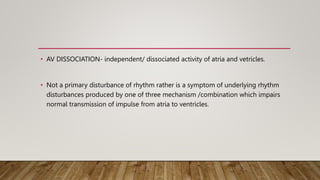
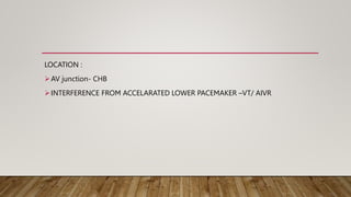
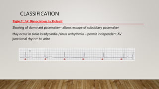
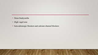
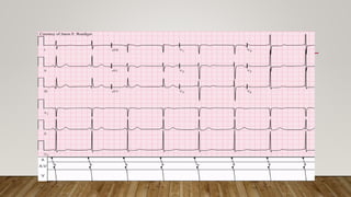
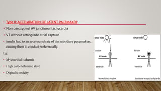
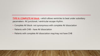
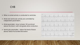
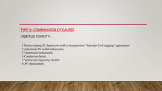
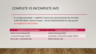
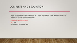
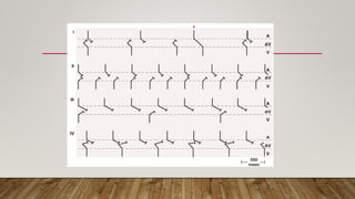
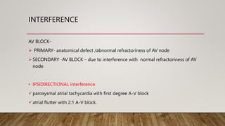
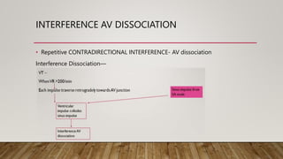
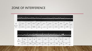
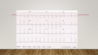
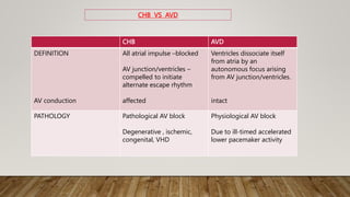
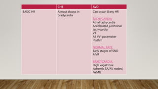
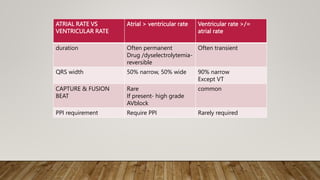
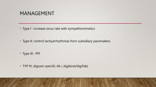
AV DISSOCIATION
BY D.ANISHAA
• AV DISSOCIATION- independent/ dissociated activity of atria and vetricles.
• Not a primary disturbance of rhythm rather is a symptom of underlying rhythm
disturbances produced by one of three mechanism /combination which impairs
normal transmission of impulse from atria to ventricles.
LOCATION :
AV junction- CHB
INTERFERENCE FROM ACCELARATED LOWER PACEMAKER –VT/ AIVR
CLASSIFICATION
Type 1: AV Dissociation by Default
Slowing of dominant pacemaker– allows escape of subsidiary pacemaker
May occur in sinus bradycardia /sinus arrhythmia – permit independent AV
junctional rhythm to arise
• Sinus bradycardia
• High vagal tone
• beta-adrenergic blockers and calcium channel blockers
• Type II: ACCELARATION OF LATENT PACEMAKER:
Non paroxysmal AV junctional tachycardia
VT without retrograde atrial capture
• insults lead to an accelerated rate of the subsidiary pacemakers,
causing them to conduct preferentially.
Eg:
• Myocardial ischemia
• High catecholamine state
• Digitalis toxicity
TYPE III: COMPLETE AV block – which allows ventricles to beat under subsidiary
pacemakers- AV junctional / ventricular escape rhythm.
• Complete AV block- not synonymous with complete AV dissociation
• Patients with CHB – have AV dissociation
• Patients with complete AV dissociation may/may not have CHB
CHB
 When no atrial activity is conducted to ventricles.
 Atrial and ventricular activity are controlled by
independent pacemakers.
 Atrial pacemaker- sinus/ ectopic/ AV junctional
(above the block ) with retrograde atrial conduction.
 Ventricular pacemaker – below the level of block-
above/ below his bundle bifurcation.
• Block @ level of AV node- usually congenital
• Block within bundle of His / distal to His bundle in purkinje – usually acquired.
TYPE IV: COMBINATION OF CAUSES
DIGITALIS TOXICITY-
1.Down sloping ST depression with a characteristic “Salvador Dali sagging” appearance
2.Junctional AV nodal tachycardia
3.Ventricular tachycardia
4.Conduction block
5.Ventricular bigeminy rhythm
6.AV dissociation
COMPLETE VS INCOMPLETE AVD
• If a single pacemaker – establish control over atria/ventricles for one beat
(CAPTURE BEAT)/ Series of beats – NO AV DISSOCIATION for that period–
incomplete AV dissociation
Isorhythmic AV dissociation Interference AV dissociation
Severe sinus bradycardia Lower level pacemaker
Junctional escape rhythm Junctional / ventricular escape rhythm
Sinus rate = junctional rate Faster intrinsic rate
COMPLETE AV DISSOCIATION
When atria/ventricle –fails to respond to a single impulse for 1 beat /series of beats– AV
DISSOCIATION occurs for that period.
COMPLETE AV dissociation-
Eg: CHB,
Atrial rate > ventricular rate
ISORHYTHMIC AV DISSOCIATION
• When sinus rate is slowed & junctional rate is accelerated
• Slowed sinus rate = accelerated junctional rhythm
• Atria captured by sinus impulse
• Ventricles by junctional impulse.
INTERFERENCE
AV BLOCK-
 PRIMARY- anatomical defect /abnormal refractoriness of AV node
SECONDARY -AV BLOCK – due to interference with normal refractoriness of AV
node
• IPSIDIRECTIONAL interference
paroxysmal atrial tachycardia with first degree A-V block
atrial flutter with 2:1 A-V block.
INTERFERENCE AV DISSOCIATION
• Repetitive CONTRADIRECTIONAL INTERFERENCE- AV dissociation
Interference Dissociation—
ZONE OF INTERFERENCE
CHB AVD
DEFINITION
AV conduction
All atrial impulse –blocked
AV junction/ventricles –
compelled to initiate
alternate escape rhythm
affected
Ventricles dissociate itself
from atria by an
autonomous focus arising
from AV junction/ventricles.
intact
PATHOLOGY Pathological AV block
Degenerative , ischemic,
congenital, VHD
Physiological AV block
Due to ill-timed accelerated
lower pacemaker activity
CHB VS AVD
CHB AVD
BASIC HR Almost always in
bradycardia
Can occur @any HR
TACHYCARDIA:
Atrial tachycardia
Accelerated junctional
tachycardia
VT
All VVI pacemaker
rhythm
NORMAL RATE:
Early stages of SND
AIVR
BRADYCARDIA;
High vagal tone
Ischemic SA/AV nodes(
IWMI)
ATRIAL RATE VS
VENTRICULAR RATE
Atrial > ventricular rate Ventricular rate >/=
atrial rate
duration Often permanent
Drug /dyselectrolytemia-
reversible
Often transient
QRS width 50% narrow, 50% wide 90% narrow
Except VT
CAPTURE & FUSION
BEAT
Rare
If present- high grade
AVblock
common
PPI requirement Require PPI Rarely required
If VT is persistent – suppress SA node
Atrial depolarization & contraction during VT – complex
Hence P wave in VT can be
Totally absent
Occur antegrade
On QRS
Over T waves
• AV dissociation in VT – rarely manifested.
• In intact VA conduction
• VT – traverse AV junction– reset SA node/ set in semi depolarized state.
CLINICAL FEATURES
1.VARYING PULSE VOLUME:
Some atrial beats– contribute to ventricular filling--- varying ventricular volumes
2.REVERSE PULSUS PARADOXUS (IN ISORHYTHMIC AV DISSOCIATION):
Pulse volume decrease during expiration
Inspiration--- increase sinus rate – AV synchrony
Expiration – decrease sinus rate – junctional rhythm takes over--AV asynchrony
2.JVP-Cannon waves
3.MR/TR:
Simultaneous contraction of atria & ventricle
MANAGEMENT
• Type I : increase sinus rate with sympathomimetics
• Type II: control tachyarrhythmias from subsidiary pacemakers
• Type III : PPI
• TYP IV: digoxin specific Ab ( digibind/digiFab)
THANK YOU

AV dissociation.pptx

  • 1.
  • 2.
    • AV DISSOCIATION-independent/ dissociated activity of atria and vetricles. • Not a primary disturbance of rhythm rather is a symptom of underlying rhythm disturbances produced by one of three mechanism /combination which impairs normal transmission of impulse from atria to ventricles.
  • 3.
    LOCATION : AV junction-CHB INTERFERENCE FROM ACCELARATED LOWER PACEMAKER –VT/ AIVR
  • 4.
    CLASSIFICATION Type 1: AVDissociation by Default Slowing of dominant pacemaker– allows escape of subsidiary pacemaker May occur in sinus bradycardia /sinus arrhythmia – permit independent AV junctional rhythm to arise
  • 5.
    • Sinus bradycardia •High vagal tone • beta-adrenergic blockers and calcium channel blockers
  • 7.
    • Type II:ACCELARATION OF LATENT PACEMAKER: Non paroxysmal AV junctional tachycardia VT without retrograde atrial capture • insults lead to an accelerated rate of the subsidiary pacemakers, causing them to conduct preferentially. Eg: • Myocardial ischemia • High catecholamine state • Digitalis toxicity
  • 9.
    TYPE III: COMPLETEAV block – which allows ventricles to beat under subsidiary pacemakers- AV junctional / ventricular escape rhythm. • Complete AV block- not synonymous with complete AV dissociation • Patients with CHB – have AV dissociation • Patients with complete AV dissociation may/may not have CHB
  • 10.
    CHB  When noatrial activity is conducted to ventricles.  Atrial and ventricular activity are controlled by independent pacemakers.  Atrial pacemaker- sinus/ ectopic/ AV junctional (above the block ) with retrograde atrial conduction.  Ventricular pacemaker – below the level of block- above/ below his bundle bifurcation.
  • 11.
    • Block @level of AV node- usually congenital • Block within bundle of His / distal to His bundle in purkinje – usually acquired.
  • 12.
    TYPE IV: COMBINATIONOF CAUSES DIGITALIS TOXICITY- 1.Down sloping ST depression with a characteristic “Salvador Dali sagging” appearance 2.Junctional AV nodal tachycardia 3.Ventricular tachycardia 4.Conduction block 5.Ventricular bigeminy rhythm 6.AV dissociation
  • 13.
    COMPLETE VS INCOMPLETEAVD • If a single pacemaker – establish control over atria/ventricles for one beat (CAPTURE BEAT)/ Series of beats – NO AV DISSOCIATION for that period– incomplete AV dissociation Isorhythmic AV dissociation Interference AV dissociation Severe sinus bradycardia Lower level pacemaker Junctional escape rhythm Junctional / ventricular escape rhythm Sinus rate = junctional rate Faster intrinsic rate
  • 14.
    COMPLETE AV DISSOCIATION Whenatria/ventricle –fails to respond to a single impulse for 1 beat /series of beats– AV DISSOCIATION occurs for that period. COMPLETE AV dissociation- Eg: CHB, Atrial rate > ventricular rate
  • 17.
    ISORHYTHMIC AV DISSOCIATION •When sinus rate is slowed & junctional rate is accelerated • Slowed sinus rate = accelerated junctional rhythm • Atria captured by sinus impulse • Ventricles by junctional impulse.
  • 18.
    INTERFERENCE AV BLOCK-  PRIMARY-anatomical defect /abnormal refractoriness of AV node SECONDARY -AV BLOCK – due to interference with normal refractoriness of AV node • IPSIDIRECTIONAL interference paroxysmal atrial tachycardia with first degree A-V block atrial flutter with 2:1 A-V block.
  • 19.
    INTERFERENCE AV DISSOCIATION •Repetitive CONTRADIRECTIONAL INTERFERENCE- AV dissociation Interference Dissociation—
  • 20.
  • 22.
    CHB AVD DEFINITION AV conduction Allatrial impulse –blocked AV junction/ventricles – compelled to initiate alternate escape rhythm affected Ventricles dissociate itself from atria by an autonomous focus arising from AV junction/ventricles. intact PATHOLOGY Pathological AV block Degenerative , ischemic, congenital, VHD Physiological AV block Due to ill-timed accelerated lower pacemaker activity CHB VS AVD
  • 23.
    CHB AVD BASIC HRAlmost always in bradycardia Can occur @any HR TACHYCARDIA: Atrial tachycardia Accelerated junctional tachycardia VT All VVI pacemaker rhythm NORMAL RATE: Early stages of SND AIVR BRADYCARDIA; High vagal tone Ischemic SA/AV nodes( IWMI)
  • 24.
    ATRIAL RATE VS VENTRICULARRATE Atrial > ventricular rate Ventricular rate >/= atrial rate duration Often permanent Drug /dyselectrolytemia- reversible Often transient QRS width 50% narrow, 50% wide 90% narrow Except VT CAPTURE & FUSION BEAT Rare If present- high grade AVblock common PPI requirement Require PPI Rarely required
  • 25.
    If VT ispersistent – suppress SA node Atrial depolarization & contraction during VT – complex Hence P wave in VT can be Totally absent Occur antegrade On QRS Over T waves
  • 26.
    • AV dissociationin VT – rarely manifested. • In intact VA conduction • VT – traverse AV junction– reset SA node/ set in semi depolarized state.
  • 27.
    CLINICAL FEATURES 1.VARYING PULSEVOLUME: Some atrial beats– contribute to ventricular filling--- varying ventricular volumes 2.REVERSE PULSUS PARADOXUS (IN ISORHYTHMIC AV DISSOCIATION): Pulse volume decrease during expiration Inspiration--- increase sinus rate – AV synchrony Expiration – decrease sinus rate – junctional rhythm takes over--AV asynchrony
  • 28.
  • 29.
    MANAGEMENT • Type I: increase sinus rate with sympathomimetics • Type II: control tachyarrhythmias from subsidiary pacemakers • Type III : PPI • TYP IV: digoxin specific Ab ( digibind/digiFab)
  • 30.