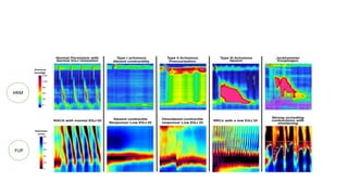
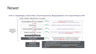
Approach to Dysphagia
Introduction
• Dysphagia is a Greek term, stands for difficulty in swallowing, due to
disruption of swallowing process.
• Dysphagia increase risk of aspiration pneumonia, malnutrition,
dehydration, weight loss, and airway obstruction, and it exerts a large
influence on rehabilitation.
Dysphagia can be secondary to defects in any of the 3 phases of
swallowing, which are as follows :
• Oral phase: Which involves the oral preparatory phase and the oral
transit phase
• Pharyngeal phase
• Esophageal phase
Dysphagia should be differentiated from disorders that prevent transfer
of food to the mouth or beyond the stomach but that are not
characterized by difficulty swallowing eg.- feeding disorders, GOO.
Paediatric Dysphagia
Factors related
• Neurodevelopmental skills involving sensory systems, cognition,
communication, and gross and fine motor behaviors.
• Cerebral Palsy--- Liquid > Solid
• Choanal atresia, cleft lip and palate, craniofacial syndromes
• Gastroesophageal reflux disease (GERD)
• Childhood achalasia--- 18% patients develops symptoms in infancy.
• Management is unique
Geriatric patients
Factors
• Increased oral and pharyngeal transit times, poor bolus control and
coordination, increased magnitude and duration of pharyngeal
pressures, and increased incidence of pharyngeal residue after
swallowing.
• Poor dentition, Atrophy of the tongue and alveolar ridge, Diminished
taste and smell sensitivity, Decreased muscle tone, Increased
ligamentous laxity, Limited laryngeal elevation.
Anatomy
• Input from the base of the tongue, as well as in the soft palate, faucial
arches, tonsils, and posterior pharyngeal wall through the facial (VII),
glossopharyngeal (IX), and vagus (X) cranial nerves to swallowing
center in pontine reticular system.
• Output back to the muscles that help in swallowing through
trigeminal (V), facial (VII), glossopharyngeal (IX), vagus (X), and
hypoglossal (XII) cranial nerves, with the trigeminal, hypoglossal, and
nucleus ambiguus constituting the efferent levels.
• Act of swallowing interrupts expiration.
Oral phase
• Oral preparatory phase: The processing of the bolus to render it
swallowable--- Tongue and Striated muscles of mastication.
• Oral propulsive (or transit) phase: The propelling of food from the oral
cavity into the oropharynx--- Muscles of tongue and labial seal.
• For single liquid swallow time is 1 sec, whereas for solid it is 5-10 secs.
Pharyngeal phase
• Important for preventing aspiration.
• The soft palate rises, the hyoid bone and larynx move upward and
forward, the vocal folds move to the midline, the epiglottis folds
backward to protect the airway, and the tongue pushes backward and
downward into the pharynx to propel the bolus downward.
• The upper esophageal sphincter relaxes during the pharyngeal phase.
• This is a totally reflexive phage of about 1 sec controlled by cranial
nerves IX (glossopharyngeal) and X (vagus).
Esophageal phase
• In the esophageal phase, the bolus is propelled downward by a
peristaltic movement.
• The lower esophageal sphincter relaxes at initiation of the swallow,
and this relaxation persists until the food bolus has been propelled
into the stomach.
• An interval of 8-20 seconds may be required for contractions to drive
the bolus into the stomach.
Pathophysiology
• A lesion in the cerebral cortex or the brainstem can cause swallowing
disorders as a result of the following:
• Decrease in range of motion (ROM) of muscles of mastication and
bolus propulsion, especially those responsible for buccal, labial, and
lingual strength and the cricopharyngeus
• Decreased sensation
• Delayed or absent pharyngeal swallowing and reductions in
pharyngeal peristalsis
• Delayed or absent laryngeal adduction and elevation
Oral-phase disorders
• Mainly assessed by Videofluorographic Study.
• Pocketing of food in the mouth, circumoral leakage, and early
pharyngeal spill can occur with weakness and poor coordination of
the lips, cheeks, and tongue. Weak posterior tongue can lead to
abnormal tongue thrusting.
Pharyngeal-phase disorders
• Dysfunction of soft palate.
• Lack of coordination of pharyngeal muscles.
• Inability of UES to relax.
Esophageal-phase disorders
• Mechanical obstruction, a motility disorder, or an impairment of the
opening of the lower esophageal sphincter.
Etiologies
Presentation
Signs and symptoms of oral or pharyngeal dysphagia include:
• Coughing or choking with swallowing
• Difficulty initiating swallowing Food sticking in the throat
• Sialorrhea
• Unexplained weight loss
• Change in dietary habits
• Recurrent pneumonia
• Change in voice or speech (wet voice)
• Nasal regurgitation
Signs and symptoms of esophageal dysphagia:
• Sensation of food sticking in the chest or throat
• Change in dietary habits
• Recurrent pneumonia
• Symptoms of gastroesophageal reflux disease (GERD), including
heartburn, belching, sour regurgitation, and water brash--- Associated
Odynophagia.
Relevant associated History:
• Recent stroke
• Neuromuscular disease
• Hypertension
• Diabetes mellitus (DM)
• Thyroid disease
• Cancer
• Dementia
• Recent injection of botulinum toxin
• Traumatic brain injury (TBI)
Relations
• With food--- Progression from Solid to liquid shows mechanical
etiology while dysmotility disorder shows dysphagia for both.
Time frame---
• Non-progressive and long-standing intermittent dysphagia often is
caused by a lower-esophageal mucosal ring (Schatzki’s ring).
• In contrast, discrete esophageal strictures cause progressive
dysphagia and if weight loss is prominent malignancy should be
considered.
Investigations
1st - Barium esophagogram or upper endoscopy?
Video swallow examination is a technique which allows video recording
of the patient swallowing barium mixed solids of varying consistencies
as well as liquids: with special attention to the pharyngeal phase of
swallowing. This would be the first investigation of choice in patients
with history suggestive of “transfer dysphagia”: disordered
oropharyngeal phase of swallowing.
Upper endoscopy is the initial investigation of choice in patients with
esophageal dysphagia as it can be both diagnostic and therapeutic:
identifying mucosal lesions, biopsy specimens can be obtained, and
dilatation can be performed.
• Barium evaluation may be more sensitive than routine endoscopy in
detecting subtle esophageal narrowing caused by mucosal rings and
is recommended as the primary test when there is a high suspicion
for achalasia or proximal esophageal lesions.
• If the upper endoscopic and barium examinations are normal, mid
and distal esophageal biopsy examinations, as well as esophageal
manometry, may be indicated.
Esophageal biopsy
• Routine mid and distal esophageal biopsy examinations in patients
with dysphagia and a normalappearing esophagus would seem
reasonable to exclude EE.
• The diagnosis of eosinophilic esophagitis is based on the presence of
>/= 20 eosinophils per high-power field.
Esophageal manometry
• The most important technology focused on assessing esophageal
motor function in endoscopy-negative dysphagia is HRM.
• The current classification scheme used to categorize esophageal
motor dysfunction is the Chicago Classification--- Pressor tracing to
pressure topography.
• The integrated relaxation pressure (IRP) provides a measure of the
resistance forces to flow through the EGJ created by contact pressure when
the lower esophageal sphincter (LES) is closed and the intrabolus pressure
when the LES is open.
• The distal latency (DL) interval assesses whether deglutitive inhibition is
intact by measuring the timing of smooth muscle contraction below the
transition zone---- Pre-mature contraction <4.5sec.
• Propulsive activity monitored by ability of esophagus to maintain 5cm
antegrade lumina closure.
• Strength of contraction measured with DCI.
• Peristaltic break below 20mmHg.
Caution
• EGJOO is a heterogeneous diagnostic group composed of patients
with evolving achalasia, mechanical obstruction, or an artifact related
to inherent issues with the IRP measurement.
• Further techniques like Timed barium, FLIP etc can be used before
treatment ensue.
• For Jackhammer esophagus--- D/D of GERD, EoE, prior SM relaxant
therapy to be considered before myotomy.
Newer techniques
High-resolution impedance manometry
• The automated impedance manometry (AIM) platform--- Impedance
signal as marker of bolus distension and intrabolus pressurization–
Showed better differentiation b/w functional and organic dysphagia.
• Esophageal impedance integral (EII) and the bolus flow time (BFT).
• EII was derived by developing a calculation of the cumulative
impedance signal within the space–time domain of the swallow wave.
• BFT as a time measurement of EGJ opening by using impedance drops
more than 90% of baseline as a marker of bolus presence and
determining the time where a preferential flow gradient was present
Functional luminal imaging probe panometry
• Adaptation of Impedance manometry with HR sensors for 3D reconstruction of
esophageal lumen.
• Assessing diameter/volume pressure changes.
Interpretation-
• Antegrade contractions occurring in a repetitive sequence spaced about 6 to 8
seconds apart are considered to be a normal response to sustained volumetric
distention.
• Contractions can occur in a retrograde direction and this pattern has been seen in
the context of spastic disorders, EGJ obstruction, and chronic opioids.
• Failure to elicit contractions is associated with aperistalsis and weak peristalsis
and may represent a myogenic dysfunction related to dilatation and atrophy or a
neurogenic dysfunction related to impaired triggering.
HRM
FLIP
Commonly followed
Newer
Thank you…

Dysphagia

  • 1.
  • 2.
    Introduction • Dysphagia isa Greek term, stands for difficulty in swallowing, due to disruption of swallowing process. • Dysphagia increase risk of aspiration pneumonia, malnutrition, dehydration, weight loss, and airway obstruction, and it exerts a large influence on rehabilitation.
  • 3.
    Dysphagia can besecondary to defects in any of the 3 phases of swallowing, which are as follows : • Oral phase: Which involves the oral preparatory phase and the oral transit phase • Pharyngeal phase • Esophageal phase Dysphagia should be differentiated from disorders that prevent transfer of food to the mouth or beyond the stomach but that are not characterized by difficulty swallowing eg.- feeding disorders, GOO.
  • 4.
    Paediatric Dysphagia Factors related •Neurodevelopmental skills involving sensory systems, cognition, communication, and gross and fine motor behaviors. • Cerebral Palsy--- Liquid > Solid • Choanal atresia, cleft lip and palate, craniofacial syndromes • Gastroesophageal reflux disease (GERD) • Childhood achalasia--- 18% patients develops symptoms in infancy. • Management is unique
  • 5.
    Geriatric patients Factors • Increasedoral and pharyngeal transit times, poor bolus control and coordination, increased magnitude and duration of pharyngeal pressures, and increased incidence of pharyngeal residue after swallowing. • Poor dentition, Atrophy of the tongue and alveolar ridge, Diminished taste and smell sensitivity, Decreased muscle tone, Increased ligamentous laxity, Limited laryngeal elevation.
  • 6.
    Anatomy • Input fromthe base of the tongue, as well as in the soft palate, faucial arches, tonsils, and posterior pharyngeal wall through the facial (VII), glossopharyngeal (IX), and vagus (X) cranial nerves to swallowing center in pontine reticular system. • Output back to the muscles that help in swallowing through trigeminal (V), facial (VII), glossopharyngeal (IX), vagus (X), and hypoglossal (XII) cranial nerves, with the trigeminal, hypoglossal, and nucleus ambiguus constituting the efferent levels. • Act of swallowing interrupts expiration.
  • 7.
    Oral phase • Oralpreparatory phase: The processing of the bolus to render it swallowable--- Tongue and Striated muscles of mastication. • Oral propulsive (or transit) phase: The propelling of food from the oral cavity into the oropharynx--- Muscles of tongue and labial seal. • For single liquid swallow time is 1 sec, whereas for solid it is 5-10 secs.
  • 8.
    Pharyngeal phase • Importantfor preventing aspiration. • The soft palate rises, the hyoid bone and larynx move upward and forward, the vocal folds move to the midline, the epiglottis folds backward to protect the airway, and the tongue pushes backward and downward into the pharynx to propel the bolus downward. • The upper esophageal sphincter relaxes during the pharyngeal phase. • This is a totally reflexive phage of about 1 sec controlled by cranial nerves IX (glossopharyngeal) and X (vagus).
  • 9.
    Esophageal phase • Inthe esophageal phase, the bolus is propelled downward by a peristaltic movement. • The lower esophageal sphincter relaxes at initiation of the swallow, and this relaxation persists until the food bolus has been propelled into the stomach. • An interval of 8-20 seconds may be required for contractions to drive the bolus into the stomach.
  • 10.
    Pathophysiology • A lesionin the cerebral cortex or the brainstem can cause swallowing disorders as a result of the following: • Decrease in range of motion (ROM) of muscles of mastication and bolus propulsion, especially those responsible for buccal, labial, and lingual strength and the cricopharyngeus • Decreased sensation • Delayed or absent pharyngeal swallowing and reductions in pharyngeal peristalsis • Delayed or absent laryngeal adduction and elevation
  • 11.
    Oral-phase disorders • Mainlyassessed by Videofluorographic Study. • Pocketing of food in the mouth, circumoral leakage, and early pharyngeal spill can occur with weakness and poor coordination of the lips, cheeks, and tongue. Weak posterior tongue can lead to abnormal tongue thrusting.
  • 12.
    Pharyngeal-phase disorders • Dysfunctionof soft palate. • Lack of coordination of pharyngeal muscles. • Inability of UES to relax.
  • 13.
    Esophageal-phase disorders • Mechanicalobstruction, a motility disorder, or an impairment of the opening of the lower esophageal sphincter.
  • 14.
  • 15.
    Presentation Signs and symptomsof oral or pharyngeal dysphagia include: • Coughing or choking with swallowing • Difficulty initiating swallowing Food sticking in the throat • Sialorrhea • Unexplained weight loss • Change in dietary habits • Recurrent pneumonia • Change in voice or speech (wet voice) • Nasal regurgitation
  • 16.
    Signs and symptomsof esophageal dysphagia: • Sensation of food sticking in the chest or throat • Change in dietary habits • Recurrent pneumonia • Symptoms of gastroesophageal reflux disease (GERD), including heartburn, belching, sour regurgitation, and water brash--- Associated Odynophagia.
  • 17.
    Relevant associated History: •Recent stroke • Neuromuscular disease • Hypertension • Diabetes mellitus (DM) • Thyroid disease • Cancer • Dementia • Recent injection of botulinum toxin • Traumatic brain injury (TBI)
  • 18.
    Relations • With food---Progression from Solid to liquid shows mechanical etiology while dysmotility disorder shows dysphagia for both. Time frame--- • Non-progressive and long-standing intermittent dysphagia often is caused by a lower-esophageal mucosal ring (Schatzki’s ring). • In contrast, discrete esophageal strictures cause progressive dysphagia and if weight loss is prominent malignancy should be considered.
  • 19.
    Investigations 1st - Bariumesophagogram or upper endoscopy? Video swallow examination is a technique which allows video recording of the patient swallowing barium mixed solids of varying consistencies as well as liquids: with special attention to the pharyngeal phase of swallowing. This would be the first investigation of choice in patients with history suggestive of “transfer dysphagia”: disordered oropharyngeal phase of swallowing. Upper endoscopy is the initial investigation of choice in patients with esophageal dysphagia as it can be both diagnostic and therapeutic: identifying mucosal lesions, biopsy specimens can be obtained, and dilatation can be performed.
  • 20.
    • Barium evaluationmay be more sensitive than routine endoscopy in detecting subtle esophageal narrowing caused by mucosal rings and is recommended as the primary test when there is a high suspicion for achalasia or proximal esophageal lesions. • If the upper endoscopic and barium examinations are normal, mid and distal esophageal biopsy examinations, as well as esophageal manometry, may be indicated.
  • 21.
    Esophageal biopsy • Routinemid and distal esophageal biopsy examinations in patients with dysphagia and a normalappearing esophagus would seem reasonable to exclude EE. • The diagnosis of eosinophilic esophagitis is based on the presence of >/= 20 eosinophils per high-power field.
  • 22.
    Esophageal manometry • Themost important technology focused on assessing esophageal motor function in endoscopy-negative dysphagia is HRM. • The current classification scheme used to categorize esophageal motor dysfunction is the Chicago Classification--- Pressor tracing to pressure topography.
  • 23.
    • The integratedrelaxation pressure (IRP) provides a measure of the resistance forces to flow through the EGJ created by contact pressure when the lower esophageal sphincter (LES) is closed and the intrabolus pressure when the LES is open. • The distal latency (DL) interval assesses whether deglutitive inhibition is intact by measuring the timing of smooth muscle contraction below the transition zone---- Pre-mature contraction <4.5sec. • Propulsive activity monitored by ability of esophagus to maintain 5cm antegrade lumina closure. • Strength of contraction measured with DCI. • Peristaltic break below 20mmHg.
  • 24.
    Caution • EGJOO isa heterogeneous diagnostic group composed of patients with evolving achalasia, mechanical obstruction, or an artifact related to inherent issues with the IRP measurement. • Further techniques like Timed barium, FLIP etc can be used before treatment ensue. • For Jackhammer esophagus--- D/D of GERD, EoE, prior SM relaxant therapy to be considered before myotomy.
  • 25.
    Newer techniques High-resolution impedancemanometry • The automated impedance manometry (AIM) platform--- Impedance signal as marker of bolus distension and intrabolus pressurization– Showed better differentiation b/w functional and organic dysphagia. • Esophageal impedance integral (EII) and the bolus flow time (BFT). • EII was derived by developing a calculation of the cumulative impedance signal within the space–time domain of the swallow wave. • BFT as a time measurement of EGJ opening by using impedance drops more than 90% of baseline as a marker of bolus presence and determining the time where a preferential flow gradient was present
  • 26.
    Functional luminal imagingprobe panometry • Adaptation of Impedance manometry with HR sensors for 3D reconstruction of esophageal lumen. • Assessing diameter/volume pressure changes. Interpretation- • Antegrade contractions occurring in a repetitive sequence spaced about 6 to 8 seconds apart are considered to be a normal response to sustained volumetric distention. • Contractions can occur in a retrograde direction and this pattern has been seen in the context of spastic disorders, EGJ obstruction, and chronic opioids. • Failure to elicit contractions is associated with aperistalsis and weak peristalsis and may represent a myogenic dysfunction related to dilatation and atrophy or a neurogenic dysfunction related to impaired triggering.
  • 27.
  • 28.
  • 29.
  • 30.