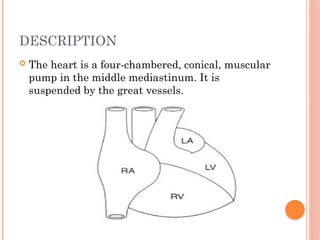
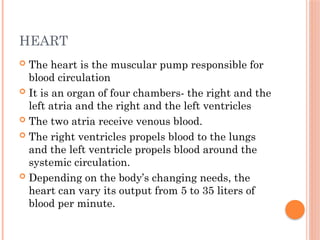
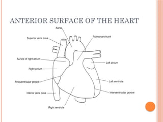
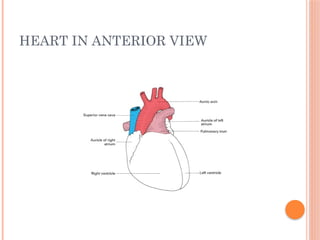
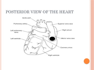
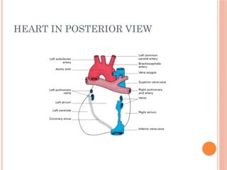
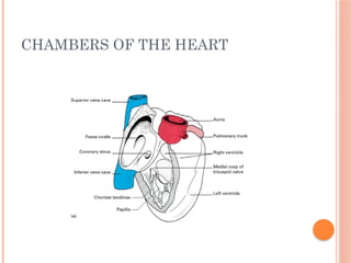
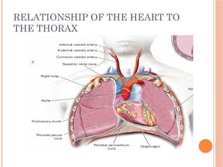
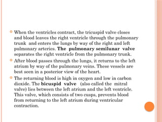
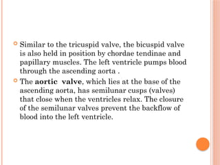
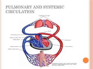
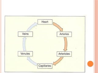
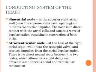
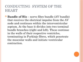
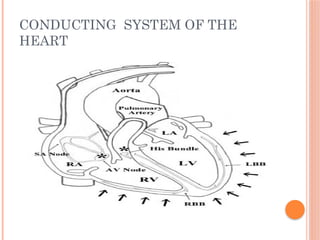
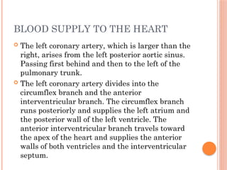
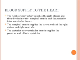
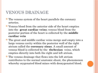
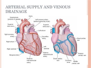
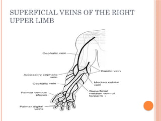
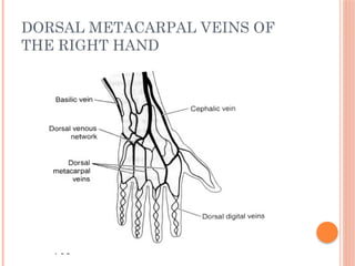
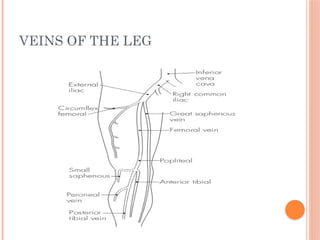
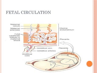
CARDIOVASCULAR
ANATOMY
KCHS/KRNA M 2016 CLASS
Hosea C.
OBJECTIVES
๏‚ข AT THE END OF THE LESSON THE
LEARNER SHOULD BE ABLE TO:
๏ƒ˜ Describe the structure and function of the heart
๏ƒ˜ Definition of cardiac circle
๏ƒ˜ Describe the conducting system of the heart
๏ƒ˜ Describe the blood flow through the heart
๏ƒ˜ Describe the blood supply to the heart
๏ƒ˜ Appreciate the location of the major great
vessels.
๏ƒ˜ Developmental anatomy of the heart (fetal
circulation)
DESCRIPTION
๏‚ข The heart is a four-chambered, conical, muscular
pump in the middle mediastinum. It is
suspended by the great vessels.
HEART
๏‚ข The heart is the muscular pump responsible for
blood circulation
๏‚ข It is an organ of four chambers- the right and the
left atria and the right and the left ventricles
๏‚ข The two atria receive venous blood.
๏‚ข The right ventricles propels blood to the lungs
and the left ventricle propels blood around the
systemic circulation.
๏‚ข Depending on the bodyโ€™s changing needs, the
heart can vary its output from 5 to 35 liters of
blood per minute.
ANTERIOR SURFACE OF THE HEART
HEART IN ANTERIOR VIEW
POSTERIOR VIEW OF THE HEART
HEART IN POSTERIOR VIEW
CHAMBERS OF THE HEART
SURFACE MARKINGS
๏‚ข The surface markings follow a quadrilateral
shape (distances from midline): third right costal
cartilage (2 cm), second left costal cartilage (3
cm), fifth left intercostal space (7 cm) and sixth
right costal cartilage (2 cm).
SURFACE MARKINGS
RELATIONSHIP OF THE HEART TO
THE THORAX
STRUCTURE
๏‚ข The heart consists of four chambers:
๏‚ข 1. Right atrium โ€“ receives deoxygenated blood from the
body via the venae cavae (inferior and superior). The
outflow of blood occurs through the tricuspid valve into the
right ventricle. The sino-atrial node is situated in the
upper part of the right atrium, and the atrioventricular
node lies near the base of the tricuspid valve.
๏‚ข 2.Right ventricle โ€“ receives blood from the right atrium
and expels it through the pulmonary valve and trunk.
๏‚ข Some of the rough internal wall muscle fibres (trabeculae)
specialize into papillary muscles, which attach to the
tricuspid valve cusps (in a similar fashion to the mitral
valve on the left side of the heart). The pulmonary valve is
tricuspid and leads into the pulmonary trunk
STRUCTURE
๏‚ข 3. Left atrium โ€“ receives oxygenated blood from
the lungs via the four pulmonary veins, which
open superoposteriorly. The blood then passes
through the mitral (bicuspid) valve into the left
ventricle
๏‚ข 4. Left ventricle โ€“ thickest-walled chamber that
distributes blood to the body via the aorta. The
aortic valve is tricuspid โ€“ with right, left and
posterior cusps. Small sinuses lie above the cusps
that give rise to the two coronary arteries โ€“ right
and left respectively
FLOW OF BLOOD THROUGH THE
HEART
INTERNAL CHAMBERS OF THE
HEART
๏‚ข As shown in the diagram, the right atrium
receives venous blood from the inferior vena cava
and superior vena cava. A small amount of
cardiac venous blood enters the right atrium by
means of the thebesian veins. This blood is low in
oxygen and high in carbon dioxide.
๏‚ข A one-way valve, the tricuspid valve, lies
between the right atrium and the right ventricle.
The tricuspid valve gets its name from its three
valve leaflets, or cusps. The tricuspid leaflets are
held in place by tendinous cords called chordae
tendinae ,which are secured to the ventricular
wall by the papillary muscles.
๏‚ข When the ventricles contract, the tricuspid valve closes
and blood leaves the right ventricle through the pulmonary
trunk and enters the lungs by way of the right and left
pulmonary arteries. The pulmonary semilunar valve
separates the right ventricle from the pulmonary trunk.
๏‚ข After blood passes through the lungs, it returns to the left
atrium by way of the pulmonary veins. These vessels are
best seen in a posterior view of the heart.
๏‚ข The returning blood is high in oxygen and low in carbon
dioxide. The bicuspid valve (also called the mitral
valve) lies between the left atrium and the left ventricle.
This valve, which consists of two cusps, prevents blood
from returning to the left atrium during ventricular
contraction.
๏‚ข Similar to the tricuspid valve, the bicuspid valve
is also held in position by chordae tendinae and
papillary muscles. The left ventricle pumps blood
through the ascending aorta .
๏‚ข The aortic valve, which lies at the base of the
ascending aorta, has semilunar cusps (valves)
that close when the ventricles relax. The closure
of the semilunar valves prevent the backflow of
blood into the left ventricle.
PULMONARY AND SYSTEMIC
CIRCULATION
CONDUCTING SYSTEM OF THE
HEART
๏‚ข Sino-atrial node โ€“ in the superior right atrial
wall (near the superior vena caval opening) and
initiates conduction impulse. The node is in direct
contact with the atrial cells and causes a wave of
depolarisation, resulting in contraction of both
atria.
๏‚ข Atrioventricular node โ€“ at the base of the right
atrial septal wall (near the tricuspid valve) and
receives impulses from the atrial depolarization.
There is no direct neural route between the two
nodes, which allows for a slight delay and
prevents simultaneous atrial and ventricular
contraction
CONDUCTING SYSTEM OF THE
HEART
๏‚ข Bundle of His โ€“ nerve fibre bundle (AV bundle)
that receives the electrical impulse from the AV
node and continues within the interventricular
septum. At the base it divides into two terminal
bundle branches (right and left). These continue
in the walls of their respective ventricles,
terminating in Purkinje fibres, which penetrate
the muscular walls and initiate ventricular
contraction.
CONDUCTING SYSTEM OF THE
HEART
๏‚ข The afferent fibers from the aortic arch baroreceptors
travel with the vagus nerve (tenth cranial).
๏‚ข The baroreceptors regulate the arterial blood pressure by
initiating reflex adjustments to changes in blood
pressure. For example, when the arterial pressure
decreases, the neural impulses transmitted from the
baroreceptors to the vasomotor and cardiac centers in
the medulla also decrease. This causes the medulla to
increase its sympathetic activity, which in turn causes
an increase in the following:
โ€ข Heart rate
โ€ข Myocardial force of contraction
โ€ข Arterial constriction
โ€ข Venous constriction.
PERICARDIUM
๏‚ข Heart is enclosed within a conical fibroserous sac
โ€“ the pericardium.
๏‚ข The outer layer is attached to the following
structures:
- Adventitia of the great vessels
-Sternopericardial ligament โ€“ to the
posterior sternum
-Central tendon of diaphragm โ€“
where it is fused inferiorly
PERICARDIUM
๏‚ข Outer fibrous layer is a tough fibrous structure
with openings to allow the aorta, pulmonary
trunk and superior vena cava to pass through
๏‚ข Serous pericardium has two
๏‚ข components:
-Outer parietal pericardium โ€“ lines the inner
surface of the fibrous sac and becomes continuous
with the visceral layer around the great vessels
๏‚ข -Inner visceral pericardium โ€“ in direct
contact with the heart and forms a potential
space between the pericardial layers.
LAYERS OF PERICARDIUM
BLOOD SUPPLY TO THE HEART
๏‚ข The blood supply of the heart originates directly from the
aorta by means of two arteries: the left coronary artery
and the right coronary artery.
๏‚ข The right coronary artery arises from the anterior aortic
sinus and passes forwards between the pulmonary trunk
and the right atrium to descend in the right part of the
atrioventricular groove.
๏‚ข At the inferior border of the heart it continues along the
atrioventricular groove to anastomose with the left
coronary at the inferior interventricular groove. It gives off
a marginal branch along the lower border of the heart and
an interventricular branch that runs forwards in the
inferior interventricular groove to anastomose near the
apex of the heart with the corresponding branch of the left
coronary artery.
BLOOD SUPPLY TO THE HEART
๏‚ข The left coronary artery, which is larger than the
right, arises from the left posterior aortic sinus.
Passing first behind and then to the left of the
pulmonary trunk.
๏‚ข The left coronary artery divides into the
circumflex branch and the anterior
interventricular branch. The circumflex branch
runs posteriorly and supplies the left atrium and
the posterior wall of the left ventricle. The
anterior interventricular branch travels toward
the apex of the heart and supplies the anterior
walls of both ventricles and the interventricular
septum.
BLOOD SUPPLY TO THE HEART
๏‚ข The right coronary artery supplies the right atrium and
then divides into the marginal branch and the posterior
inter ventricular branch .
๏‚ข The marginal branch supplies the lateral walls of the right
atrium and right ventricle.
๏‚ข The posterior interventricular branch supplies the
posterior wall of both ventricles
VENOUS DRAINAGE
๏‚ข The venous system of the heart parallels the coronary
arteries.
๏‚ข Venous blood from the anterior side of the heart empties
into the great cardiac veins; venous blood from the
posterior portion of the heart is collected by the middle
cardiac vein
๏‚ข The great and middle cardiac veins merge and empty into a
large venous cavity within the posterior wall of the right
atrium called the coronary sinus. A small amount of
venous blood is collected by the thebesian veins, which
empties directly into both the right and left atrium.
๏‚ข The venous drainage that flows into the left atrium
contributes to the normal anatomic shunt, the phenomenon
whereby oxygenated blood mixes with deoxygenated blood.
ARTERIAL SUPPLY AND VENOUS
DRAINAGE
NERVE SUPPLY
๏‚ข Autonomic supply:
โ— Parasympathetic โ€“ from the vagus nerve
(cardio-inhibitory)
โ— Sympathetic โ€“ cervical (C1โ€“4, C5 and C6, C7โ€“
T1) and upper thoracic (T2โ€“5) ganglia (cardio-
accelerator) via the superficial and deep cardiac
plexuses
โ— Phrenic nerve (C3โ€“5) supplies the pericardium
THE GREAT VESSELS
๏‚ข AORTA
๏‚ข Commences at the aortic valve and terminates at its
bifurcation into the common iliac arteries (L4 level). There
are four parts:
๏‚ข 1. Ascending โ€“ 5 cm long, posterior to sternum. It gives off:
โ— Right coronary artery
โ— Left coronary artery
o 2.Arch โ€“ runs upwards, backwards and to the left. It gives
off:
โ— Brachiocephalic, which divides into:
o โ— Right common carotid
o โ— Right subclavian
o โ— Left common carotid
o โ— Left subclavian
THE GREAT VESSELS
๏‚ข 3. Descending thoracic aorta โ€“ starts at level T4
and runs down to the aortic opening in the
diaphragm (T12). It gives off:
โ— Visceral branches โ€“ pericardial, bronchial,
esophageal, mediastinal and phrenic
๏‚ข โ— Somatic branches โ€“ posterior intercostals,
dorsal, muscular, lateral cutaneous and
mammary
THE GREAT VESSELS
๏‚ข 4. Abdominal aorta โ€“ starts at the aortic opening in the
diaphragm and ends at the common iliac bifurcation. It gives
off:
โ— Lumbar arteries (paired)
โ— Visceral arteries (paired) โ€“ inferior phrenic,
suprarenal, renal and gonadal
โ— Midline (unpaired) arteries:
โ— Celiac trunk โ€“ supplies the foregut (via left gastric,
common hepatic and splenic)
โ— Superior mesenteric โ€“ supplies the midgut (via inferior
pancreaticoduodenal, jejunal ileal, ileocolic, right and middle
colic)
โ— Inferior mesenteric โ€“ supplies the hindgut (via left
colic,sigmoid and superior rectal)
THE GREAT VESSELS
SCHEMATIC DIAGRAM OF THE
INFERIOR VENA CAVA AND ITS
BRANCHES
PRINCIPAL RELATIONS OF THE
ARCH OF THE AORTA
MAJOR ARTERIES OF THE HEAD
AND NECK
ARTERIES OF UPPER LIMB
ARTERIES OF THE LOWER LIMB
MAJOR ARTERIES
MAJOR VEINS OF HEAD AND NECK
SUPERFICIAL VEINS OF THE RIGHT
UPPER LIMB
DORSAL METACARPAL VEINS OF
THE RIGHT HAND
VEINS OF THE LEG
FETAL CIRCULATION
FETAL CIRCULATION
๏‚ข Umbilical vein โ€“ oxygenated blood enters the
body via the umbilical vein. After mixing with
deoxygenated blood in the ductus venosus, it
reaches the right atrium (via inferior vena cava
that receives blood from trunk and limbs)
๏‚ข Right atrium โ€“ anatomical relationship of the
venae cavae ensures that most of the blood in the
right atrium (from the inferior vena cava)
bypasses the right ventricle and goes directly to
the left atrium via the patent foramen ovale.
FETAL CIRCULATION
๏‚ข Left atrium โ€“ blood from the left atrium mixes
with (deoxygenated) blood from the lungs and is
expelled via the left ventricle into the aorta, and
ultimately around the body.
๏‚ข Mixing โ€“ some blood does not flow directly from
the right into the left atrium, but instead it is
directed to the right ventricle (mainly blood from
the superior vena cava). This deoxygenated blood
flows through the lungs and pulmonary trunk,
and then via the ductus arteriosus into the aorta
FETAL CIRCULATION
FETAL CIRCULATION
๏‚ข Transitional circulation โ€“ following the clamping
of the umbilical cord at birth, and with the large
decrease in pulmonary vessel pressure with
inspiration, significant pressure and flow
changes occur. The fall in right atrial pressure
and increase in left atrial pressure causes the
foramen ovale to close, as the septum secundum
and septum primum oppose.
FETAL CIRCULATION
๏‚ข This is an immediate functional closure only. As
a result, all blood from the right atrium is now
forced into the right ventricle. The ductus
arteriosus also constricts due to the high partial
pressure of oxygen (functionally complete by 12
hours). The change to adult circulation is
complete by 3 months, by which time the
foramen ovale is anatomically fused (fossa ovalis)
and the ductus arteriosus is obliterated.
SUMMARY
๏‚ข Cardiovascular anatomy is vital aspect in
anesthesia care.
๏‚ข Understanding the normal cardiac anatomy is
important so as to guide in understanding the
abnormal
๏‚ข Conduction defects and coronary heart disease
have an impact in geriatric anesthesia
๏‚ข Fetal circulation is important in understanding
abnormal defects
๏‚ข Supply and demand in cardiovascular system
must be understood as it relates to all aspects of
anesthesia care

Cardiovascular anatomy and physiology .ppx

  • 1.
  • 2.
    OBJECTIVES ๏‚ข AT THEEND OF THE LESSON THE LEARNER SHOULD BE ABLE TO: ๏ƒ˜ Describe the structure and function of the heart ๏ƒ˜ Definition of cardiac circle ๏ƒ˜ Describe the conducting system of the heart ๏ƒ˜ Describe the blood flow through the heart ๏ƒ˜ Describe the blood supply to the heart ๏ƒ˜ Appreciate the location of the major great vessels. ๏ƒ˜ Developmental anatomy of the heart (fetal circulation)
  • 3.
    DESCRIPTION ๏‚ข The heartis a four-chambered, conical, muscular pump in the middle mediastinum. It is suspended by the great vessels.
  • 4.
    HEART ๏‚ข The heartis the muscular pump responsible for blood circulation ๏‚ข It is an organ of four chambers- the right and the left atria and the right and the left ventricles ๏‚ข The two atria receive venous blood. ๏‚ข The right ventricles propels blood to the lungs and the left ventricle propels blood around the systemic circulation. ๏‚ข Depending on the bodyโ€™s changing needs, the heart can vary its output from 5 to 35 liters of blood per minute.
  • 5.
  • 6.
  • 7.
  • 8.
  • 9.
  • 10.
    SURFACE MARKINGS ๏‚ข Thesurface markings follow a quadrilateral shape (distances from midline): third right costal cartilage (2 cm), second left costal cartilage (3 cm), fifth left intercostal space (7 cm) and sixth right costal cartilage (2 cm).
  • 11.
  • 12.
    RELATIONSHIP OF THEHEART TO THE THORAX
  • 13.
    STRUCTURE ๏‚ข The heartconsists of four chambers: ๏‚ข 1. Right atrium โ€“ receives deoxygenated blood from the body via the venae cavae (inferior and superior). The outflow of blood occurs through the tricuspid valve into the right ventricle. The sino-atrial node is situated in the upper part of the right atrium, and the atrioventricular node lies near the base of the tricuspid valve. ๏‚ข 2.Right ventricle โ€“ receives blood from the right atrium and expels it through the pulmonary valve and trunk. ๏‚ข Some of the rough internal wall muscle fibres (trabeculae) specialize into papillary muscles, which attach to the tricuspid valve cusps (in a similar fashion to the mitral valve on the left side of the heart). The pulmonary valve is tricuspid and leads into the pulmonary trunk
  • 14.
    STRUCTURE ๏‚ข 3. Leftatrium โ€“ receives oxygenated blood from the lungs via the four pulmonary veins, which open superoposteriorly. The blood then passes through the mitral (bicuspid) valve into the left ventricle ๏‚ข 4. Left ventricle โ€“ thickest-walled chamber that distributes blood to the body via the aorta. The aortic valve is tricuspid โ€“ with right, left and posterior cusps. Small sinuses lie above the cusps that give rise to the two coronary arteries โ€“ right and left respectively
  • 15.
    FLOW OF BLOODTHROUGH THE HEART
  • 16.
  • 17.
    ๏‚ข As shownin the diagram, the right atrium receives venous blood from the inferior vena cava and superior vena cava. A small amount of cardiac venous blood enters the right atrium by means of the thebesian veins. This blood is low in oxygen and high in carbon dioxide. ๏‚ข A one-way valve, the tricuspid valve, lies between the right atrium and the right ventricle. The tricuspid valve gets its name from its three valve leaflets, or cusps. The tricuspid leaflets are held in place by tendinous cords called chordae tendinae ,which are secured to the ventricular wall by the papillary muscles.
  • 18.
    ๏‚ข When theventricles contract, the tricuspid valve closes and blood leaves the right ventricle through the pulmonary trunk and enters the lungs by way of the right and left pulmonary arteries. The pulmonary semilunar valve separates the right ventricle from the pulmonary trunk. ๏‚ข After blood passes through the lungs, it returns to the left atrium by way of the pulmonary veins. These vessels are best seen in a posterior view of the heart. ๏‚ข The returning blood is high in oxygen and low in carbon dioxide. The bicuspid valve (also called the mitral valve) lies between the left atrium and the left ventricle. This valve, which consists of two cusps, prevents blood from returning to the left atrium during ventricular contraction.
  • 19.
    ๏‚ข Similar tothe tricuspid valve, the bicuspid valve is also held in position by chordae tendinae and papillary muscles. The left ventricle pumps blood through the ascending aorta . ๏‚ข The aortic valve, which lies at the base of the ascending aorta, has semilunar cusps (valves) that close when the ventricles relax. The closure of the semilunar valves prevent the backflow of blood into the left ventricle.
  • 20.
  • 22.
    CONDUCTING SYSTEM OFTHE HEART ๏‚ข Sino-atrial node โ€“ in the superior right atrial wall (near the superior vena caval opening) and initiates conduction impulse. The node is in direct contact with the atrial cells and causes a wave of depolarisation, resulting in contraction of both atria. ๏‚ข Atrioventricular node โ€“ at the base of the right atrial septal wall (near the tricuspid valve) and receives impulses from the atrial depolarization. There is no direct neural route between the two nodes, which allows for a slight delay and prevents simultaneous atrial and ventricular contraction
  • 23.
    CONDUCTING SYSTEM OFTHE HEART ๏‚ข Bundle of His โ€“ nerve fibre bundle (AV bundle) that receives the electrical impulse from the AV node and continues within the interventricular septum. At the base it divides into two terminal bundle branches (right and left). These continue in the walls of their respective ventricles, terminating in Purkinje fibres, which penetrate the muscular walls and initiate ventricular contraction.
  • 24.
  • 25.
    ๏‚ข The afferentfibers from the aortic arch baroreceptors travel with the vagus nerve (tenth cranial). ๏‚ข The baroreceptors regulate the arterial blood pressure by initiating reflex adjustments to changes in blood pressure. For example, when the arterial pressure decreases, the neural impulses transmitted from the baroreceptors to the vasomotor and cardiac centers in the medulla also decrease. This causes the medulla to increase its sympathetic activity, which in turn causes an increase in the following: โ€ข Heart rate โ€ข Myocardial force of contraction โ€ข Arterial constriction โ€ข Venous constriction.
  • 26.
    PERICARDIUM ๏‚ข Heart isenclosed within a conical fibroserous sac โ€“ the pericardium. ๏‚ข The outer layer is attached to the following structures: - Adventitia of the great vessels -Sternopericardial ligament โ€“ to the posterior sternum -Central tendon of diaphragm โ€“ where it is fused inferiorly
  • 27.
    PERICARDIUM ๏‚ข Outer fibrouslayer is a tough fibrous structure with openings to allow the aorta, pulmonary trunk and superior vena cava to pass through ๏‚ข Serous pericardium has two ๏‚ข components: -Outer parietal pericardium โ€“ lines the inner surface of the fibrous sac and becomes continuous with the visceral layer around the great vessels ๏‚ข -Inner visceral pericardium โ€“ in direct contact with the heart and forms a potential space between the pericardial layers.
  • 28.
  • 29.
    BLOOD SUPPLY TOTHE HEART ๏‚ข The blood supply of the heart originates directly from the aorta by means of two arteries: the left coronary artery and the right coronary artery. ๏‚ข The right coronary artery arises from the anterior aortic sinus and passes forwards between the pulmonary trunk and the right atrium to descend in the right part of the atrioventricular groove. ๏‚ข At the inferior border of the heart it continues along the atrioventricular groove to anastomose with the left coronary at the inferior interventricular groove. It gives off a marginal branch along the lower border of the heart and an interventricular branch that runs forwards in the inferior interventricular groove to anastomose near the apex of the heart with the corresponding branch of the left coronary artery.
  • 30.
    BLOOD SUPPLY TOTHE HEART ๏‚ข The left coronary artery, which is larger than the right, arises from the left posterior aortic sinus. Passing first behind and then to the left of the pulmonary trunk. ๏‚ข The left coronary artery divides into the circumflex branch and the anterior interventricular branch. The circumflex branch runs posteriorly and supplies the left atrium and the posterior wall of the left ventricle. The anterior interventricular branch travels toward the apex of the heart and supplies the anterior walls of both ventricles and the interventricular septum.
  • 31.
    BLOOD SUPPLY TOTHE HEART ๏‚ข The right coronary artery supplies the right atrium and then divides into the marginal branch and the posterior inter ventricular branch . ๏‚ข The marginal branch supplies the lateral walls of the right atrium and right ventricle. ๏‚ข The posterior interventricular branch supplies the posterior wall of both ventricles
  • 32.
    VENOUS DRAINAGE ๏‚ข Thevenous system of the heart parallels the coronary arteries. ๏‚ข Venous blood from the anterior side of the heart empties into the great cardiac veins; venous blood from the posterior portion of the heart is collected by the middle cardiac vein ๏‚ข The great and middle cardiac veins merge and empty into a large venous cavity within the posterior wall of the right atrium called the coronary sinus. A small amount of venous blood is collected by the thebesian veins, which empties directly into both the right and left atrium. ๏‚ข The venous drainage that flows into the left atrium contributes to the normal anatomic shunt, the phenomenon whereby oxygenated blood mixes with deoxygenated blood.
  • 33.
    ARTERIAL SUPPLY ANDVENOUS DRAINAGE
  • 34.
    NERVE SUPPLY ๏‚ข Autonomicsupply: โ— Parasympathetic โ€“ from the vagus nerve (cardio-inhibitory) โ— Sympathetic โ€“ cervical (C1โ€“4, C5 and C6, C7โ€“ T1) and upper thoracic (T2โ€“5) ganglia (cardio- accelerator) via the superficial and deep cardiac plexuses โ— Phrenic nerve (C3โ€“5) supplies the pericardium
  • 35.
    THE GREAT VESSELS ๏‚ขAORTA ๏‚ข Commences at the aortic valve and terminates at its bifurcation into the common iliac arteries (L4 level). There are four parts: ๏‚ข 1. Ascending โ€“ 5 cm long, posterior to sternum. It gives off: โ— Right coronary artery โ— Left coronary artery o 2.Arch โ€“ runs upwards, backwards and to the left. It gives off: โ— Brachiocephalic, which divides into: o โ— Right common carotid o โ— Right subclavian o โ— Left common carotid o โ— Left subclavian
  • 36.
    THE GREAT VESSELS ๏‚ข3. Descending thoracic aorta โ€“ starts at level T4 and runs down to the aortic opening in the diaphragm (T12). It gives off: โ— Visceral branches โ€“ pericardial, bronchial, esophageal, mediastinal and phrenic ๏‚ข โ— Somatic branches โ€“ posterior intercostals, dorsal, muscular, lateral cutaneous and mammary
  • 37.
    THE GREAT VESSELS ๏‚ข4. Abdominal aorta โ€“ starts at the aortic opening in the diaphragm and ends at the common iliac bifurcation. It gives off: โ— Lumbar arteries (paired) โ— Visceral arteries (paired) โ€“ inferior phrenic, suprarenal, renal and gonadal โ— Midline (unpaired) arteries: โ— Celiac trunk โ€“ supplies the foregut (via left gastric, common hepatic and splenic) โ— Superior mesenteric โ€“ supplies the midgut (via inferior pancreaticoduodenal, jejunal ileal, ileocolic, right and middle colic) โ— Inferior mesenteric โ€“ supplies the hindgut (via left colic,sigmoid and superior rectal)
  • 38.
  • 39.
    SCHEMATIC DIAGRAM OFTHE INFERIOR VENA CAVA AND ITS BRANCHES
  • 40.
    PRINCIPAL RELATIONS OFTHE ARCH OF THE AORTA
  • 41.
    MAJOR ARTERIES OFTHE HEAD AND NECK
  • 42.
  • 43.
    ARTERIES OF THELOWER LIMB
  • 44.
  • 45.
    MAJOR VEINS OFHEAD AND NECK
  • 46.
    SUPERFICIAL VEINS OFTHE RIGHT UPPER LIMB
  • 47.
    DORSAL METACARPAL VEINSOF THE RIGHT HAND
  • 48.
  • 49.
  • 50.
    FETAL CIRCULATION ๏‚ข Umbilicalvein โ€“ oxygenated blood enters the body via the umbilical vein. After mixing with deoxygenated blood in the ductus venosus, it reaches the right atrium (via inferior vena cava that receives blood from trunk and limbs) ๏‚ข Right atrium โ€“ anatomical relationship of the venae cavae ensures that most of the blood in the right atrium (from the inferior vena cava) bypasses the right ventricle and goes directly to the left atrium via the patent foramen ovale.
  • 51.
    FETAL CIRCULATION ๏‚ข Leftatrium โ€“ blood from the left atrium mixes with (deoxygenated) blood from the lungs and is expelled via the left ventricle into the aorta, and ultimately around the body. ๏‚ข Mixing โ€“ some blood does not flow directly from the right into the left atrium, but instead it is directed to the right ventricle (mainly blood from the superior vena cava). This deoxygenated blood flows through the lungs and pulmonary trunk, and then via the ductus arteriosus into the aorta
  • 52.
  • 53.
    FETAL CIRCULATION ๏‚ข Transitionalcirculation โ€“ following the clamping of the umbilical cord at birth, and with the large decrease in pulmonary vessel pressure with inspiration, significant pressure and flow changes occur. The fall in right atrial pressure and increase in left atrial pressure causes the foramen ovale to close, as the septum secundum and septum primum oppose.
  • 54.
    FETAL CIRCULATION ๏‚ข Thisis an immediate functional closure only. As a result, all blood from the right atrium is now forced into the right ventricle. The ductus arteriosus also constricts due to the high partial pressure of oxygen (functionally complete by 12 hours). The change to adult circulation is complete by 3 months, by which time the foramen ovale is anatomically fused (fossa ovalis) and the ductus arteriosus is obliterated.
  • 55.
    SUMMARY ๏‚ข Cardiovascular anatomyis vital aspect in anesthesia care. ๏‚ข Understanding the normal cardiac anatomy is important so as to guide in understanding the abnormal ๏‚ข Conduction defects and coronary heart disease have an impact in geriatric anesthesia ๏‚ข Fetal circulation is important in understanding abnormal defects ๏‚ข Supply and demand in cardiovascular system must be understood as it relates to all aspects of anesthesia care