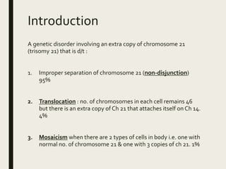
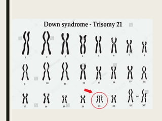
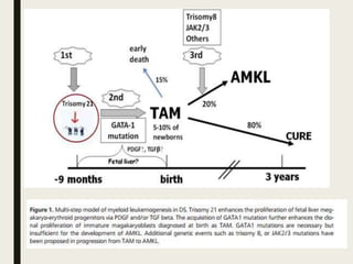
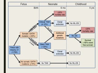
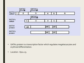
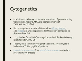
DOWN SYNDROME
RELATED
LEUKEMIAS
Introduction
A genetic disorder involving an extra copy of chromosome 21
(trisomy 21) that is d/t :
1. Improper separation of chromosome 21 (non-disjunction)
95%
2. Translocation : no. of chromosomes in each cell remains 46
but there is an extra copy of Ch 21 that attaches itself on Ch 14.
4%
3. Mosaicism when there are 2 types of cells in body i.e. one with
normal no. of chromosome 21 & one with 3 copies of ch 21. 1%
■ Incidence 1 in 1000 - 1100 live births worldwide.
■ Symptoms include intellectual disabilities, distinct facial
features, developmental delay, congenital heart diseases,
alzheimers disease, GI malformations , poor immune system &
leukemias.
DS related leukemias
■ TAMTransient abnormal myelopoiesis
■ MDS (prior to leukemia)
■ AML : 20% childhood leukemia, 40% DS leukemia
■ ALL : 80% childhood leukemia, 60% DS leukemia
Transient abnormal myelopoeisis
■ A disorder of DS newborns that presents with clinical and morphologic
findings indistinguishable from AML.
■ Characterized by increased peripheral blood blasts, usually
megakaryoblasts, with or without clinical symptoms.
■ Underlying genetics demonstrate somatic mutations in
the GATA1 gene.
■ majority of newborns are asymptomatic, with spontaneous resolution
by 2-3 months (median 54 days), although some develop severe
disease.
■ Most of the antenatal cases die before birth with fetal hydrops
■ The cell of origin is fetal liver hematopoietic stem cell/progenitor cell.
EPIDEMIOLOGY
■ It occurs in approximately 10% of DS newborns.
Clinical features
■ Median presentation within 3 – 7 days of birth
■ Male : female ratio of 1.2-1.5 : 1
■ Thrombocytopenia is common. (thrombocytosis may be seen)
■ There may be marked leucocytosis and % of in BM may exceed
the BM.
■ Hb & ANC usually within normal limits.
■ most common clinical symptom is hepatomegaly, which is a
result of megakaryoblast infiltration and/or hepatic fibrosis (
PDGF &TGF-β by megakaryoblasts)
■ Splenomegaly, exudative effusions maybe present.
■ In utero presentations include hydrops fetalis and anemia.
MORPHOLOGYAND
IMMUNOPHENOTYPE
■ Are similar to those of blasts in most cases of DS AML.
■ In most cases blasts are positive for
CD34,CD56,CD117,CD13,CD33,CD7,CD4,CD41,CD42,TPO-R,IL-
3R,CD36,CD61,CD71 and are
■ negative for MPO,CD15,CD14AND Glycophorin a.
Peripheral blood smear reveals increased blasts which are medium to large in size with smooth chromatin, prominent
nucleoli (sometimes multiple) and basophilic cytoplasm. Some of these blasts may demonstrate cytoplasmic blebs.
Giant platelets may often be seen.
■ Usually a BM exam does not need to be performed as the
peripheral blood blast count is equal to or exceeds the bone
marrow blast percentage.
■ If performed, the bone marrow may demonstrate variable
numbers of megakaryocytes, including frequent dysplastic
forms such as micromegakaryocytes.
■ Variable numbers of blasts may be present, in one study
ranging from 0-87%, with a mean of 26% (Massey et al., 2006).
Dyserythropoiesis may be seen in up to 25% of patients
(Massey et al., 2006).
■ Liver biopsies may demonstrate fibrosis with sinusoidal
infiltration by megakaryoblasts (Massey et al., 2006).
BM biopsy : increased blasts characterized as large cells with round to irregular nuclear
contours, prominent nuclei, and mild amounts of cytoplasm. Background maturing
erythroid and myeloid precursors are identified.
CYTOGENETICS
■ Cytogenetics on PB mostly demonstrates trisomy 21.
■ about 20% of affected infants have additional cytogenetic
aberrations including MLL gene rearrangements and complex
karyotypes.
Genes
■ TAM is characterized by somatic mutations in the transcription
factor gene GATA1 on chromosome Xp11.23.These mutations
are predominantly within exon 2 (97%), with the remaining in
exon 3.1.
■ GATA1 mutations result in N-terminal truncating mutations
leading to a shortened GATA1 protein, GATA1s.
• GATA1 protein is a transcription factor which regulates megakaryocytes and
erythroid differentiation.
• Location : Xp11.23.
ACUTE MYELOID LEUKEMIA & MDS
Individuals with DS have a 50 fold increase in incidence of acute
leukemia during the first 5 yrs of life compared to non- DS
individuals.
AML in DS is usually an acute megakaryoblastic leukemia(AMKL-
M7) ,accounting for 50% of acute leukemia in DS individuals
beyond the neonatal period.
■ There are no biological differences in DS individuals between
MDS andAML,therefore a comparable diagnostic
differentiation is not relevant and would have no prognostic or
therapeutic consequences.
EPIDEMIOLOGY
■ Majority of children with DS having myeloid leukemia are
under 5 yrs of life.
■ DS children account for 20% of all paediatric patients with
AML/MDS .
■ Myeloid leukemia of DS occurs in 20-30% of children with a
prior history ofTAM and leukemia usually occurs 1-3 yrs after
TAM.
SITES OF INVOLVEMENT
■ Blood & BM are principle sites of involvement.
■ Extramedullary involvement of liver & spleen is almost always
present.
Clinical Features
■ Disorder mainly manifests in first 3 yrs of life.
■ It may occur after remission period fromTAM or as overt
progression ofTAM without remission period.
■ Clinical course in children with <20% blast cells in BM appear to
be indolent & presents initially with a period of
thrombocytopenia with or without leukopenia & anemia & low
or absent blasts.
■ Preleukemic phase comparable to refractory cytopenia of
childhood generally preceeds MDS with excess blasts or overt
leukemia.
■ In 70% of patients, MDS precedes acute leukemia,
■ Hepatosplenomegaly may be present.
■ AML-DS cases are less likely to have lymphadenopathy & CNS
blast involvement
Morphology
■ Peripheral blood and bone marrow blasts in ML-DS are similar
to those inTAM, both morphologically and
immunophenotypically.
■ The bone marrow aspirate may be hemodilute and/or
aparticulate due to marrow fibrosis hampering aspiration.
■ During the myelodysplastic syndrome phase, dysplastic
changes of megakaryocytes and erythroid cells may also be
identified along with low levels of blasts in the bone marrow.
Morphology
In preleukemic phase the disease has features of RCC,lacking
significant inc of blasts.
Erythroid cells are macrocytic.
Dysplastic features may be more pronounced.
In c/o AML,blasts and occasionally erythroid precursors are usually
present in peripheral blood.
Erythrocytes often show considerable anisopoikilocytosis.
■ Platelet count is usually low and giant platelets may be
observed.
■ In BM aspirate ,morphology of leukemic blasts show particular
features with round to slightly irregular nuclei and a moderate
amount of basophilic cytoplasm.
■ Cytoplasmic blebs may be present also.
■ Cytoplasm of a variable numb of blasts contains coarse
granules ,that are generally MPO negative.
■ Erythroid precursors often show megaloblastic changes as well
as dysplastic forms,including bi-or tri nucleated cells and
nuclear fragments.
■ BM core may show dense network of reticulin fibres making
aspiration difficult.
■ Maturing cells of neutrophil lineage are dec usually.
Increased blasts may be identified on a bm aspirate if it does contain particles.These blasts are
characterized as medium to large sized cells with round to irregular nuclear contours, smooth
chromatin, prominent nucleoli (sometimes multiple), and basophilic cytoplasm often with cytoplasmic
blebs. Rare blasts may demonstrate cytoplasmic granules such as the blast in the bottom center.
Dysplastic megakaryocytes may also be identified such as the small megakaryocyte at the top center.
In the BM biopsy obtained from the same patient demonstrates
increased blasts with prominent nucleoli in a background of maturing
erythroid and myeloid precursors.
BM trephine biopsy demonstrates :
marked fibrosis, as demonstrated by the bottom panel stained for reticulin.The core biopsy can demonstrate
a marked increase in atypical and dysplastic megakaryocytes, many of which are blasts.The inset panel
demonstrates CD117 immunohistochemical staining of the megakaryoblasts.
IMMUNOPHENOTYPE
■ Mostly blasts are positive for
CD117,CD13,CD33,CD7,CD4,CD42,TPO-R,IL-
3R,CD36,CD41,CD61,CD71,and
■ negative for MPO ,CD15,CD14 and glycophorin.
■ Antibodies to CD41 and CD61 may be present.
Cytogenetics
■ In addition to trisomy 21, somatic mutations of gene encoding
transcription factor GATA1 are pathognomonic of
TAM,AML/MDS of DS.
■ Recurrent genetic abnormalities such as t(8;21), t(15;17),
and inv(16) are underrepresented in this cohort compared to
those without DS.
■ t(1;22) often found in infant megakaryoblastic leukemia is only
rarely found in AML-DS
■ Trisomy 8 is a common cytogenetic abnormality in myeloid
leukemia of DS in 13-36% of patients.
■ Loss of chromosome 5 &/or loss of chromosome 7 material is
present in 23% of cases
Treatment
■ UnlikeTAM, ML-DS does require chemotherapy to achieve
complete remission (CR).
■ However, compared to non-Down syndrome pediatric acute
leukemia, they can be treated with reduced intensity
chemotherapy protocols without stem cell transplantation.
■ Reduced chemotherapy intensity is encouraged due to high
rates of treatment toxicity in DS children.
Prognostic factors
■ Clinical outcome for young children with DS & AML with GATA1
is associated with better response to chemotherapy and is
favourable prognosis compared to non-DS childrenAML.
■ However, ML-DS children are still at high risk for treatment-
related deaths, often due to infection.
■ WBC count >20K/uL & age >3 years are independent predictors
for poor event-free survival
Acute lymphoblastic leukemia
(ALL)
■ The risk of developing acute lymphoblastic leukemia (ALL) is
approximately 10 to 20 times higher in DS compared with
children without DS and accounts for 1 to 3 percent of all
patients with ALL.The clinical presentation is similar to that in
children without DS.
■ A report comparingALL in children with and without DS, the
following findings were noted at presentation
■ Leukocyte count and leukemic cell mass were similar
■ Age distribution and immunophenotype Clinically were
indistinguishable
■ Mediastinal mass (1.6 versus 8.9 percent) and CNS leukemia (0
versus 2.7 percent) were less common in DS, both favorable
prognostic sign.
■ LessT-cell leukemia or translocation of (9;22) or t(4;11) were
seen in DS, both unfavorable prognostic signs
■ Cytogenetic differences occurred, including less hyperdiploidy
in DS, an unfavorable prognostic sign
■ B-precursor ALL is the most common type of leukemia among
patients with Down syndrome (DS).Children with DS who
develop ALL are usually younger than 10 years of age and often
respond to chemotherapy as well as do children without DS.
However, children with DS are more likely to experience severe
toxicity with standard chemotherapy regimens, particularly
those requiring methotrexate, and often require reduced doses
of chemotherapy.
■ Because children with DS have a higher incidence of treatment-
related toxicity, their survival rate is generally inferior to that of
children without DS .
The most common genetic abnormality seen in ALL that develops
in children with DS is the overexpression of CRLF2 and JAK2
mutations .Although JAK2 mutations and CRLF2
overexpression have clearly been associated with poor
prognosis in patients with non-DSALL, the prognostic
significance of these molecular changes in DS-ALL remains
unclear.
■ Immunophenotype is typical for B-cell precursor ALL.
■ Blasts are positive for CD10,CD19,CD79a.
Treatment of leukemia in ds
■ Leukemia of patients with DS are often treated suboptimally
and many patients don’t receive cytoreductiveT/T at all.
■ Their several physical abnormalities including potentially life
threatening cardiac and intestinal malformations alongwith
mental retardation and associated psychosocial issues as well
as inc chemotherapy related toxicity make the use of standard
chemotherapy difficult for the physician
■ Fibroblasts and lymphocytes of DS individuals have increased
chromosomal sensitivity to mutagenic agents and abnormal
DNA repair.
■ Patients are therefore disposed to more severe toxicity of
irradiation or alkylating agents.
■ Despite these obstacles ,modern intensive schedules including
methotrexate have been used.
■ With improved supportive careT/T is acceptably tolerated.
Thank you

Down Syndrome related leukemias

  • 1.
  • 2.
    Introduction A genetic disorderinvolving an extra copy of chromosome 21 (trisomy 21) that is d/t : 1. Improper separation of chromosome 21 (non-disjunction) 95% 2. Translocation : no. of chromosomes in each cell remains 46 but there is an extra copy of Ch 21 that attaches itself on Ch 14. 4% 3. Mosaicism when there are 2 types of cells in body i.e. one with normal no. of chromosome 21 & one with 3 copies of ch 21. 1%
  • 4.
    ■ Incidence 1in 1000 - 1100 live births worldwide. ■ Symptoms include intellectual disabilities, distinct facial features, developmental delay, congenital heart diseases, alzheimers disease, GI malformations , poor immune system & leukemias.
  • 5.
    DS related leukemias ■TAMTransient abnormal myelopoiesis ■ MDS (prior to leukemia) ■ AML : 20% childhood leukemia, 40% DS leukemia ■ ALL : 80% childhood leukemia, 60% DS leukemia
  • 7.
    Transient abnormal myelopoeisis ■A disorder of DS newborns that presents with clinical and morphologic findings indistinguishable from AML. ■ Characterized by increased peripheral blood blasts, usually megakaryoblasts, with or without clinical symptoms. ■ Underlying genetics demonstrate somatic mutations in the GATA1 gene. ■ majority of newborns are asymptomatic, with spontaneous resolution by 2-3 months (median 54 days), although some develop severe disease. ■ Most of the antenatal cases die before birth with fetal hydrops ■ The cell of origin is fetal liver hematopoietic stem cell/progenitor cell.
  • 8.
    EPIDEMIOLOGY ■ It occursin approximately 10% of DS newborns.
  • 10.
    Clinical features ■ Medianpresentation within 3 – 7 days of birth ■ Male : female ratio of 1.2-1.5 : 1 ■ Thrombocytopenia is common. (thrombocytosis may be seen) ■ There may be marked leucocytosis and % of in BM may exceed the BM. ■ Hb & ANC usually within normal limits. ■ most common clinical symptom is hepatomegaly, which is a result of megakaryoblast infiltration and/or hepatic fibrosis ( PDGF &TGF-β by megakaryoblasts) ■ Splenomegaly, exudative effusions maybe present. ■ In utero presentations include hydrops fetalis and anemia.
  • 11.
    MORPHOLOGYAND IMMUNOPHENOTYPE ■ Are similarto those of blasts in most cases of DS AML. ■ In most cases blasts are positive for CD34,CD56,CD117,CD13,CD33,CD7,CD4,CD41,CD42,TPO-R,IL- 3R,CD36,CD61,CD71 and are ■ negative for MPO,CD15,CD14AND Glycophorin a.
  • 12.
    Peripheral blood smearreveals increased blasts which are medium to large in size with smooth chromatin, prominent nucleoli (sometimes multiple) and basophilic cytoplasm. Some of these blasts may demonstrate cytoplasmic blebs. Giant platelets may often be seen.
  • 13.
    ■ Usually aBM exam does not need to be performed as the peripheral blood blast count is equal to or exceeds the bone marrow blast percentage. ■ If performed, the bone marrow may demonstrate variable numbers of megakaryocytes, including frequent dysplastic forms such as micromegakaryocytes. ■ Variable numbers of blasts may be present, in one study ranging from 0-87%, with a mean of 26% (Massey et al., 2006). Dyserythropoiesis may be seen in up to 25% of patients (Massey et al., 2006). ■ Liver biopsies may demonstrate fibrosis with sinusoidal infiltration by megakaryoblasts (Massey et al., 2006).
  • 14.
    BM biopsy :increased blasts characterized as large cells with round to irregular nuclear contours, prominent nuclei, and mild amounts of cytoplasm. Background maturing erythroid and myeloid precursors are identified.
  • 17.
    CYTOGENETICS ■ Cytogenetics onPB mostly demonstrates trisomy 21. ■ about 20% of affected infants have additional cytogenetic aberrations including MLL gene rearrangements and complex karyotypes.
  • 18.
    Genes ■ TAM ischaracterized by somatic mutations in the transcription factor gene GATA1 on chromosome Xp11.23.These mutations are predominantly within exon 2 (97%), with the remaining in exon 3.1. ■ GATA1 mutations result in N-terminal truncating mutations leading to a shortened GATA1 protein, GATA1s.
  • 19.
    • GATA1 proteinis a transcription factor which regulates megakaryocytes and erythroid differentiation. • Location : Xp11.23.
  • 20.
    ACUTE MYELOID LEUKEMIA& MDS Individuals with DS have a 50 fold increase in incidence of acute leukemia during the first 5 yrs of life compared to non- DS individuals. AML in DS is usually an acute megakaryoblastic leukemia(AMKL- M7) ,accounting for 50% of acute leukemia in DS individuals beyond the neonatal period.
  • 21.
    ■ There areno biological differences in DS individuals between MDS andAML,therefore a comparable diagnostic differentiation is not relevant and would have no prognostic or therapeutic consequences.
  • 22.
    EPIDEMIOLOGY ■ Majority ofchildren with DS having myeloid leukemia are under 5 yrs of life. ■ DS children account for 20% of all paediatric patients with AML/MDS . ■ Myeloid leukemia of DS occurs in 20-30% of children with a prior history ofTAM and leukemia usually occurs 1-3 yrs after TAM.
  • 23.
    SITES OF INVOLVEMENT ■Blood & BM are principle sites of involvement. ■ Extramedullary involvement of liver & spleen is almost always present.
  • 24.
    Clinical Features ■ Disordermainly manifests in first 3 yrs of life. ■ It may occur after remission period fromTAM or as overt progression ofTAM without remission period. ■ Clinical course in children with <20% blast cells in BM appear to be indolent & presents initially with a period of thrombocytopenia with or without leukopenia & anemia & low or absent blasts. ■ Preleukemic phase comparable to refractory cytopenia of childhood generally preceeds MDS with excess blasts or overt leukemia. ■ In 70% of patients, MDS precedes acute leukemia, ■ Hepatosplenomegaly may be present. ■ AML-DS cases are less likely to have lymphadenopathy & CNS blast involvement
  • 25.
    Morphology ■ Peripheral bloodand bone marrow blasts in ML-DS are similar to those inTAM, both morphologically and immunophenotypically. ■ The bone marrow aspirate may be hemodilute and/or aparticulate due to marrow fibrosis hampering aspiration. ■ During the myelodysplastic syndrome phase, dysplastic changes of megakaryocytes and erythroid cells may also be identified along with low levels of blasts in the bone marrow.
  • 26.
    Morphology In preleukemic phasethe disease has features of RCC,lacking significant inc of blasts. Erythroid cells are macrocytic. Dysplastic features may be more pronounced. In c/o AML,blasts and occasionally erythroid precursors are usually present in peripheral blood. Erythrocytes often show considerable anisopoikilocytosis.
  • 27.
    ■ Platelet countis usually low and giant platelets may be observed. ■ In BM aspirate ,morphology of leukemic blasts show particular features with round to slightly irregular nuclei and a moderate amount of basophilic cytoplasm. ■ Cytoplasmic blebs may be present also. ■ Cytoplasm of a variable numb of blasts contains coarse granules ,that are generally MPO negative.
  • 28.
    ■ Erythroid precursorsoften show megaloblastic changes as well as dysplastic forms,including bi-or tri nucleated cells and nuclear fragments. ■ BM core may show dense network of reticulin fibres making aspiration difficult. ■ Maturing cells of neutrophil lineage are dec usually.
  • 29.
    Increased blasts maybe identified on a bm aspirate if it does contain particles.These blasts are characterized as medium to large sized cells with round to irregular nuclear contours, smooth chromatin, prominent nucleoli (sometimes multiple), and basophilic cytoplasm often with cytoplasmic blebs. Rare blasts may demonstrate cytoplasmic granules such as the blast in the bottom center. Dysplastic megakaryocytes may also be identified such as the small megakaryocyte at the top center.
  • 30.
    In the BMbiopsy obtained from the same patient demonstrates increased blasts with prominent nucleoli in a background of maturing erythroid and myeloid precursors.
  • 31.
    BM trephine biopsydemonstrates : marked fibrosis, as demonstrated by the bottom panel stained for reticulin.The core biopsy can demonstrate a marked increase in atypical and dysplastic megakaryocytes, many of which are blasts.The inset panel demonstrates CD117 immunohistochemical staining of the megakaryoblasts.
  • 32.
    IMMUNOPHENOTYPE ■ Mostly blastsare positive for CD117,CD13,CD33,CD7,CD4,CD42,TPO-R,IL- 3R,CD36,CD41,CD61,CD71,and ■ negative for MPO ,CD15,CD14 and glycophorin. ■ Antibodies to CD41 and CD61 may be present.
  • 33.
    Cytogenetics ■ In additionto trisomy 21, somatic mutations of gene encoding transcription factor GATA1 are pathognomonic of TAM,AML/MDS of DS. ■ Recurrent genetic abnormalities such as t(8;21), t(15;17), and inv(16) are underrepresented in this cohort compared to those without DS. ■ t(1;22) often found in infant megakaryoblastic leukemia is only rarely found in AML-DS ■ Trisomy 8 is a common cytogenetic abnormality in myeloid leukemia of DS in 13-36% of patients. ■ Loss of chromosome 5 &/or loss of chromosome 7 material is present in 23% of cases
  • 34.
    Treatment ■ UnlikeTAM, ML-DSdoes require chemotherapy to achieve complete remission (CR). ■ However, compared to non-Down syndrome pediatric acute leukemia, they can be treated with reduced intensity chemotherapy protocols without stem cell transplantation. ■ Reduced chemotherapy intensity is encouraged due to high rates of treatment toxicity in DS children.
  • 35.
    Prognostic factors ■ Clinicaloutcome for young children with DS & AML with GATA1 is associated with better response to chemotherapy and is favourable prognosis compared to non-DS childrenAML. ■ However, ML-DS children are still at high risk for treatment- related deaths, often due to infection. ■ WBC count >20K/uL & age >3 years are independent predictors for poor event-free survival
  • 36.
    Acute lymphoblastic leukemia (ALL) ■The risk of developing acute lymphoblastic leukemia (ALL) is approximately 10 to 20 times higher in DS compared with children without DS and accounts for 1 to 3 percent of all patients with ALL.The clinical presentation is similar to that in children without DS.
  • 37.
    ■ A reportcomparingALL in children with and without DS, the following findings were noted at presentation ■ Leukocyte count and leukemic cell mass were similar ■ Age distribution and immunophenotype Clinically were indistinguishable ■ Mediastinal mass (1.6 versus 8.9 percent) and CNS leukemia (0 versus 2.7 percent) were less common in DS, both favorable prognostic sign.
  • 38.
    ■ LessT-cell leukemiaor translocation of (9;22) or t(4;11) were seen in DS, both unfavorable prognostic signs ■ Cytogenetic differences occurred, including less hyperdiploidy in DS, an unfavorable prognostic sign
  • 39.
    ■ B-precursor ALLis the most common type of leukemia among patients with Down syndrome (DS).Children with DS who develop ALL are usually younger than 10 years of age and often respond to chemotherapy as well as do children without DS. However, children with DS are more likely to experience severe toxicity with standard chemotherapy regimens, particularly those requiring methotrexate, and often require reduced doses of chemotherapy.
  • 40.
    ■ Because childrenwith DS have a higher incidence of treatment- related toxicity, their survival rate is generally inferior to that of children without DS . The most common genetic abnormality seen in ALL that develops in children with DS is the overexpression of CRLF2 and JAK2 mutations .Although JAK2 mutations and CRLF2 overexpression have clearly been associated with poor prognosis in patients with non-DSALL, the prognostic significance of these molecular changes in DS-ALL remains unclear.
  • 41.
    ■ Immunophenotype istypical for B-cell precursor ALL. ■ Blasts are positive for CD10,CD19,CD79a.
  • 42.
    Treatment of leukemiain ds ■ Leukemia of patients with DS are often treated suboptimally and many patients don’t receive cytoreductiveT/T at all. ■ Their several physical abnormalities including potentially life threatening cardiac and intestinal malformations alongwith mental retardation and associated psychosocial issues as well as inc chemotherapy related toxicity make the use of standard chemotherapy difficult for the physician
  • 43.
    ■ Fibroblasts andlymphocytes of DS individuals have increased chromosomal sensitivity to mutagenic agents and abnormal DNA repair. ■ Patients are therefore disposed to more severe toxicity of irradiation or alkylating agents. ■ Despite these obstacles ,modern intensive schedules including methotrexate have been used. ■ With improved supportive careT/T is acceptably tolerated.
  • 44.