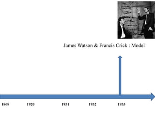
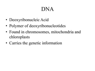
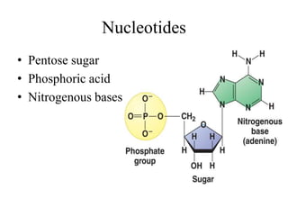
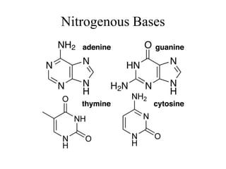
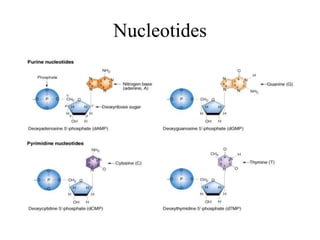
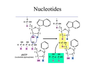
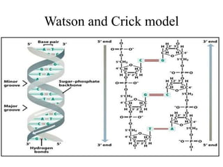
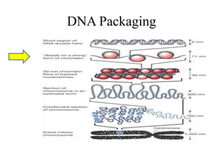
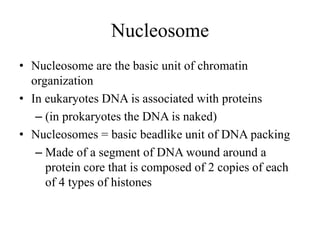
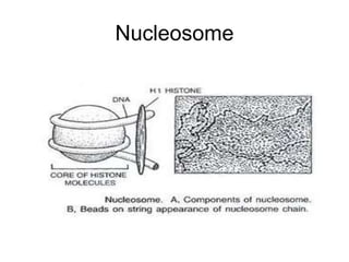
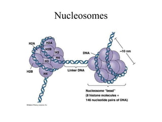
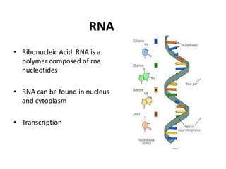
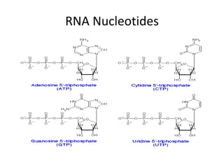
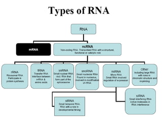
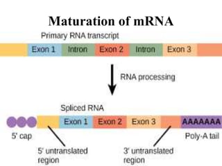
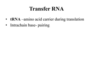
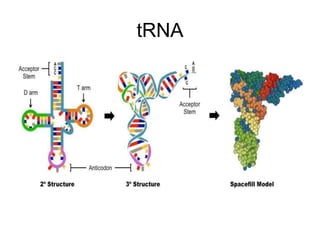
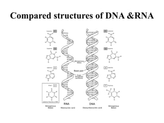
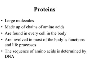
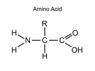
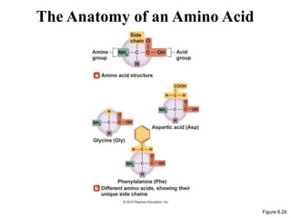
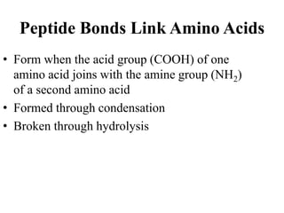
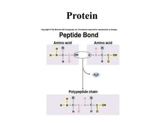
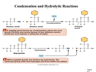
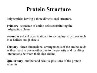
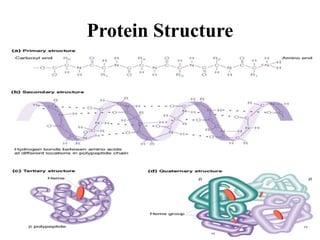
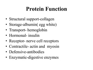
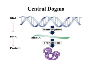
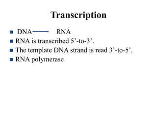
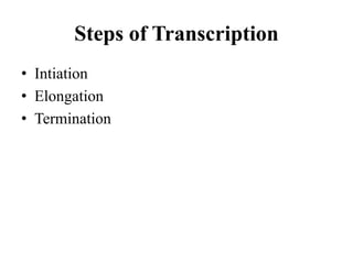
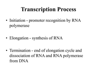
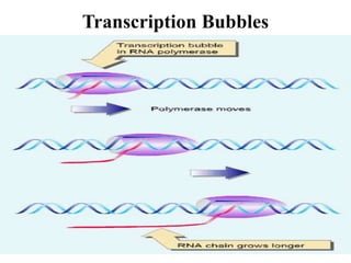
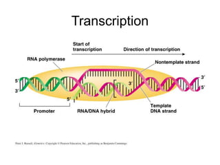
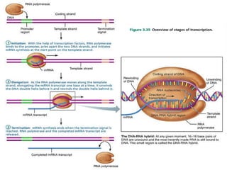
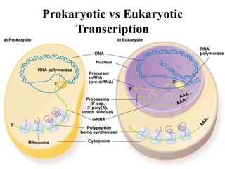
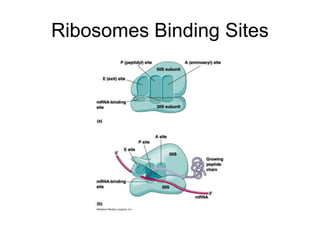
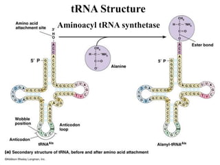
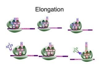
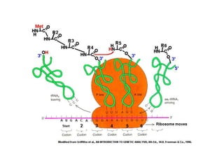
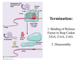
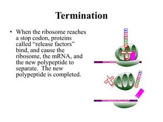
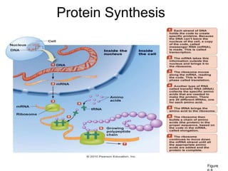
KEY FEATURES OF DNA,RNA AND PROTEIN
AND THEIR INTER-RELATIONSHIP
JUNAID
&
ISRA DURRANI
Faculty of Life Sciences and Informatics, BUITEMS, Quetta
OVERVIEW
• DNA
• RNA
• PROTEIN
• CENTRAL DOGMA
BRIEF HISTORY
Friedrich Miescher : nuclein
1868 1920 1951 1952 1953
P A Levene : Basic structure of nucleic acid
1868 1920 1951 1952 1953
Erwin Chargaff : Chargaff rule A=T , G=C
1868 1920 1951 1952 1953
Wilkin & Rosalind: Xray diffraction analysis of DNA
1868 1920 1951 1952 1953
James Watson & Francis Crick : Model
1868 1920 1951 1952 1953
DNA STRUCTURE
DNA
• Deoxyribonucleic Acid
• Polymer of deoxyribonucleotides
• Found in chromosomes, mitochondria and
chloroplasts
• Carries the genetic information
Nucleotides
• Pentose sugar
• Phosphoric acid
• Nitrogenous bases
Nitrogenous Bases
Nucleotides
Nucleotides
Watson and Crick model
DNA PACKAGING
Prokaryotic DNA Packing
DNA Packaging
Nucleosome
• Nucleosome are the basic unit of chromatin
organization
• In eukaryotes DNA is associated with proteins
– (in prokaryotes the DNA is naked)
• Nucleosomes = basic beadlike unit of DNA packing
– Made of a segment of DNA wound around a
protein core that is composed of 2 copies of each
of 4 types of histones
Nucleosome
Nucleosomes
DNA Packaging
Solenoid
Nucleosome+ H1=
30nm spiral
DNA Packaging
Loop Domains
• Chromatin fibers forms loop which attach to
nonhistone proteins
• Non histone proteins form a scaffold
• 300nm in diameter
DNA Packaging
Metaphase Chromosome
• Looped domains coil and fold, further
compacting chromatin
• Result in the characteristic metaphase
chromsome seen in karyotypes
DNA REPLICATION
DNA Replication Model
• In the late 1950s, three different mechanisms were proposed for the
replication of DNA
– Conservative model
• Both parental strands stay together after DNA replication
– Semiconservative model
• The daughter double-stranded DNA contains one parental and one
daughter strand following replication
– Dispersive model
• The daughter DNA are interspersed in both parental and new
strands following replication
DNA Replication Model
Replication of DNA
• The process of copying a DNA molecule
• Each old DNA strand serves as a template
• Replication involves 3 main steps
1. Unwinding
2. Complementary base pairing
3. Joining
Prokaryotic replication
• Bacteria have a single circular loop of DNA
• Replication moves around the circular DNA
molecule in both directions
• The process begins at the origin of replication
and always occur in the 5 to 3
• Replication stop when the 2 DNA polymerases
meet at a termination region
• 40 min to replicate
Prokaryotic Replication
Eukaryotic DNA Replication
• DNA replication begins at numerous points
along linear chromosome
• Replication bubbles spread bi directionally
until they meet
• Replication fork v shape formed during dna
replication
Starts at origin
Initiator proteins identify specific base sequences on DNA
called sites of origin
Prokaryotes – single origin site
Eukaryotes – multiple sites of origin
Prokaryotes Eukaryotes
DNA Replication
RNA primers required
Semi-discontinuous replication
Anti parallel strands replicated simultaneously
 Leading strand synthesis continuously in 5’– 3’
 Lagging strand synthesis in fragments in 5’-3’
DNA Replication
Core proteins at the replication fork
Topoisomerases
Helicases
Primase
Single strand
binding proteins
DNA polymerase
DNA ligase
- Prevents torsion by DNA breaks
- separates 2 strands
- RNA primer synthesis
- prevent reannealing
of single strands
- synthesis of new strand
- seals nick via phosphodiester linkage
RNA STRUCTURE
RNA
• Ribonucleic Acid RNA is a
polymer composed of rna
nucleotides
• RNA can be found in nucleus
and cytoplasm
• Transcription
RNA Nucleotides
Messenger RNA
• mRNA contains the genetic information from DNA
(Template for protein synthesis).
• Synthesized from its 5’ to 3’ end
• Single-stranded
• Generally mRNAs are linear (although some
prokaryotic RNA viruses are circular and act as
mRNAs)
Maturation of mRNA
Transfer RNA
• tRNA –amino acid carrier during translation
• Intrachain base- pairing
tRNA
Ribosomal RNA
• rRNA is a structural component of ribosomes
Compared structures of DNA &RNA
PROTEIN
Proteins
• Large molecules
• Made up of chains of amino acids
• Are found in every cell in the body
• Are involved in most of the body’s functions
and life processes
• The sequence of amino acids is determined by
DNA
Amino Acid
The Anatomy of an Amino Acid
Figure 6.2b
Peptide Bonds Link Amino Acids
• Form when the acid group (COOH) of one
amino acid joins with the amine group (NH2)
of a second amino acid
• Formed through condensation
• Broken through hydrolysis
Protein
Condensation and Hydrolytic Reactions
Figure
Protein Structure
Polypeptides having a three dimensional structure.
Primary–sequence of amino acids constituting the
polypeptide chain
Secondary–local organization into secondary structures such
as  helices and  sheets
Tertiary –three dimensional arrangements of the amino acids
as they react to one another due to the polarity and resulting
interactions between their side chains
Quaternary–number and relative positions of the protein
subunits
Protein Structure
Protein Function
• Structural support-collagen
• Storage-albumin( egg white)
• Transport- hemoglobin
• Hormonal- insulin
• Receptor- nerve cell receptors
• Contractile- actin and myosin
• Defensive-antibodies
• Enzymatic-digestive enzymes
CENTRAL DOGMA
Central Dogma
Genes
• Genes=units of genetic information (hereditary information)
• Order of nucleotides make up the genetic code
• Genes can contain the information for one polypeptide
• Genes can also regulate how other genes are expressed
Gene
• The gene has three regions, each with a
function in transcription:
a. promoter
b. RNA-coding sequence
c. terminator
TRANSCRIPTION
Transcription
 DNA RNA
 RNA is transcribed 5’-to-3’.
 The template DNA strand is read 3’-to-5’.
 RNA polymerase
Steps of Transcription
• Intiation
• Elongation
• Termination
Transcription Process
• Initiation - promoter recognition by RNA
polymerase
• Elongation - synthesis of RNA
• Termination - end of elongation cycle and
dissociation of RNA and RNA polymerase
from DNA
Transcription Bubbles
Transcription
Termination of transcription
 Termination can be rho-independent or rho-
dependent (rho is a protein termination factor)
 Most termination is rho-independent
Rho independent termination
Mechanism of Action of Rho
Prokaryotic vs Eukaryotic
Transcription
TRANSLATION
Ribosome
• made of proteins and rRNA
• each has a large and small subunit
• each has three binding sites for tRNA on its
surface
• components of ribosomes are made in the
nucleus and exported to the cytoplasm where
they join to form one functional unit
Ribosome Binding Sites
• A site (aminoacyl site)
– Holds tRNA carrying next amino acid
• P site (peptidyl-tRNA site)
– holds tRNA carrying growing polypeptide chain
• E site (exit site)
– uncharged tRNA leaves ribosome from exit site
Ribosomes Binding Sites
tRNA Structure
Aminoacyl tRNA synthetase
Aminoacyl tRNA Synthetases
• Enzymes which bond specific amino acids to their
particular tRNAs.
• There are 20 different aminoacyl tRNA
synthetases, one for each amino acid.
• Covalent linkage through an ester bond
• tRNA linked to amino acid is charged tRNA.
Translation has 3 Steps, Each Requiring
Different Supporting Proteins
• Initiation
– Requires Initiation Factors
• Elongation
– Requires Elongation Factors
• Termination
– Requires Termination Factor
Initiation
The initiation process involves first
joining the mRNA, the initiator
methionine-tRNA, and the small
ribosomal subunit. Several “initiation
factors”--additional proteins--are also
involved. The large ribosomal subunit
then joins the complex.
Initiation
Ribosome + mRNA + tRNA +ribosome
Initiation:
1. Binding of initiation factors
to small subunit.
2. Binding of first tRNA and
mRNA to small subunit.
3. Binding of large subunit.
Elongation:
1. Binding of next tRNA
using EFs at
A site.
2. Peptide Bond
formation between 2
amino acids.
3. Translocation of
ribosome.
E P A
E PA
E PA
E PA
E PA
Elongation
Termination:
1. Binding of Release
Factor to Stop Codon
UGA, UAA, UAG.
2. Disassembly
Termination
• When the ribosome reaches
a stop codon, proteins
called “release factors”
bind, and cause the
ribosome, the mRNA, and
the new polypeptide to
separate. The new
polypeptide is completed.
Protein Synthesis
Figure
SUMMARY
• Key features of DNA, RNA and Protein
• Inter relation between them
o Central dogma
o Structure formation
o Gene regulation
REFERENCES

DNA RNA PROTEIN Central Dogma