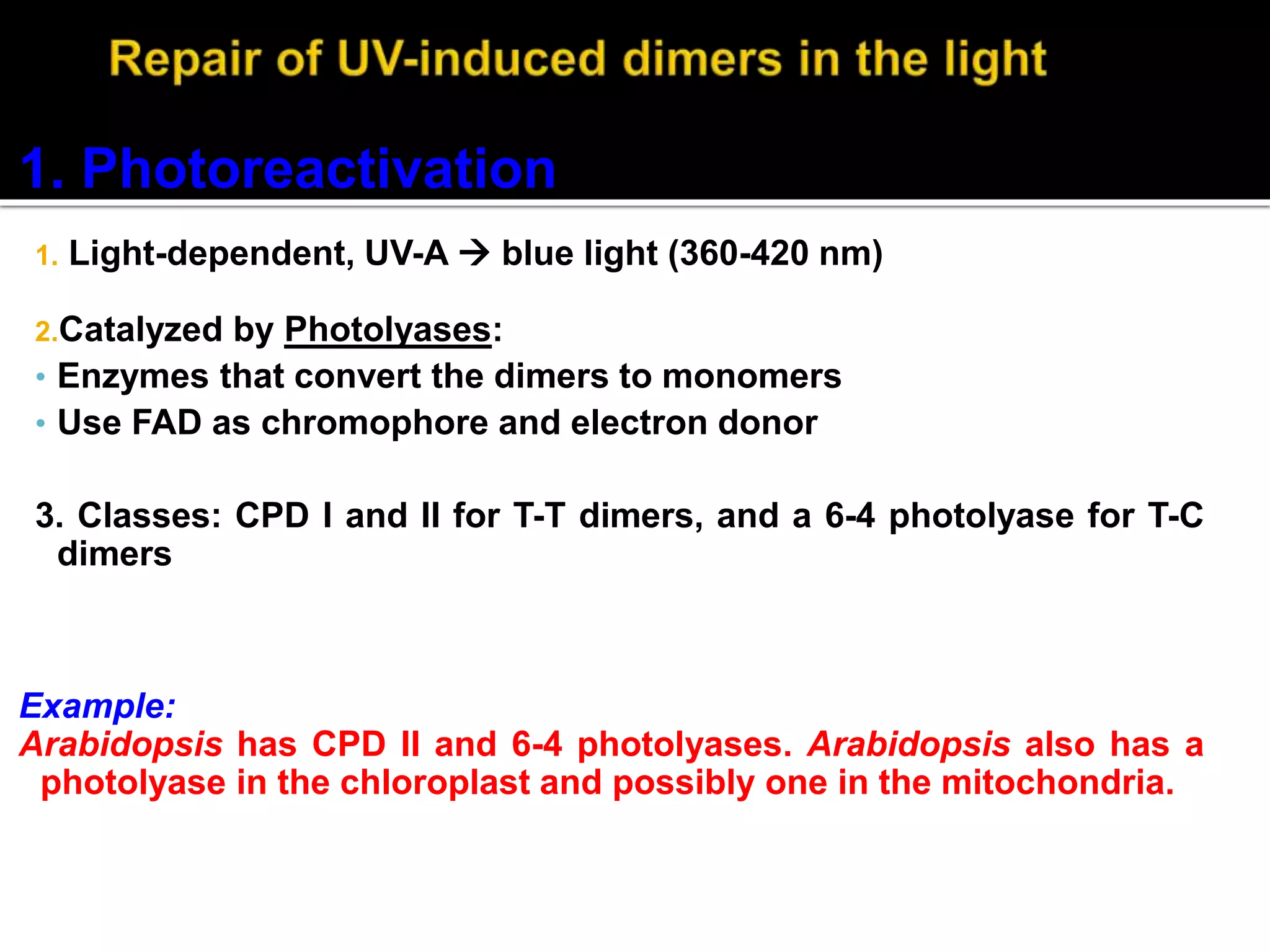
 Prepared by:
Prashant VC
Department of Zoology
Gulbarga University,
Kalaburagi
Introduction
DNA Polymerase as Self Correcting Enzyme
Correct nucleotide has greater affinity for moving polymerase than incorrect
nucleotide
Exonucleolytic proofreading of DNA polymerase
DNA molecules w/ mismatched 3’ OH end are not
effective templates; polymerase cannot extend when
3’ OH is not base paired
DNA polymerase has separate catalytic site that
removes unpaired residues at terminus
 When DNA repair fails, fewer mutations corrected  increase in
number of mutations in the genome.
 The protein p53 monitors repair of damaged DNA.
 If damage too severe, p53 protein promotes programmed cell
death (apoptosis)
 Mutations in genes encoding DNA repair proteins can be
inherited  overall increase in mutations as errors or damage to
DNA no longer repaired efficiently.
1. Deamination: (C  U and A hypoxanthine)
2. Depurination: purine base (A or G) lost
3. T-T and T-C dimers: bases become cross-linked, T-T more
prominent, caused by UV light (UV-C (<280 nm) and UV-B
(280-320 nm)
4. Alkylation: an alkyl group (e.g., CH3) gets added to bases;
chemical induced; some harmless, some cause mutations by
mispairing during replication or stop polymerase altogether
5. Oxidative damage: guanine oxidizes to 8-oxo-guanine,
also cause SS and DS breaks, very important for
organelles
6. Replication errors: wrong nucleotide (or modified nt)
inserted
7. Double-strand breaks (DSB): induced by ionizing
radiation, transposons, topoisomerases, homing
endonucleases, and mechanical stress on chromosomes
, oxidative free radical formation
Depurination
Chemical Modification Photodamage thymine dimer
Chemical Modification by O2 free radicals
Deamination
G A C
T
ds DNA Break Mismatch
Thymidine dimer
AP site
Covalent X-linking
ss Break
C-U deamination
1. Photoreactivation
1. Light-dependent, UV-A  blue light (360-420 nm)
2.Catalyzed by Photolyases:
• Enzymes that convert the dimers to monomers
• Use FAD as chromophore and electron donor
3. Classes: CPD I and II for T-T dimers, and a 6-4 photolyase for T-C
dimers
Example:
Arabidopsis has CPD II and 6-4 photolyases. Arabidopsis also has a
photolyase in the chloroplast and possibly one in the mitochondria.
Fig. 6.12 in Buchanan et al.
Photolyase
gene
expression
also induced
or increased
by light.
Fig. 6.12 in Buchanan et al.
2. Removal of Alkylation
 Problem: how do cells know which is the right template strand?
 In E. coli, new DNA not methylated right away
 Mismatch recognized by mutS, then mutL binds and attracts
mutH (endonuclease that cleaves mismatch and nearest CTAG
that is not methylated)
 Eucaryotes (including Arabidopsis) have mutS and mutL
homologues, but no mutH
 Also have the requisite exonucleases, but not clear how the
strand specificity is determined
Strand Directed Mismatch Repair System
 Removes replication errors not recognized by
replication machine
 Detects distortion in DNA helix
 Distinguishes newly replicated strand from parental
strand by methylation of A residues in GATC in
bacteria
 Methylation occurs shortly after replication occurs
 Reduces error rate 100X
 3 Step Process
recognition of mismatch
excision of segment of DNA containing mismatch
resynthesis of excised fragment
Strand Directed Mismatch Repair in Mammals
 Newly synthesized strand is preferentially nicked and
can be distinguish in this manner from parental
strand
 Defective copy of mismatch repair gene predisposed to
cancer
Mut H:
Endonuclease
Mut H:
Endonuclease
In E.coli, A of each GATC is
methylated.
Mismatch Repair
mutH is endonuclease
The bases cytosine, adenine and guanine can undergo spontaneous
depurination to respectively form uracil, hypoxanthine and
xanthine.
These altered bases do not exist in the normal DNA, and therefore
need to be removed.
This is carried out by base excision repair
A defective DNA in which cytosine is deaminated to uracil is acted
upon by the enzyme Uracil DNA glycosylase.
This results in the removal of the defective base Uracil.
An endonuclease cuts the backbone of DNA strand near the defect
and removes a few bases.
The gap so created is filled up by the action of repair DNA
Base Excision Repair (BER)
Variety of DNA glycosylases,
for different types of damaged
bases.
AP endonuclease recognizes
sites with a missing base;
cleaves sugar-phosphate
backbone.
Deoxyribose
phosphodiesterase removes
the sugar-phosphate lacking
the base.
Deaminated C
Fig. 6.15
Base Excision Repair
a. DNA glycosylase recognizes damaged base
b. Removes base leaving deoxyribose sugar
c. AP endonuclease cuts phosphodiester backbone
d. DNA polymerase replaces missing nucleotide
e. DNA ligase seals nick
Nucleotide Excision Repair
(in E. coli of a T-T dimer)
Endonuclease cuts on either
side of damage (~20 nt
altogether).
Strands unwound by helicase.
Fig. 6.14 in Buchanan et al.
1. UvrA,B
2. UvrC
3. UvrD
2 general ways to repair DSBs:
1. Homologous recombination (HR) - repair of broken
DNA using the intact homologue. Very accurate.
2. Non-homologous end joining (NHEJ) - ligating non-
homologous ends. Prone to errors, ends can be
damaged before ligation (genetic material lost), or get
translocations.
Usage: NHEJ >> HR in plants and animals (in the cells’ nucleus)
RecA/Rad51
Resolvase (recG)
DSBR by HR
Modified from Fig. 6.18 in
Buchanan et al.
3’ SS extensions
RecA binds preferentially
to SS DNA and will
catalyze invasion of a DS
DNA molecule by a SS
homologue.
Important for many types
of homologous
recombination, such as
during meoisis (in yeast).
Fig. 6.19
 Arabidopsis has rad51, resolvase (recG), and repA (SS DNA binding in
animals) homologues, all needed for HR.
 Also has homologues of key genes required for NHEJ (e.g., Ku70 and
Ku80).
 Processing of DSBs very important – they can block cell cycle
progression and trigger apoptosis (programmed cell death).
Disorder Frequency Defect
Fanconi’s anaemia 1/22,000 in some
popns.
Deficient excision
repair
Hereditary nonpolyposis
colon cance
1/200 Deficient mismatch
repair
Werner’s syndrome 3/1,000,000 Deficient helicase
Xeroderma pigmentosum 1/250,000 Deficient excision
repair
 This is a very good illustration of the devastating
effects of even tiny changes to the DNA
 Red Blood Cells
 Hemoglobin -
 Has a large protein component
 2 beta globin chains
 A single base change -substitution causes the disease
DNA Repair .pptx

DNA Repair .pptx

  • 1.
     Prepared by: PrashantVC Department of Zoology Gulbarga University, Kalaburagi Introduction DNA Polymerase as Self Correcting Enzyme Correct nucleotide has greater affinity for moving polymerase than incorrect nucleotide Exonucleolytic proofreading of DNA polymerase
  • 2.
    DNA molecules w/mismatched 3’ OH end are not effective templates; polymerase cannot extend when 3’ OH is not base paired DNA polymerase has separate catalytic site that removes unpaired residues at terminus
  • 4.
     When DNArepair fails, fewer mutations corrected  increase in number of mutations in the genome.  The protein p53 monitors repair of damaged DNA.  If damage too severe, p53 protein promotes programmed cell death (apoptosis)  Mutations in genes encoding DNA repair proteins can be inherited  overall increase in mutations as errors or damage to DNA no longer repaired efficiently.
  • 6.
    1. Deamination: (C U and A hypoxanthine) 2. Depurination: purine base (A or G) lost 3. T-T and T-C dimers: bases become cross-linked, T-T more prominent, caused by UV light (UV-C (<280 nm) and UV-B (280-320 nm) 4. Alkylation: an alkyl group (e.g., CH3) gets added to bases; chemical induced; some harmless, some cause mutations by mispairing during replication or stop polymerase altogether
  • 7.
    5. Oxidative damage:guanine oxidizes to 8-oxo-guanine, also cause SS and DS breaks, very important for organelles 6. Replication errors: wrong nucleotide (or modified nt) inserted 7. Double-strand breaks (DSB): induced by ionizing radiation, transposons, topoisomerases, homing endonucleases, and mechanical stress on chromosomes , oxidative free radical formation
  • 8.
    Depurination Chemical Modification Photodamagethymine dimer Chemical Modification by O2 free radicals Deamination
  • 9.
    G A C T dsDNA Break Mismatch Thymidine dimer AP site Covalent X-linking ss Break C-U deamination
  • 10.
    1. Photoreactivation 1. Light-dependent,UV-A  blue light (360-420 nm) 2.Catalyzed by Photolyases: • Enzymes that convert the dimers to monomers • Use FAD as chromophore and electron donor 3. Classes: CPD I and II for T-T dimers, and a 6-4 photolyase for T-C dimers Example: Arabidopsis has CPD II and 6-4 photolyases. Arabidopsis also has a photolyase in the chloroplast and possibly one in the mitochondria.
  • 11.
    Fig. 6.12 inBuchanan et al. Photolyase gene expression also induced or increased by light.
  • 12.
    Fig. 6.12 inBuchanan et al. 2. Removal of Alkylation
  • 13.
     Problem: howdo cells know which is the right template strand?  In E. coli, new DNA not methylated right away  Mismatch recognized by mutS, then mutL binds and attracts mutH (endonuclease that cleaves mismatch and nearest CTAG that is not methylated)  Eucaryotes (including Arabidopsis) have mutS and mutL homologues, but no mutH  Also have the requisite exonucleases, but not clear how the strand specificity is determined
  • 14.
    Strand Directed MismatchRepair System  Removes replication errors not recognized by replication machine  Detects distortion in DNA helix  Distinguishes newly replicated strand from parental strand by methylation of A residues in GATC in bacteria  Methylation occurs shortly after replication occurs  Reduces error rate 100X  3 Step Process recognition of mismatch excision of segment of DNA containing mismatch resynthesis of excised fragment
  • 15.
    Strand Directed MismatchRepair in Mammals  Newly synthesized strand is preferentially nicked and can be distinguish in this manner from parental strand  Defective copy of mismatch repair gene predisposed to cancer
  • 16.
  • 17.
    In E.coli, Aof each GATC is methylated. Mismatch Repair mutH is endonuclease
  • 18.
    The bases cytosine,adenine and guanine can undergo spontaneous depurination to respectively form uracil, hypoxanthine and xanthine. These altered bases do not exist in the normal DNA, and therefore need to be removed. This is carried out by base excision repair
  • 19.
    A defective DNAin which cytosine is deaminated to uracil is acted upon by the enzyme Uracil DNA glycosylase. This results in the removal of the defective base Uracil. An endonuclease cuts the backbone of DNA strand near the defect and removes a few bases. The gap so created is filled up by the action of repair DNA
  • 20.
    Base Excision Repair(BER) Variety of DNA glycosylases, for different types of damaged bases. AP endonuclease recognizes sites with a missing base; cleaves sugar-phosphate backbone. Deoxyribose phosphodiesterase removes the sugar-phosphate lacking the base. Deaminated C Fig. 6.15
  • 21.
    Base Excision Repair a.DNA glycosylase recognizes damaged base b. Removes base leaving deoxyribose sugar c. AP endonuclease cuts phosphodiester backbone d. DNA polymerase replaces missing nucleotide e. DNA ligase seals nick
  • 24.
    Nucleotide Excision Repair (inE. coli of a T-T dimer) Endonuclease cuts on either side of damage (~20 nt altogether). Strands unwound by helicase. Fig. 6.14 in Buchanan et al. 1. UvrA,B 2. UvrC 3. UvrD
  • 25.
    2 general waysto repair DSBs: 1. Homologous recombination (HR) - repair of broken DNA using the intact homologue. Very accurate. 2. Non-homologous end joining (NHEJ) - ligating non- homologous ends. Prone to errors, ends can be damaged before ligation (genetic material lost), or get translocations. Usage: NHEJ >> HR in plants and animals (in the cells’ nucleus)
  • 26.
    RecA/Rad51 Resolvase (recG) DSBR byHR Modified from Fig. 6.18 in Buchanan et al. 3’ SS extensions
  • 27.
    RecA binds preferentially toSS DNA and will catalyze invasion of a DS DNA molecule by a SS homologue. Important for many types of homologous recombination, such as during meoisis (in yeast). Fig. 6.19
  • 28.
     Arabidopsis hasrad51, resolvase (recG), and repA (SS DNA binding in animals) homologues, all needed for HR.  Also has homologues of key genes required for NHEJ (e.g., Ku70 and Ku80).  Processing of DSBs very important – they can block cell cycle progression and trigger apoptosis (programmed cell death).
  • 29.
    Disorder Frequency Defect Fanconi’sanaemia 1/22,000 in some popns. Deficient excision repair Hereditary nonpolyposis colon cance 1/200 Deficient mismatch repair Werner’s syndrome 3/1,000,000 Deficient helicase Xeroderma pigmentosum 1/250,000 Deficient excision repair
  • 30.
     This isa very good illustration of the devastating effects of even tiny changes to the DNA  Red Blood Cells  Hemoglobin -  Has a large protein component  2 beta globin chains  A single base change -substitution causes the disease