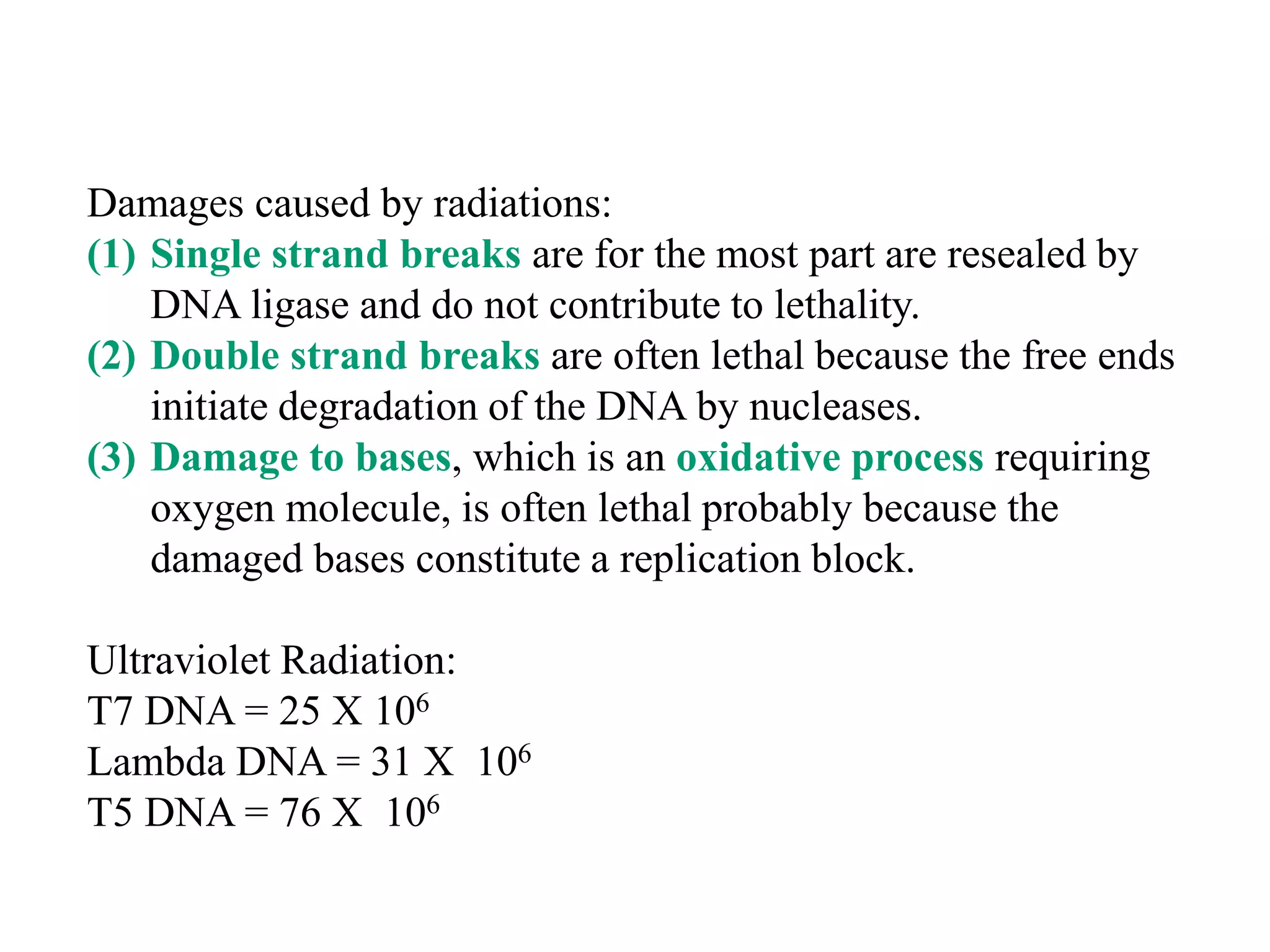
(I) DNA can be damaged by radiation, chemicals, and other environmental factors which cells have developed mechanisms to repair. (II) There are direct repair systems like photoreactivation and base excision repair that remove damaged bases. (III) Nucleotide excision repair and mismatch repair pathways cut out the damaged DNA section and resynthesize the correct sequence. (IV) Double strand breaks are repaired by nonhomologous end joining or homologous recombination.


![[I] Direct repair systems fill in nicks and correct some types of
nucleotide modification:
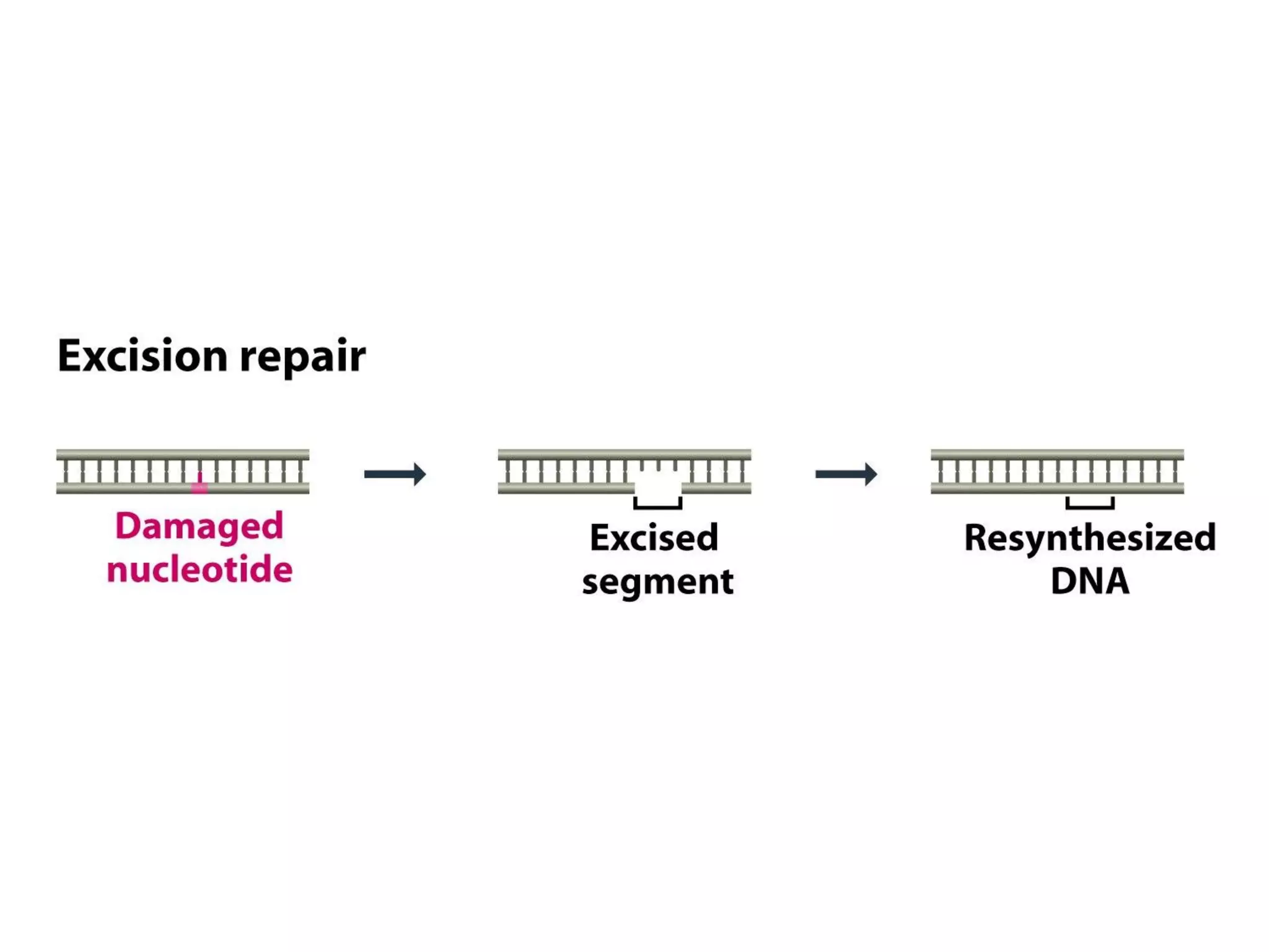
- Most of the DNA damages are repaired by excision of damaged nucleotide
followed by resynthesis of a new stretch of DNA.
- A few types can be repaired directly
(i) Nicks can be repaired by
DNA ligase, where
phosphodiester bond has
been broken
- Nicks are produced by
ionizing radiations.](https://image.slidesharecdn.com/dnadamagerepairmechanisms-230330082502-1dc8589d/75/DNA-Damage-Repair-mechanisms-pdf-41-2048.jpg)

![[II] Excision Repair:
- Pathway: includes many components
(a) Base Excision Repair
- removal of damaged nucleotide base, creating an AP site
- Excision of a short piece of polynucleotide around AP site
- Resynthesis with DNA polymerase and ligation
(b) Nucleotide Excision Repair
- similar but does not remove the damaged nucleotide base
- Excision of a short piece of polynucleotide around damaged site
- Resynthesis with DNA polymerase and ligation
- Can repair severely damaged areas of DNA](https://image.slidesharecdn.com/dnadamagerepairmechanisms-230330082502-1dc8589d/75/DNA-Damage-Repair-mechanisms-pdf-43-2048.jpg)


![[b] Nucleotide Excision Repair
It can repair extensive type of damages, such as cross links and
attachment of large chemical groups.
It can also repair cyclobutyl dimers by a Dark Repair Process.
Difference with Base Excision:
(i) It is not preceded by removal of selective base
(ii) A longer stretch of polynucleotide is removed.
Two types is E. coli
• “Short Patch” process: 12 nucleotides long
• “Long Patch” process: up to 2 kb](https://image.slidesharecdn.com/dnadamagerepairmechanisms-230330082502-1dc8589d/75/DNA-Damage-Repair-mechanisms-pdf-50-2048.jpg)


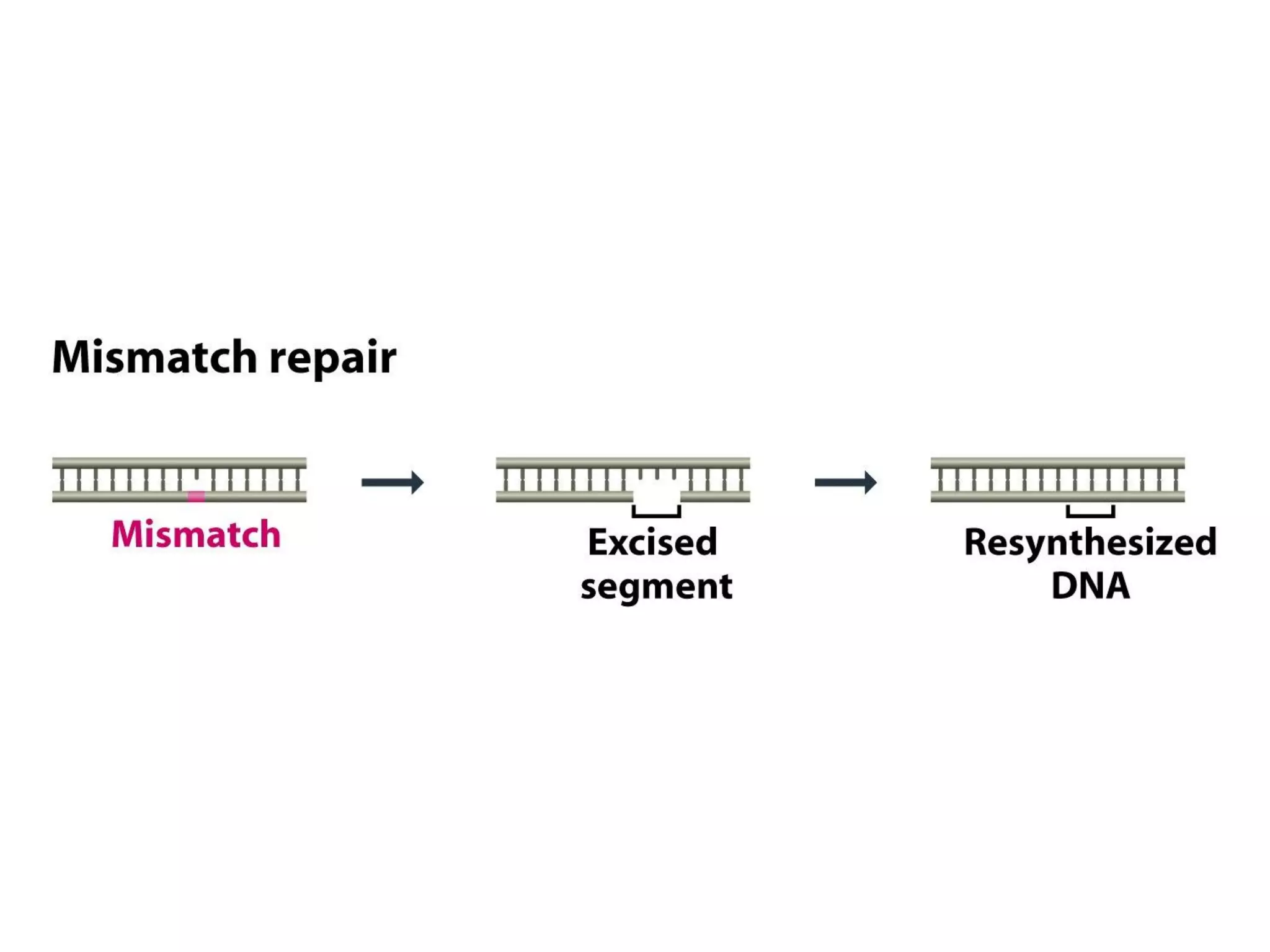
![[III] Mismatch Repair
- Mainly for correcting error of replication.
- Earlier discussed methods search for abnormal chemical
structures, but can not identify mismatch.
- As it is normal nucleotide, A, T, C or G.
- This repair system looks for absence of base pairing between
parent and daughter strands.
- Then it excises part of daughter polynucleotide and fills in gap.
- Important: repair must be there in newly synthesized strand
which has acquired mismatch and parent has correct sequence.
- How is it identified?](https://image.slidesharecdn.com/dnadamagerepairmechanisms-230330082502-1dc8589d/75/DNA-Damage-Repair-mechanisms-pdf-55-2048.jpg)


![[IV] Repair of DNA Breaks:
* Single Strand Break Repair
- produced by some type of oxidative damage
- exposed single strand is coated with PARP1 proteins, which
protects it from breaking and taking part in unwanted
recombination.
- Break is filled by enzymes involved in excision repair
pathways.](https://image.slidesharecdn.com/dnadamagerepairmechanisms-230330082502-1dc8589d/75/DNA-Damage-Repair-mechanisms-pdf-67-2048.jpg)


![[V] BYPASSING DNA DAMAGE DURING GENOME
REPLICATION
- Extensive damage cell faces a tough choice between dying or
attempting to replicate the damaged region though it is error-
prone causing mutations.
- Uses one of the emergency procedures
* SOS Response:
- Best studied process
- Enables cell to replicate DNA even if AP sites/ cyclobutyl dimers/
other photoproducts are present.
- Mutasome (DNA Pol V) : A trimer - UmuD’2C : 2 UmuD’ &
1 UmuC and several copies of Rec A proteins
- Rec A protein - single strand binding protein – role in repair and
recombination pathways
- Rec A binding enables mutasome to replace DNA Pol III, carries
out replication until damaged site is bypassed → DNA Pol III
takes over.](https://image.slidesharecdn.com/dnadamagerepairmechanisms-230330082502-1dc8589d/75/DNA-Damage-Repair-mechanisms-pdf-72-2048.jpg)