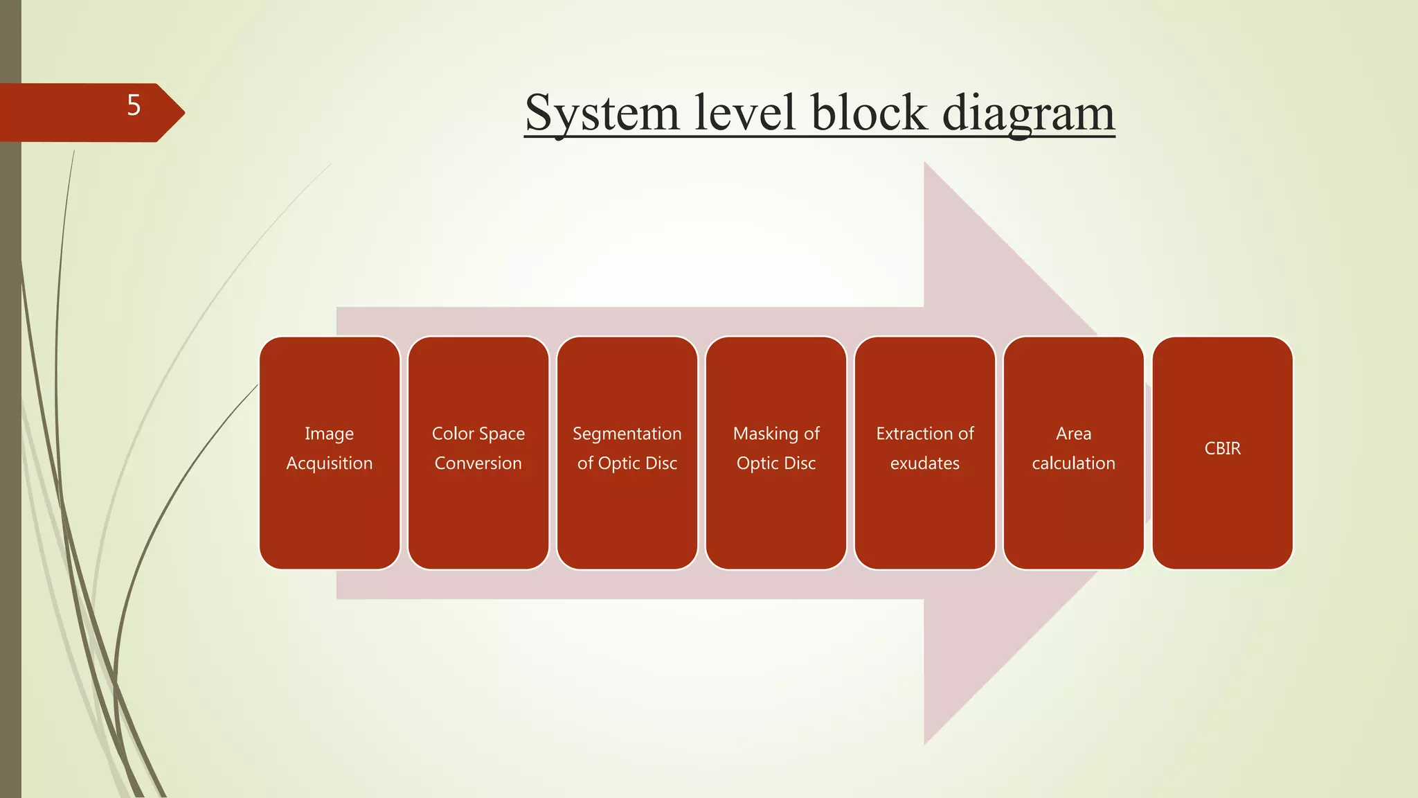
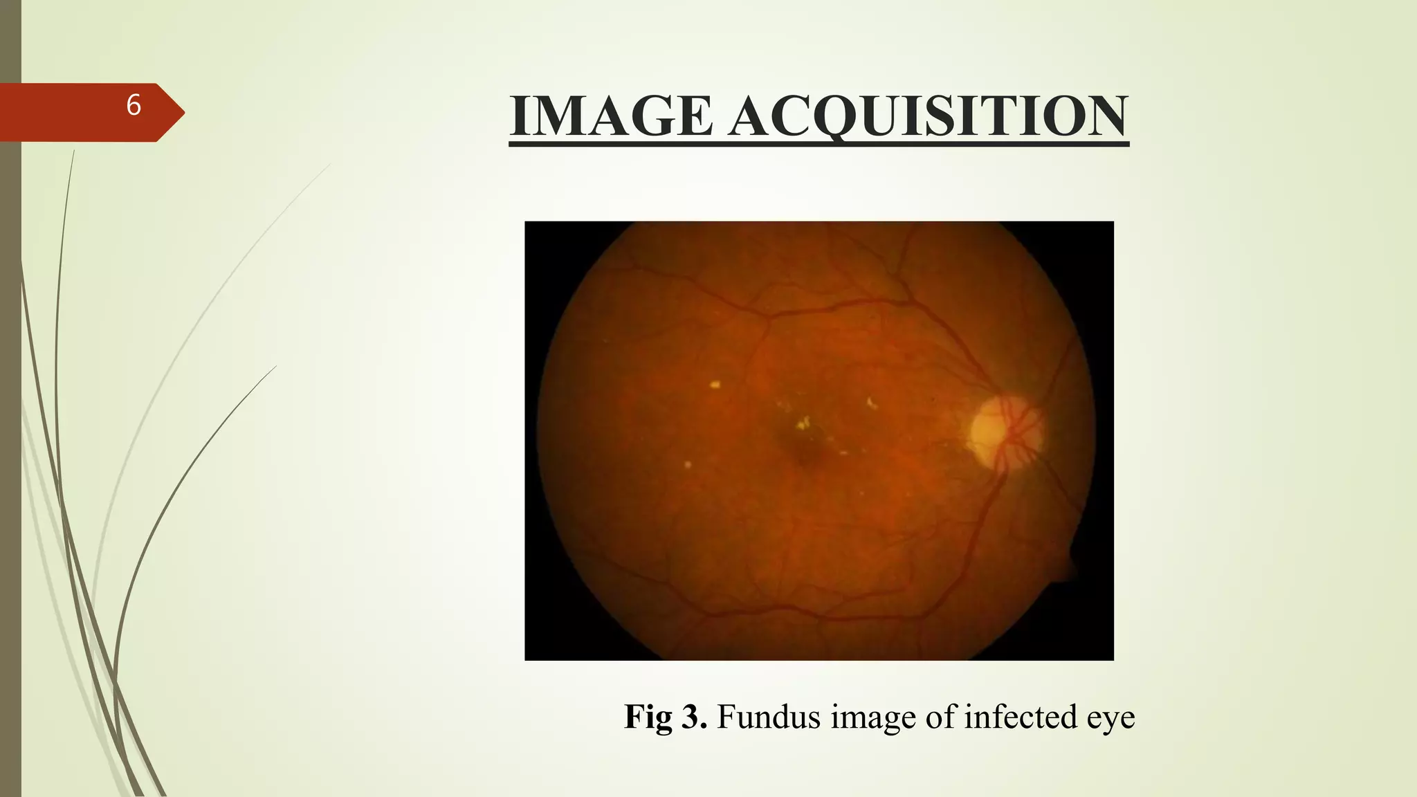
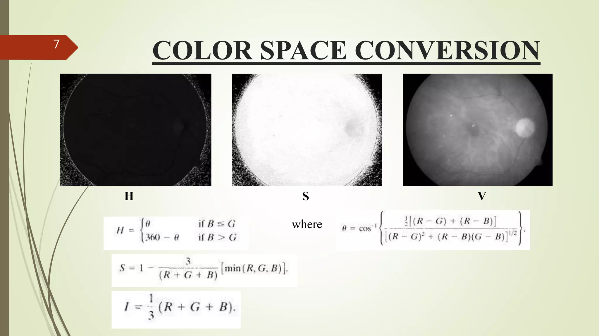
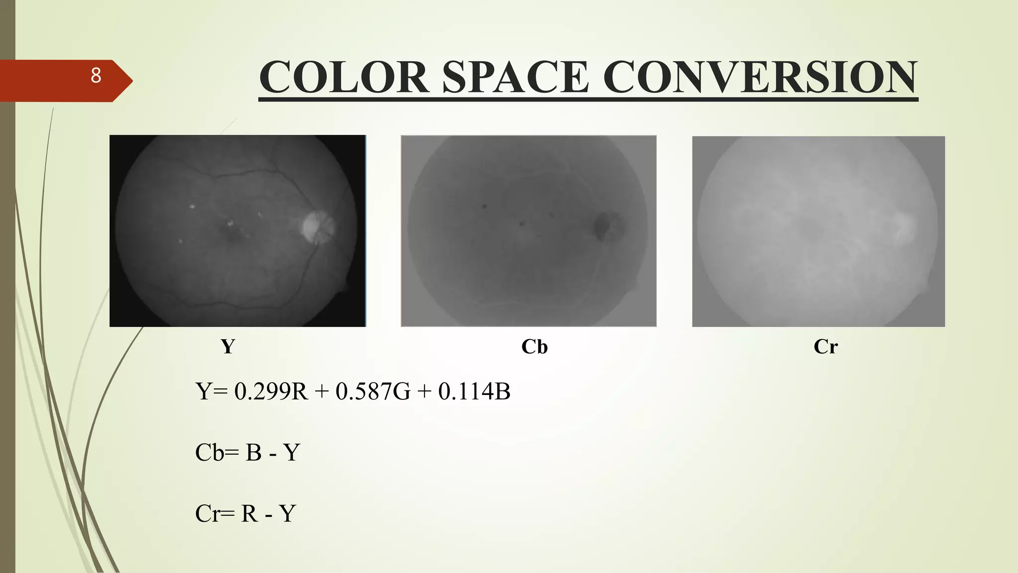
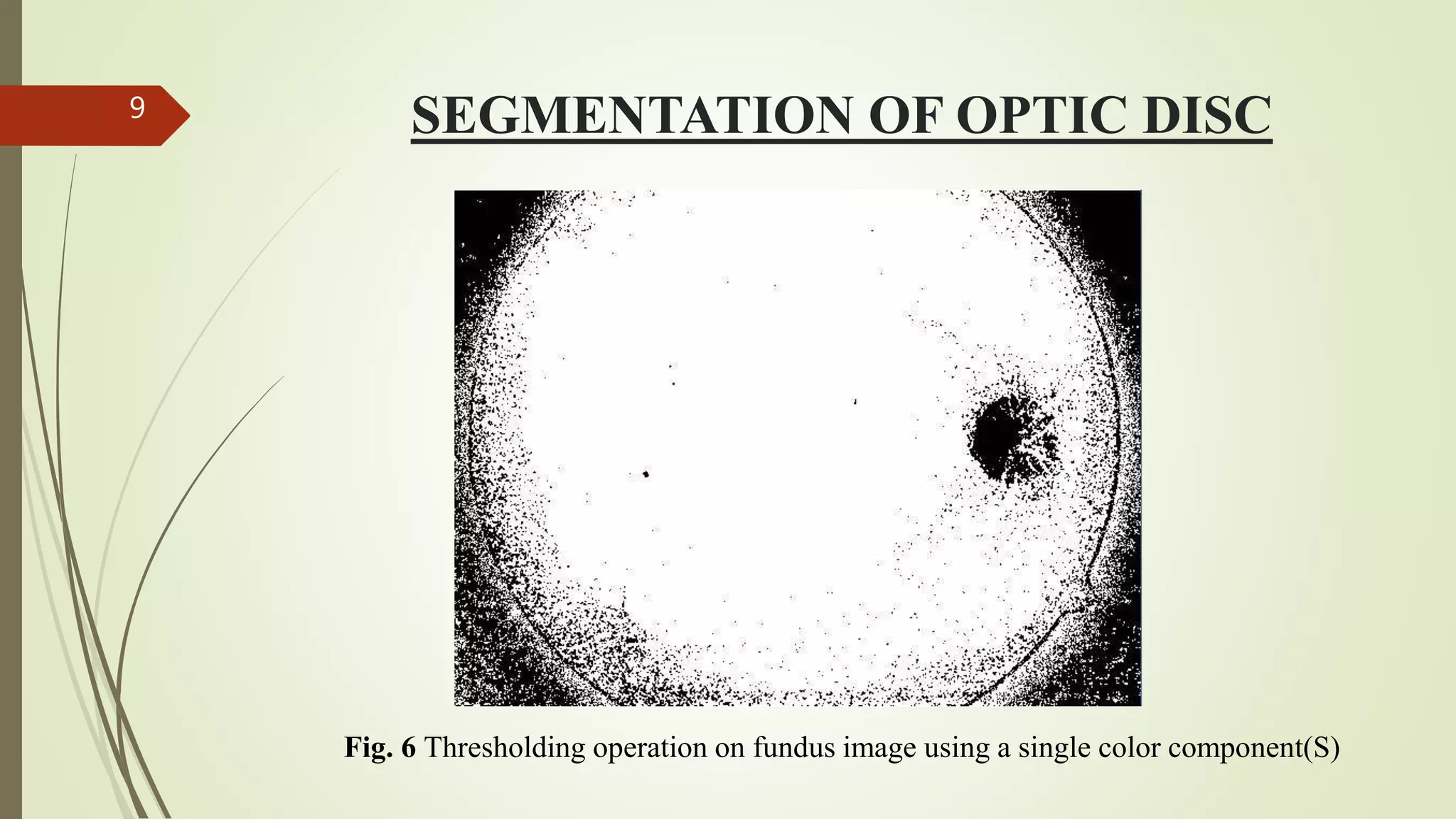
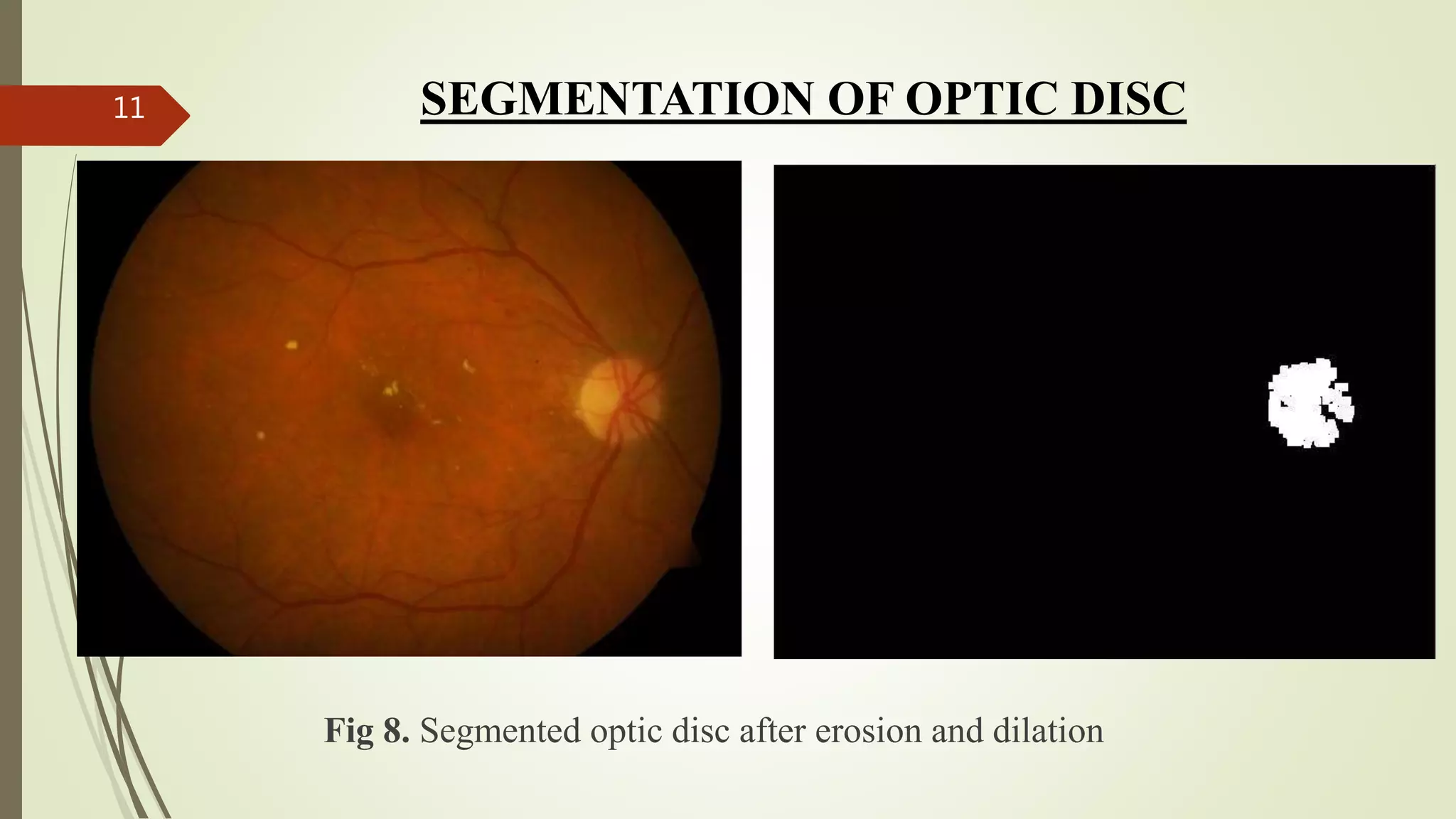
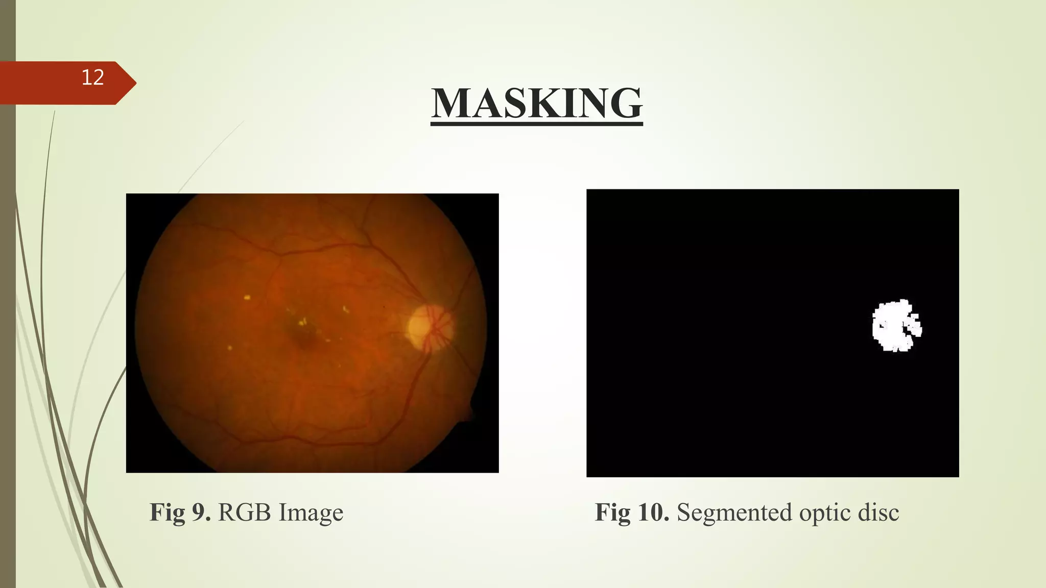
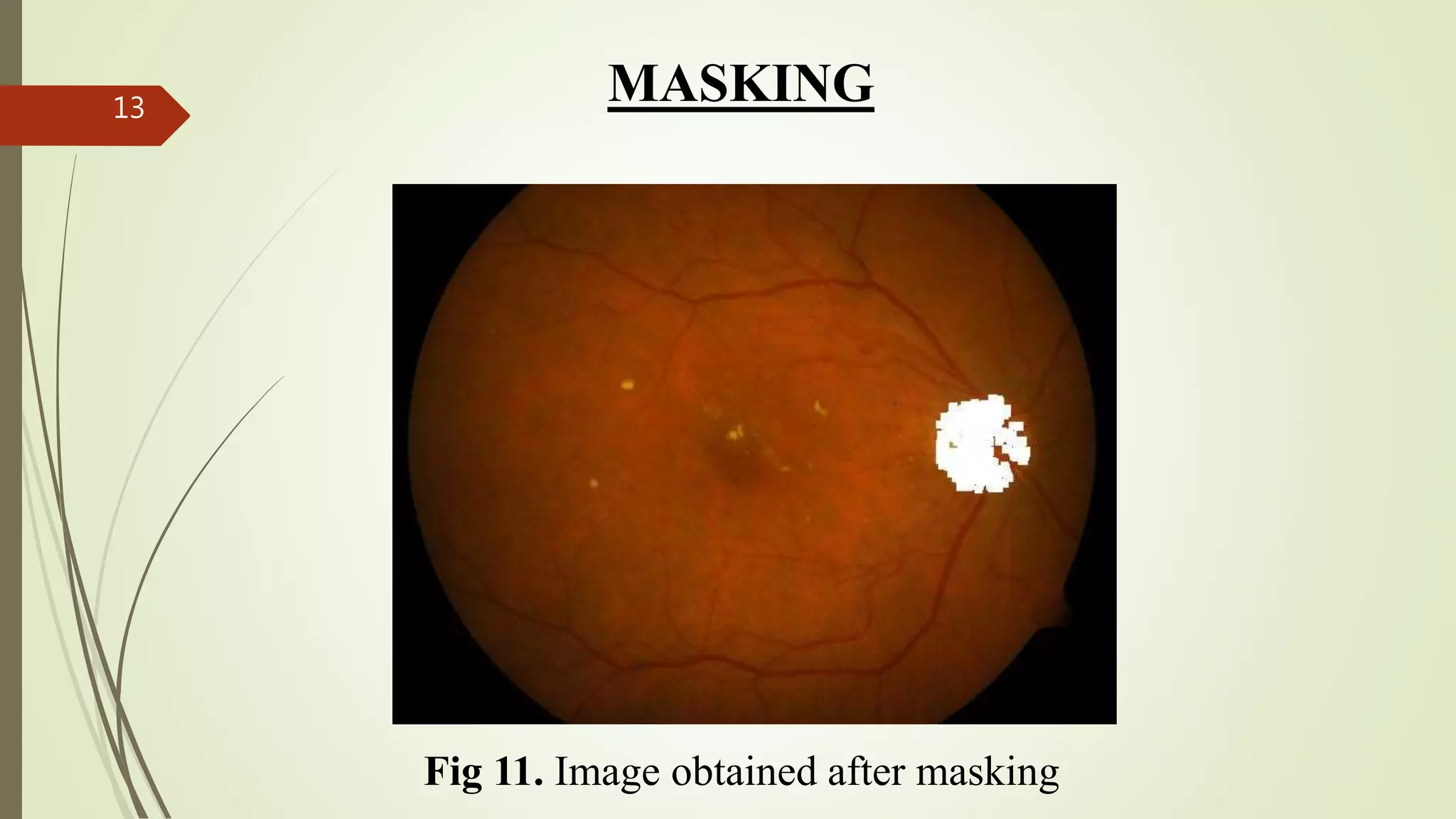
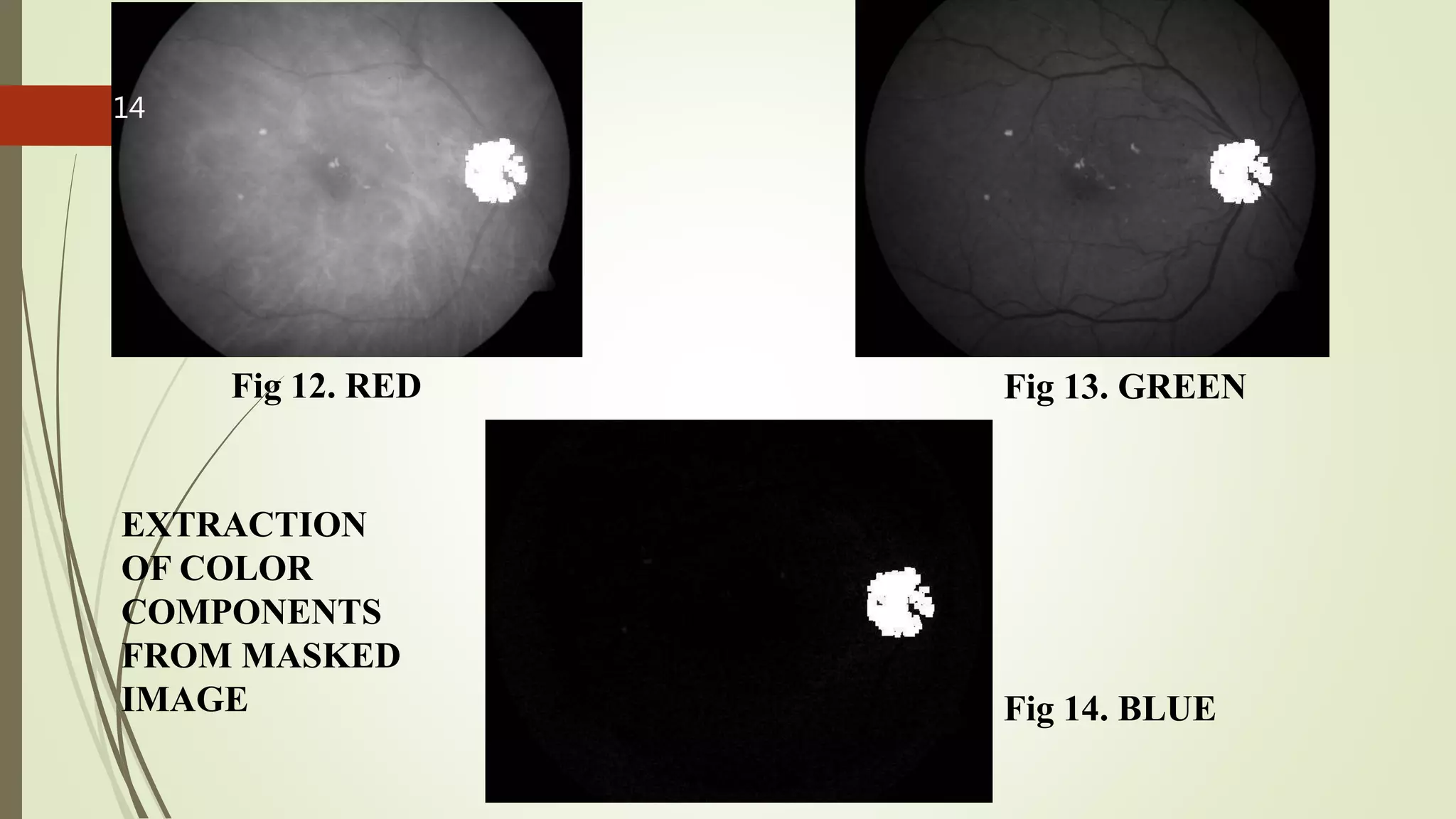
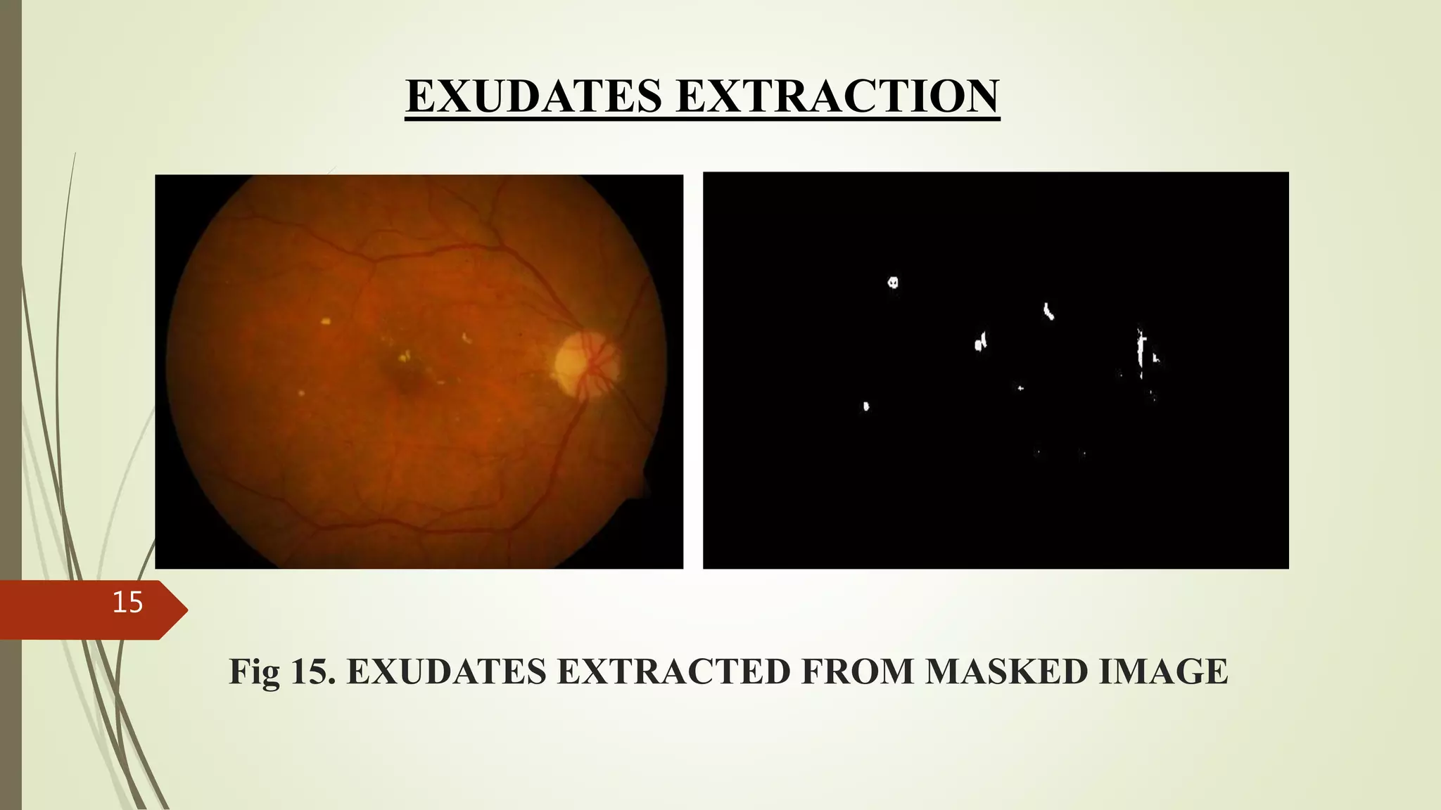
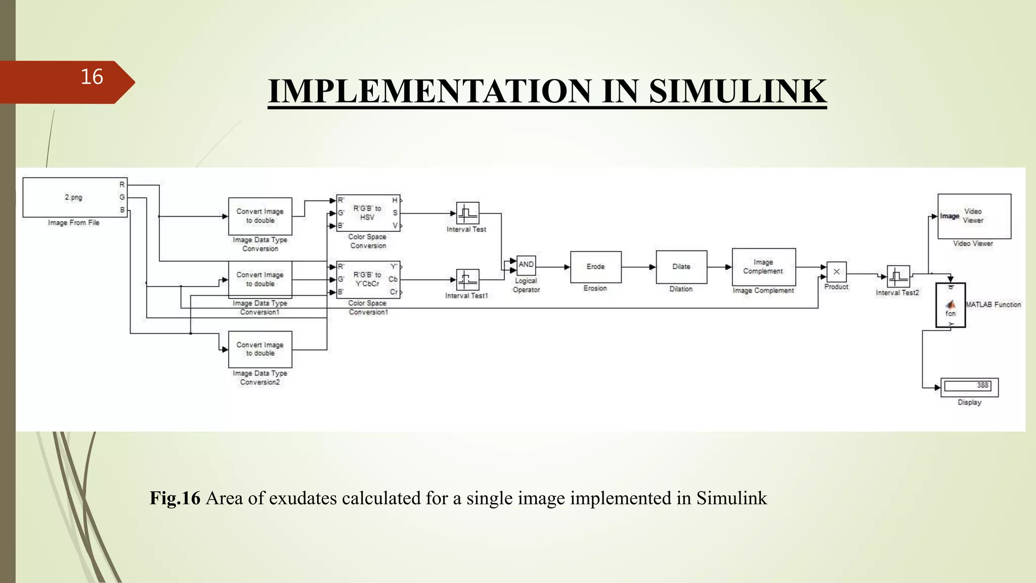
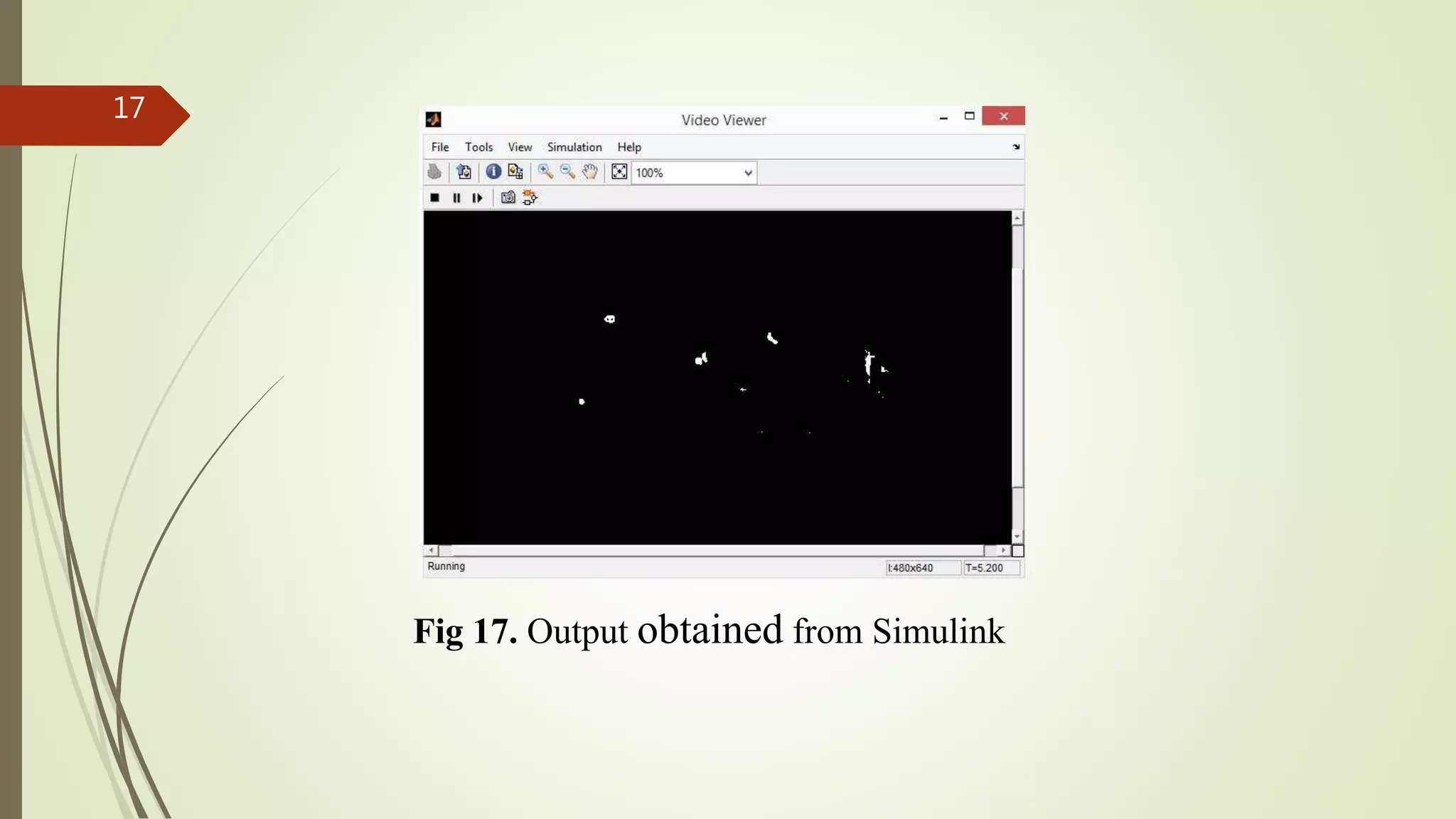
DESIGN AND DEVELOPMENT OF AN
EARLY OPTICAL DISEASE
RECOGNITION SYSTEM USING
FUNDUS IMAGING ON FPGA
1
NEED FOR THE PROJECT
 Individuals with untreated polygenic disorder are twenty five times
more in danger for vision defect than the final population.
 The longer an individual has had polygenic disorder, the higher the
chance of developing diabetic retinopathy.
 But with regular, correct eye care and treatment at the proper time,
the incidence of severe vision loss are often greatly reduced.
2
PROBLEM STATEMENT
 The fundus image of the healthy eye has only the optic disk in it.
Whereas the fundus image of an infected eye has optic disk along
with spots with the same intensity level as that of the optic disk.
 These spots are called as exudates OR cotton wool spots and are
characteristic of diabetic retinopathy.
 We aim to extract the characteristics (exudates) obtained from the
fundus image of a person’s eye.
3
Fig.1 Healthy fundus image Fig.2 Infected fundus image
4
System level block diagram5
Image
Acquisition
Color Space
Conversion
Segmentation
of Optic Disc
Masking of
Optic Disc
Extraction of
exudates
Area
calculation
CBIR
IMAGE ACQUISITION6
Fig 3. Fundus image of infected eye
COLOR SPACE CONVERSION7
H S V
where
COLOR SPACE CONVERSION8
Y Cb Cr
Y= 0.299R + 0.587G + 0.114B
Cb= B - Y
Cr= R - Y
SEGMENTATION OF OPTIC DISC9
Fig. 6 Thresholding operation on fundus image using a single color component(S)
Fig.7 Segmented optic disc before erosion and dilation
10 SEGMENTATION OF OPTIC DISC
Fig 8. Segmented optic disc after erosion and dilation
11 SEGMENTATION OF OPTIC DISC
MASKING
Fig 9. RGB Image Fig 10. Segmented optic disc
12
Fig 11. Image obtained after masking
MASKING13
Fig 12. RED Fig 13. GREEN
Fig 14. BLUE
EXTRACTION
OF COLOR
COMPONENTS
FROM MASKED
IMAGE
14
Fig 15. EXUDATES EXTRACTED FROM MASKED IMAGE
EXUDATES EXTRACTION
15
IMPLEMENTATION IN SIMULINK
Fig.16 Area of exudates calculated for a single image implemented in Simulink
16
Fig 17. Output obtained from Simulink
17
CONTENT BASED IMAGE RETRIEVAL
 Content-based image retrieval (CBIR) is the application of computer
vision techniques to the image retrieval problem, that is, the problem
of searching for digital images in large databases.
 “Content-based" means that the search analyses the contents of the
image rather than the metadata such as keywords, tags, or
descriptions associated with the image.
 CBIR is desirable because searches that rely purely on metadata are
dependent on annotation quality and completeness.
18
CONTENT BASED IMAGE RETRIEVAL
Fig. 18 A test image with a database image
19
CONTENT BASED IMAGE RETRIEVAL
Fig.19 Multiport switch and JTAG programming along with MATLAB function block for comparison
20
FUTURE WORKS
 Better segmentation of optic disc can be achieved.
 Along with the area, the medicine to be prescribed can also be
displayed.
 Handheld ophthalmoscopes which can take the fundus image
without dilation of the pupil.
21
- VRUSHAK K(1BG11TE062)
VIKRAM(1BG11TE061)
DINESH N SHENOY(1BG11TE015)
22

Design and development of an early optical disease recognition system using fundus imaging on FPGA

  • 1.
    DESIGN AND DEVELOPMENTOF AN EARLY OPTICAL DISEASE RECOGNITION SYSTEM USING FUNDUS IMAGING ON FPGA 1
  • 2.
    NEED FOR THEPROJECT  Individuals with untreated polygenic disorder are twenty five times more in danger for vision defect than the final population.  The longer an individual has had polygenic disorder, the higher the chance of developing diabetic retinopathy.  But with regular, correct eye care and treatment at the proper time, the incidence of severe vision loss are often greatly reduced. 2
  • 3.
    PROBLEM STATEMENT  Thefundus image of the healthy eye has only the optic disk in it. Whereas the fundus image of an infected eye has optic disk along with spots with the same intensity level as that of the optic disk.  These spots are called as exudates OR cotton wool spots and are characteristic of diabetic retinopathy.  We aim to extract the characteristics (exudates) obtained from the fundus image of a person’s eye. 3
  • 4.
    Fig.1 Healthy fundusimage Fig.2 Infected fundus image 4
  • 5.
    System level blockdiagram5 Image Acquisition Color Space Conversion Segmentation of Optic Disc Masking of Optic Disc Extraction of exudates Area calculation CBIR
  • 6.
    IMAGE ACQUISITION6 Fig 3.Fundus image of infected eye
  • 7.
  • 8.
    COLOR SPACE CONVERSION8 YCb Cr Y= 0.299R + 0.587G + 0.114B Cb= B - Y Cr= R - Y
  • 9.
    SEGMENTATION OF OPTICDISC9 Fig. 6 Thresholding operation on fundus image using a single color component(S)
  • 10.
    Fig.7 Segmented opticdisc before erosion and dilation 10 SEGMENTATION OF OPTIC DISC
  • 11.
    Fig 8. Segmentedoptic disc after erosion and dilation 11 SEGMENTATION OF OPTIC DISC
  • 12.
    MASKING Fig 9. RGBImage Fig 10. Segmented optic disc 12
  • 13.
    Fig 11. Imageobtained after masking MASKING13
  • 14.
    Fig 12. REDFig 13. GREEN Fig 14. BLUE EXTRACTION OF COLOR COMPONENTS FROM MASKED IMAGE 14
  • 15.
    Fig 15. EXUDATESEXTRACTED FROM MASKED IMAGE EXUDATES EXTRACTION 15
  • 16.
    IMPLEMENTATION IN SIMULINK Fig.16Area of exudates calculated for a single image implemented in Simulink 16
  • 17.
    Fig 17. Outputobtained from Simulink 17
  • 18.
    CONTENT BASED IMAGERETRIEVAL  Content-based image retrieval (CBIR) is the application of computer vision techniques to the image retrieval problem, that is, the problem of searching for digital images in large databases.  “Content-based" means that the search analyses the contents of the image rather than the metadata such as keywords, tags, or descriptions associated with the image.  CBIR is desirable because searches that rely purely on metadata are dependent on annotation quality and completeness. 18
  • 19.
    CONTENT BASED IMAGERETRIEVAL Fig. 18 A test image with a database image 19
  • 20.
    CONTENT BASED IMAGERETRIEVAL Fig.19 Multiport switch and JTAG programming along with MATLAB function block for comparison 20
  • 21.
    FUTURE WORKS  Bettersegmentation of optic disc can be achieved.  Along with the area, the medicine to be prescribed can also be displayed.  Handheld ophthalmoscopes which can take the fundus image without dilation of the pupil. 21
  • 22.