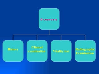
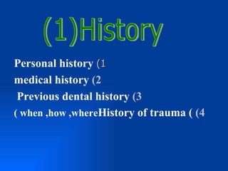
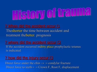
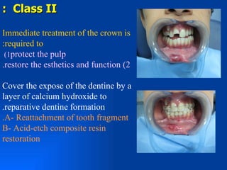
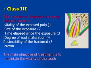
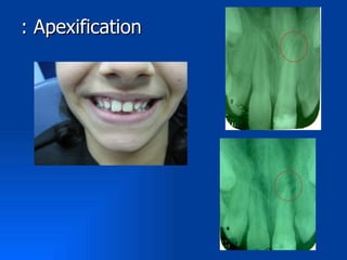
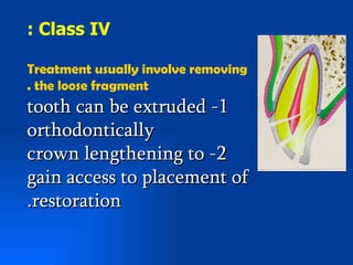
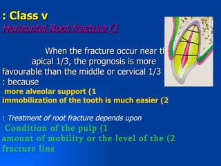
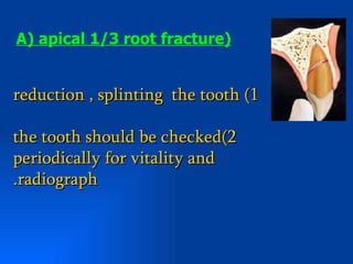
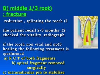
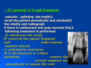
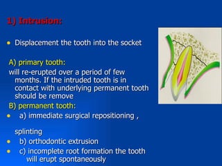
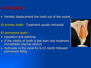
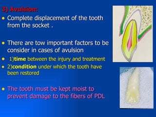
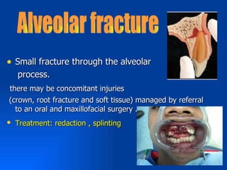
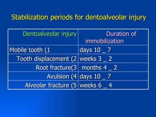
Dr. Mohammad A. Barayan
: Definition
Injury which is limited to the teeth
and supporting structures of the
.alveolus
N.B Boys are three times more
.at risk than girls


: Causes
.Traffic Accident -1
.Falls -2
.During Epileptic seizures -3
 .Sport injuries -4
D iagnos is


            Clinical
History                                  Radiographic
          examination    Vitality test
                                         Examination
Personal history (1
medical history (2
 Previous dental history (3
( when ,how ,whereHistory of trauma ( (4
? When did the accident occur (1
 Theshorter the time between accident and
 treatment thebetter. prognosis
? where did the accident occur (2
If the accident occurred indirty place prophylactic tetanus
is indicated

? how did the injury occur (3
Direct force under the chin → → condylar fracture
Direct force to teeth→ → Crown F, Root F, displacement
Extraoral Examination
Laceration ;Abrasions ;Contusions on the head and
neck can be noted visually
Anyasymmetries including deviation in mouth
.opening

Intraoral Examination
( .. Soft tissue ( tongue ; gingiva
Teeth ( displacement ; mobility ; tooth fracture ;
( colour change
Vitality test just following traumatic
injury often given false negative
response
Types of vitality test
Thermal pulp test (1
cold test


heat test

 (2Electrical pulp test
 Cavity test (3
*s o f t t is s u e in ju r ie s

1- Determination of child immunization
status:-

•If the child had received a primary
immunization activated with booster injection
of toxoid .
•Unimmunized child can be protected by
tetanus antitoxin.

2- Adequate debridment of the wound
stage of root formation -1
presence of root fractur -2
 periapical radiolucencies -3
 injury of the supporting periodontal membrane -4
(degree of intrusion or extrusion o the tooth)
 size of the pulp -5

N. B. If a jaw fracture is suspected extaoral
radiographs indicated (panoramic and lateral
 ( oblique views
:Ellis classification
 :Class I
 crack or fracture of E only
 :Class II
 fracture of E , D with out pulp exposure
 :Class III
 fracture of E , D with pulp exposure
:Class IV
Fracture line passes beneath the gingival margin
:Class V
 Root fracture
            a( vertical      b( horizontal
                               ((apical , middle , cervical
: Class I

  a crack of the -1 enamel
without loss of tooth
. structure
Do not require immediate
.treatment

fracture of enamel only -2
 smoothing the sharp edge
regular vitality test ,
 radiograph
: Class II

Immediate treatment of the crown is
:required to
 (1protect the pulp
.restore the esthetics and function (2

Cover the expose of the dentine by a
layer of calcium hydroxide to
.reparative dentine formation
.A- Reattachment of tooth fragment
B- Acid-etch composite resin
restoration
: Class III
The treatment depends on many
:factors such as
.vitality of the exposed pulp (1
.Size of the exposure (2
.Time elapsed since the exposure (3
.Degree of root maturation (4
Restorability of the fractured (5
.crown

The main objective of treatment is to
. maintain the vitality of the tooth
Small exposure                              Large exposure

   Early                    Late                    Early                 Late


Open   Close       open            closed    open       closed     open      Closed


                  pulpoto      pulpecto     Pulpoto    pulpecto   Pulpe
                    my           my           my         my       ctomy
   Direct pulp
      capping                                                             pulpec
                  pulpecto                  pulpecto              Apexifi tomy
                    my                        my                   cation
: Apexification
: Class IV

Treatment usually involve removing
. the loose fragment
tooth can be extruded -1
orthodontically
crown lengthening to -2
gain access to placement of
.restoration
: Class v
Horizontal Root fracture (1

           When the fracture occur near the
       apical 1/3, the prognosis is more
favourable than the middle or cervical 1/3
: because
 more alveolar support (1
immobilization of the tooth is much easier (2

: Treatment of root fracture depends upon
 Condition of the pulp (1
amount of mobility or the level of the (2
fracture line
A( apical 1/3 root fracture(


reduction , splinting the tooth (1

the tooth should be checked(2
periodically for vitality and
.radiograph
B( middle 1/3 root(
: fracture
reduction , splinting the tooth (1

the patient recall 2-3 months ,(2
checked the vitality ,radiograph

if the tooth non vital and no(3
healing the following treatment is
:performed
 a( R C T of both fragments
     b( apical fragment removed
             surgically
c( intraradicular pin to stabilize
: C( cervical 1/3 root fracture(
 reductin , splinting the tooth(1
recall the patient periodically and checked(2
the vitality and radiograph
if there is radiolucent and pulp necrosis the(3
 following treatment is performed
 a( extraction the tooth
b( removed the apical fragment
and                       endo-osseous
 implant placed
c( orthodontic extrusion
d( if the fracture is 1-2mm
infrabony                       remove the
                      coronal segment and
  osteoplasty to expose the root
2) vertical root fracture :

• usually the prognosis is not favorable

• treatment of V R F :
1)extraction of the tooth
2)using co2 laser and ND:YAG laser beam
Concussion *


• A mild blow to the tooth resulting in mild
  sensitivity requires little or no treatment

• Need only regular vitality test
subluxation*

• Mobility of the tooth without displacement
• Tooth may be sensitive to percussion
• If mobility is extensive    splint the tooth
 using the acid –etch splinting technique.
• Regular vitality test and radiograph
• 1) lateral luxation
• 2) intrusive luxation
• 3) extrusive luxation
• 4) avulsion
1( Lateral luxation :

• Displacement of the tooth in any
    direction other than the axial one

• If the patient comes immediately
    after trauma reposition, splinting
•   Once the tooth have solidified in
    their position orthodontic
    treatment is required
1( Intrusion:

• Displacement the tooth into the socket

 A) primary tooth:
 will re-erupted over a period of few
   months. If the intruded tooth is in
   contact with underlying permanent tooth
   should be remove
 B) permanent tooth:
• a) immediate surgical repositioning ,
    splinting
•    b) orthodontic extrusion
•    c) incomplete root formation the tooth
        will erupt spontaneously
2( Extrusion :

• Partially displacement the tooth out of the socket .

A) primary tooth: Treatment usually extracted

B) permanent tooth :
• reposition and splinting
• If the vitality of tooth is lost start root treatment
  immediately placing calcium
• hydroxide in the canal for 6-12 month followed
  permanent filling.
3( Avulsion:
• Complete displacement of the tooth
  from the socket .

• There are tow important factors to be
    consider in cases of avulsion
•    1)time between the injury and treatment
•   2)condition under which the tooth have
    been restored


• The tooth must be kept moist to
    prevent damage to the fibers of PDL
• In many cases the initial patient contact is by
    phone
•   The tooth should be handled by the crown
•   The tooth should be placed in suitable storage
    medium (milk, unsalted water, lens solution )or
    in buccal vestibule or under the tongue .
•   At the dental office :
•   a) information about tetanus immunization
    should be obtained
•   b) replantation , splinting for 1_2weeks but in
    immature apices 2-3weeks
•   c) calcium hydroxide should be placed
•   d) RCT
• Small fracture through the alveolar
   process.
there may be concomitant injuries
(crown, root fracture and soft tissue) managed by referral
  to an oral and maxillofacial surgery .
• Treatment: redaction , splinting
: Types of splinting

1) acid_etched composite splinting
2) Interdental wiring
3) ( vacuum_formed plastic) splint
4) arch bare splint

• More rigid and the longer the stabilization,
 the more root resorption , ankylosis that
 can be expected .
Stabilization periods for dentoalveolar injury

    Dentoalveolar injury             Duration of
                              immobilization
Mobile tooth (1             days 10 _ 7
  Tooth displacement (2     weeks 3 _ 2
          Root fracture(3    months 4 _ 2
              Avulsion (4   days 10 _ 7
     Alveolar fracture (5   weeks 6 _ 4
dento – alveolar injuries

dento – alveolar injuries

  • 1.
  • 2.
    : Definition Injury whichis limited to the teeth and supporting structures of the .alveolus N.B Boys are three times more .at risk than girls : Causes .Traffic Accident -1 .Falls -2 .During Epileptic seizures -3 .Sport injuries -4
  • 3.
    D iagnos is Clinical History Radiographic examination Vitality test Examination
  • 4.
    Personal history (1 medicalhistory (2 Previous dental history (3 ( when ,how ,whereHistory of trauma ( (4
  • 5.
    ? When didthe accident occur (1 Theshorter the time between accident and treatment thebetter. prognosis ? where did the accident occur (2 If the accident occurred indirty place prophylactic tetanus is indicated ? how did the injury occur (3 Direct force under the chin → → condylar fracture Direct force to teeth→ → Crown F, Root F, displacement
  • 6.
    Extraoral Examination Laceration ;Abrasions;Contusions on the head and neck can be noted visually Anyasymmetries including deviation in mouth .opening Intraoral Examination ( .. Soft tissue ( tongue ; gingiva Teeth ( displacement ; mobility ; tooth fracture ; ( colour change
  • 7.
    Vitality test justfollowing traumatic injury often given false negative response Types of vitality test Thermal pulp test (1 cold test heat test (2Electrical pulp test Cavity test (3
  • 8.
    *s o ft t is s u e in ju r ie s 1- Determination of child immunization status:- •If the child had received a primary immunization activated with booster injection of toxoid . •Unimmunized child can be protected by tetanus antitoxin. 2- Adequate debridment of the wound
  • 9.
    stage of rootformation -1 presence of root fractur -2 periapical radiolucencies -3 injury of the supporting periodontal membrane -4 (degree of intrusion or extrusion o the tooth) size of the pulp -5 N. B. If a jaw fracture is suspected extaoral radiographs indicated (panoramic and lateral ( oblique views
  • 10.
    :Ellis classification :ClassI crack or fracture of E only :Class II fracture of E , D with out pulp exposure :Class III fracture of E , D with pulp exposure :Class IV Fracture line passes beneath the gingival margin :Class V Root fracture a( vertical b( horizontal ((apical , middle , cervical
  • 11.
    : Class I a crack of the -1 enamel without loss of tooth . structure Do not require immediate .treatment fracture of enamel only -2 smoothing the sharp edge regular vitality test , radiograph
  • 12.
    : Class II Immediatetreatment of the crown is :required to (1protect the pulp .restore the esthetics and function (2 Cover the expose of the dentine by a layer of calcium hydroxide to .reparative dentine formation .A- Reattachment of tooth fragment B- Acid-etch composite resin restoration
  • 13.
    : Class III Thetreatment depends on many :factors such as .vitality of the exposed pulp (1 .Size of the exposure (2 .Time elapsed since the exposure (3 .Degree of root maturation (4 Restorability of the fractured (5 .crown The main objective of treatment is to . maintain the vitality of the tooth
  • 14.
    Small exposure Large exposure Early Late Early Late Open Close open closed open closed open Closed pulpoto pulpecto Pulpoto pulpecto Pulpe my my my my ctomy Direct pulp capping pulpec pulpecto pulpecto Apexifi tomy my my cation
  • 15.
  • 16.
    : Class IV Treatmentusually involve removing . the loose fragment tooth can be extruded -1 orthodontically crown lengthening to -2 gain access to placement of .restoration
  • 17.
    : Class v HorizontalRoot fracture (1 When the fracture occur near the apical 1/3, the prognosis is more favourable than the middle or cervical 1/3 : because more alveolar support (1 immobilization of the tooth is much easier (2 : Treatment of root fracture depends upon Condition of the pulp (1 amount of mobility or the level of the (2 fracture line
  • 18.
    A( apical 1/3root fracture( reduction , splinting the tooth (1 the tooth should be checked(2 periodically for vitality and .radiograph
  • 19.
    B( middle 1/3root( : fracture reduction , splinting the tooth (1 the patient recall 2-3 months ,(2 checked the vitality ,radiograph if the tooth non vital and no(3 healing the following treatment is :performed a( R C T of both fragments b( apical fragment removed surgically c( intraradicular pin to stabilize
  • 20.
    : C( cervical1/3 root fracture( reductin , splinting the tooth(1 recall the patient periodically and checked(2 the vitality and radiograph if there is radiolucent and pulp necrosis the(3 following treatment is performed a( extraction the tooth b( removed the apical fragment and endo-osseous implant placed c( orthodontic extrusion d( if the fracture is 1-2mm infrabony remove the coronal segment and osteoplasty to expose the root
  • 21.
    2) vertical rootfracture : • usually the prognosis is not favorable • treatment of V R F : 1)extraction of the tooth 2)using co2 laser and ND:YAG laser beam
  • 22.
    Concussion * • Amild blow to the tooth resulting in mild sensitivity requires little or no treatment • Need only regular vitality test
  • 23.
    subluxation* • Mobility ofthe tooth without displacement • Tooth may be sensitive to percussion • If mobility is extensive splint the tooth using the acid –etch splinting technique. • Regular vitality test and radiograph
  • 24.
    • 1) lateralluxation • 2) intrusive luxation • 3) extrusive luxation • 4) avulsion
  • 25.
    1( Lateral luxation: • Displacement of the tooth in any direction other than the axial one • If the patient comes immediately after trauma reposition, splinting • Once the tooth have solidified in their position orthodontic treatment is required
  • 26.
    1( Intrusion: • Displacementthe tooth into the socket A) primary tooth: will re-erupted over a period of few months. If the intruded tooth is in contact with underlying permanent tooth should be remove B) permanent tooth: • a) immediate surgical repositioning , splinting • b) orthodontic extrusion • c) incomplete root formation the tooth will erupt spontaneously
  • 27.
    2( Extrusion : •Partially displacement the tooth out of the socket . A) primary tooth: Treatment usually extracted B) permanent tooth : • reposition and splinting • If the vitality of tooth is lost start root treatment immediately placing calcium • hydroxide in the canal for 6-12 month followed permanent filling.
  • 28.
    3( Avulsion: • Completedisplacement of the tooth from the socket . • There are tow important factors to be consider in cases of avulsion • 1)time between the injury and treatment • 2)condition under which the tooth have been restored • The tooth must be kept moist to prevent damage to the fibers of PDL
  • 29.
    • In manycases the initial patient contact is by phone • The tooth should be handled by the crown • The tooth should be placed in suitable storage medium (milk, unsalted water, lens solution )or in buccal vestibule or under the tongue . • At the dental office : • a) information about tetanus immunization should be obtained • b) replantation , splinting for 1_2weeks but in immature apices 2-3weeks • c) calcium hydroxide should be placed • d) RCT
  • 30.
    • Small fracturethrough the alveolar process. there may be concomitant injuries (crown, root fracture and soft tissue) managed by referral to an oral and maxillofacial surgery . • Treatment: redaction , splinting
  • 31.
    : Types ofsplinting 1) acid_etched composite splinting 2) Interdental wiring 3) ( vacuum_formed plastic) splint 4) arch bare splint • More rigid and the longer the stabilization, the more root resorption , ankylosis that can be expected .
  • 32.
    Stabilization periods fordentoalveolar injury Dentoalveolar injury Duration of immobilization Mobile tooth (1 days 10 _ 7 Tooth displacement (2 weeks 3 _ 2 Root fracture(3 months 4 _ 2 Avulsion (4 days 10 _ 7 Alveolar fracture (5 weeks 6 _ 4

Editor's Notes

  • #7 Begins immediately when the patient enters the office . Hematoma in the fioor of the moth indicate mand F . If ther is more than 2 teeth alveolar F should be suspected . Non vital tooth often appear dis colored
  • #10 All traumatized teeth should be take a x-ray
  • #11 There are more than 2 classification for classifyng dental trauma but the ellis classifictaion is the most famous and used
  • #12 If the patient came immediately after the trauma (vitality t ,x-ry) very important to provide the basis for comparison of subsequent examination if the patient came very late (no apparent effect or dest calcification or necrosis or resoption )
  • #18 If you tack x_ray immediately following the trauma may be not see the R F , tack anther x-ray after 1-2 weeks . If the F segments close proximity and the pulp remain vital callus may reunite the two segments
  • #19 Reduce the occ surface . Digital pressure , composite splint .
  • #24 Often hemorrhage around the gingival margin . Toled don’t use the affected tooth , reduce the occlusion
  • #26 The root displaced on the opposite direction to the crown . There is mobility and tender to percussion . X-ray widening in PDL . The prognosis for tooth retention is fair and for pulp retention it is poor
  • #27 The crown appear short . Discontanus PMS . . Almost pulp is necrosis especially in mature apex . Tender to percussion no mobility . External R resoripion, loss of marginal bony support complcation of surgical reposition
  • #28 The crown appear long . Mobility
  • #30 Primary tooth: usually the treatment is extraction
  • #32 we can used the Composite with orthodontic wire or heavy nylon suture