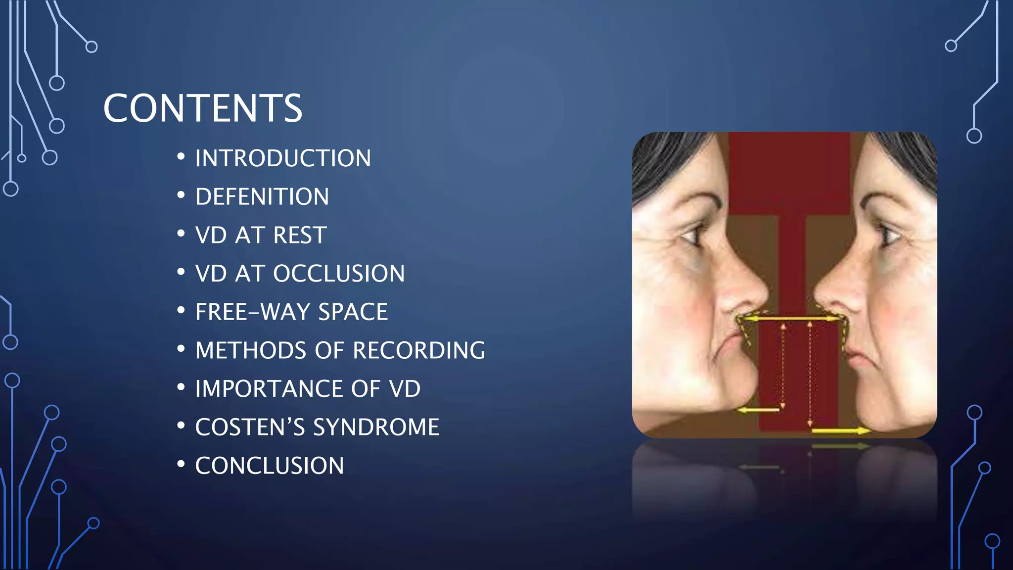
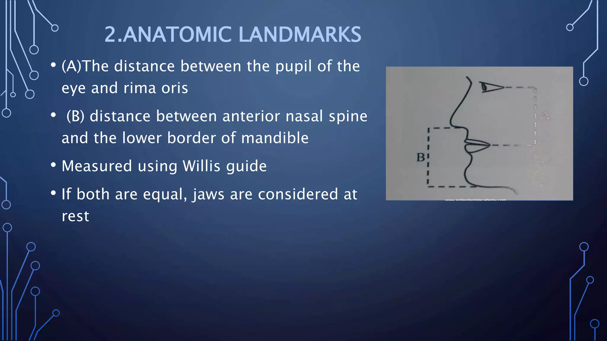
BASIL JOSE
PART 1
CONTENTS
• INTRODUCTION
• DEFENITION
• VD AT REST
• VD AT OCCLUSION
• FREE-WAY SPACE
• METHODS OF RECORDING
• IMPORTANCE OF VD
• COSTEN’S SYNDROME
• CONCLUSION
Vertical dimension is the amount of separation between maxilla
and mandible in a frontal plane which depends on the TMJ and the tone of
muscles of mastication. Even if this relation is easy to record its very
critical as any error in VD are the first to produce a discomfort and strain.
• The length of face as determined by the amount of
separation of the jaws –GPT 9
FACTORS AFFECTING:
• Teeth : act as occlusal vertical occlusal stop
• Musculature : opening and closing muscles tend to be in a state
of minimal tonic contraction
VD AT REST
• “Length of the face when the mandible is in rest position”- GPT
• Reference point during recording VD at occlusion
• VD at rest= VD at occlusion+ freeway space
• Records at physiological rest position of mandible-achieved by
functional movements
METHODS OF MEASUREMENT :
• Facial measurement after swallowing and relaxing
• Tactile sense
• Measurement of anatomic landmarks
• Speech
• Facial expression
1.FACIAL MEASUERMENTS AFTER
SWALLOWING AND RELAXING
• Patient is asked to upright and relax
• Two reference points are marked on nose and tip of chin
• Patient is asked to make functional movements
• Patient asked to relax his shoulders
• Mandible comes to physiologic rest position
• Distance between two points measured
2.ANATOMIC LANDMARKS
• (A)The distance between the pupil of the
eye and rima oris
• (B) distance between anterior nasal spine
and the lower border of mandible
• Measured using Willis guide
• If both are equal, jaws are considered at
rest
3.TACTILE SENSATION
• Paient asked to stand erect and open his mouth wide till he
feels discomfort in his muscles of mastication
• Next patient asked to close mouth slowly. Stop closing when
muscle's feels relaxed
• Distance between two reference patient point recorded
4.SPEECH
• Two methods :
• 1st METHOD
• Patient asked to repeatedly pronounce the letter ‘m’. Distance
between two reference points is measured immediately after
patient stops
• 2nd METHOD
• dentist keep talking to patient and measure the distance
between reference point immediately after patient stops
5.FACIAL EXPRESSION
•Features that indicate jaw at rest position:
• Skin around the eyes and chin should be relaxed
• Nostrils relaxed
• Upper and lower lips should have slight contact in single plane
•‘The length of the face when the teeth/
occlusal rim or any other stops are in contact
and the mandible is in centric relation or the
teeth are in centric relation’ – GPT
•Constant position
METHODS FOR DETEREMINING VD AT OCCLUSION
• MECHANICAL METHODS : do not require any functional
movement
• PHYSIOLOGICAL METHODS
a) RIDGE RELATION
-Distance from incisive papilla to mandibular incisor
-Parallelism of ridges
b) PRE-EXTRACTION RECORDS
-Profile photographs
-Profile silhouttes
-radiography
-Articulated casts
-Facial measurements
c) MEASUREMENT FROM FORMER DENTURE
-Power point
-Using wax occlusal rims
-Physiological rest position
-Phonetics
-Aesthetics
-Swallowing threshold
-Tactile sense neuromuscular perception
-Patient’s perception of comfort
RIDGE RELATIONS
• The positional relationship of the mandibular ridge to the
maxillary ridge - GPT
Measure by two methods:
• Distance from the incisive papilla to mandibular incisors
• Parallelism of ridge
Incisive Papilla to Mandibular Incisors:
• The incisive papilla is a stable landmark that changes
comparatively little with resorption of the alveolar ridge.
• The distance between papilla to maxillary incisal edge is 6 mm
.vertical overlap is 2mm usually.
• hence distance between incisive papilla and lower incisor is
approx. 4mm
• Based on this VD at occlusion calculated
RIDGE PARALLELISM
• Parallelism of the maxillary and mandibular ridges, plus a 5-
degree opening in the posterior region, often gives a clue as to
the appropriate amount of jaw separation.
• Because the clinical crowns of the anterior and posterior teeth
have nearly the same length, their removal tends to leave the
residual alveolar ridges nearly parallel to each other. This
parallelism is natural, provided there has been no abnormal
change in the alveolar process such as previous advanced
periodontal disease or gross overeruptions
PRE-EXTRACTION RECORDS
Gives an idea about the VD at occlusion of the
patient when the teeth were present
PROFILE PHOTOGRAPHS:
• Made before extraction
• Taken at maximum occlusion
• Enlarged to actual size of the patient
• anatomical landmarks are measured and compared
• Used to determine VD at occlusion
PROFILE SILHOUTTE
• Silhouette means an outline
• An accurate silhouette is made using cardboard or wire
using patients photograph
• Serves as template..gives VD at rest
• Positioned on pt’s face while recording VD at oclusion
• Chin should be at least 2mm above lower border of silhouette
RADIOGRAPHY
• Cephalometric radiographs and radiographic condylar
fossa are evaluated
ARTICULATED CASTS
• Maxillary and mandibular cast mounted are used as pre
extraction records
• After extraction the cast are articulated in a separate articulator
and inter occlusal distance compared
FACIAL MEASUREMENT
• Two tattoo points are marked on upper and
lower halves of face before extraction
• VD at occlusion is measured
• Measured using divider or caliper
MEASUREMENT FROM FORMER DENTURE
• Boley’s gauge used to measure distance
between border of maxillary and mandibular
denture when in occlusion
PHYSIOLOGICAL METHODS
POWER POINT (by Boos)
• Maxillary record base with central bearing plate
• Mandibular record base with bimeter
• Record bases are inserted in patient’s mouth
• asked to bite in different degrees of jaw separation
• Biting force transferred from central bearing plate to bimeter
• Highest reading point from bimeter is called power point
USING WAX OCCLUSAL RIMS
• Tentative VD are measured and cast are articulated in tentative
centric relation
• Tracing devices attached to rims is used for graphic tracing
PHYSIOLOGIC REST POSITION
(Niswonger’s method)
• Patient is made to sit in upright
• Occlusal rims are inserted
• Patient is asked to swallow and relax
• Lips are carefully parted to measure free way space(2-
4mm)
PHONETICS
SILVERMAN’S CLOSEST SPEAKING SPACE
• Closest speaking space measures VD in function
• Upper and lower teeth are in closest relation without
contact when c,h , s , j are pronounced
• Ideally lower incisor should touch palatal surface of
upper incisor
THE F OR V AND S SPEAKING ANTERIOR TOOTH RELATION :
• Position of anterior teeth is determined by position of maxilla
when F or V is pronounced
• Position of lower anterior teeth is determined by position of
mandible while pronouncing S
AESTHETICS
• Depends on tooth selection
SKIN
• If VD high – skin stretched , nasolabial fold
obliterated nasolabial angle increased
LIPS
• Affected by thickness of labial flange
• Occlusal rim should aid in lip support
TACTILE SENSE OR NEURO MUSCULAR PERCEPTION
• Maxillary record base with central bearing plate
• Mandibular record base with central bearing screw
• Both are inserted
• Central bearing screw is tightened until patient experiences
discomfort. This point is recorded.
• This is repeated
• Central bearing point is reduced until a comfortable jaw
relationship is attained
PATIENT PERCEPTION OF COMFORT
• Record base with extensively tall occlusal rims are inserted in
mouth
• Excessive wax is removed stepwise till comfortable occlusal
height is attained
• Depends on patient cooperation
Inter occlusal distance
The space between teeth at rest
Exist only at rest
It is the difference between VD at rest and VD at occlusion
1-8mm in natural dentition
2-4mm in CD patients
EFFECTS OF INCREASED VD
• Unattractive appearance
• Discomfort
• Soreness and ulceration
• Accelerated ridge resorption
• Impaired function
• Clicking of teeth
• Loss of freeway space
EFFECTS OF DECREASED VD
• Unattractive appearance
• TMJ problems
• Angular cheilitis
• Masticatory inefficiency
• Loss of tongue space
• Costen's Syndrome
COSTEN’S SYNDROME
• decreased vertical dimension producing a number of symptoms
of the TMJ region collectively called Costen's Syndrome
• SIGNS & SYMPTOMS
Mild catarrhal deafness and dizzy spells which are
caused by the inflation of the eustachian tube
• Tinnitus or a snapping voice in the joint, which are experienced
during chewing. The patient has painful limited joint movement
and tenderness on palpation and certain neuralgic sensation
like burning, pricking sensation of the tongue, throat and side
of nose and various forms of atypical headache
• Dryness of mouth due to disturbed salivary function
CONCLUSION
• Many methods of assessing and recording vertical jaw
relations in edentulous patients have been presented
and evaluated. Since there is no precise scientific
method of determining the correct vertical relations, the
registration of vertical relations depends upon the
clinical experience and judgment of the dental surgeon
himself. •Several methods should be used to verify the
vertical dimension and there is no one single best
method to do so
REFERENCES
• Prosthodontic treatment for edentulous patients : Boucher 9th
Edition
• ESSENTIAL OF COMPLETE DENTURE PROSTHODONTICS-WINKER
THANK YOU

Vertical jaw relation

  • 1.
  • 2.
    CONTENTS • INTRODUCTION • DEFENITION •VD AT REST • VD AT OCCLUSION • FREE-WAY SPACE • METHODS OF RECORDING • IMPORTANCE OF VD • COSTEN’S SYNDROME • CONCLUSION
  • 3.
    Vertical dimension isthe amount of separation between maxilla and mandible in a frontal plane which depends on the TMJ and the tone of muscles of mastication. Even if this relation is easy to record its very critical as any error in VD are the first to produce a discomfort and strain.
  • 4.
    • The lengthof face as determined by the amount of separation of the jaws –GPT 9 FACTORS AFFECTING: • Teeth : act as occlusal vertical occlusal stop • Musculature : opening and closing muscles tend to be in a state of minimal tonic contraction
  • 5.
    VD AT REST •“Length of the face when the mandible is in rest position”- GPT • Reference point during recording VD at occlusion • VD at rest= VD at occlusion+ freeway space • Records at physiological rest position of mandible-achieved by functional movements
  • 6.
    METHODS OF MEASUREMENT: • Facial measurement after swallowing and relaxing • Tactile sense • Measurement of anatomic landmarks • Speech • Facial expression
  • 7.
    1.FACIAL MEASUERMENTS AFTER SWALLOWINGAND RELAXING • Patient is asked to upright and relax • Two reference points are marked on nose and tip of chin • Patient is asked to make functional movements • Patient asked to relax his shoulders • Mandible comes to physiologic rest position • Distance between two points measured
  • 8.
    2.ANATOMIC LANDMARKS • (A)Thedistance between the pupil of the eye and rima oris • (B) distance between anterior nasal spine and the lower border of mandible • Measured using Willis guide • If both are equal, jaws are considered at rest
  • 9.
    3.TACTILE SENSATION • Paientasked to stand erect and open his mouth wide till he feels discomfort in his muscles of mastication • Next patient asked to close mouth slowly. Stop closing when muscle's feels relaxed • Distance between two reference patient point recorded
  • 10.
    4.SPEECH • Two methods: • 1st METHOD • Patient asked to repeatedly pronounce the letter ‘m’. Distance between two reference points is measured immediately after patient stops • 2nd METHOD • dentist keep talking to patient and measure the distance between reference point immediately after patient stops
  • 11.
    5.FACIAL EXPRESSION •Features thatindicate jaw at rest position: • Skin around the eyes and chin should be relaxed • Nostrils relaxed • Upper and lower lips should have slight contact in single plane
  • 12.
    •‘The length ofthe face when the teeth/ occlusal rim or any other stops are in contact and the mandible is in centric relation or the teeth are in centric relation’ – GPT •Constant position
  • 13.
    METHODS FOR DETEREMININGVD AT OCCLUSION • MECHANICAL METHODS : do not require any functional movement • PHYSIOLOGICAL METHODS
  • 14.
    a) RIDGE RELATION -Distancefrom incisive papilla to mandibular incisor -Parallelism of ridges b) PRE-EXTRACTION RECORDS -Profile photographs -Profile silhouttes -radiography -Articulated casts -Facial measurements c) MEASUREMENT FROM FORMER DENTURE
  • 15.
    -Power point -Using waxocclusal rims -Physiological rest position -Phonetics -Aesthetics -Swallowing threshold -Tactile sense neuromuscular perception -Patient’s perception of comfort
  • 16.
    RIDGE RELATIONS • Thepositional relationship of the mandibular ridge to the maxillary ridge - GPT Measure by two methods: • Distance from the incisive papilla to mandibular incisors • Parallelism of ridge
  • 17.
    Incisive Papilla toMandibular Incisors: • The incisive papilla is a stable landmark that changes comparatively little with resorption of the alveolar ridge. • The distance between papilla to maxillary incisal edge is 6 mm .vertical overlap is 2mm usually. • hence distance between incisive papilla and lower incisor is approx. 4mm • Based on this VD at occlusion calculated
  • 19.
    RIDGE PARALLELISM • Parallelismof the maxillary and mandibular ridges, plus a 5- degree opening in the posterior region, often gives a clue as to the appropriate amount of jaw separation.
  • 20.
    • Because theclinical crowns of the anterior and posterior teeth have nearly the same length, their removal tends to leave the residual alveolar ridges nearly parallel to each other. This parallelism is natural, provided there has been no abnormal change in the alveolar process such as previous advanced periodontal disease or gross overeruptions
  • 22.
    PRE-EXTRACTION RECORDS Gives anidea about the VD at occlusion of the patient when the teeth were present PROFILE PHOTOGRAPHS: • Made before extraction • Taken at maximum occlusion • Enlarged to actual size of the patient • anatomical landmarks are measured and compared • Used to determine VD at occlusion
  • 23.
    PROFILE SILHOUTTE • Silhouettemeans an outline • An accurate silhouette is made using cardboard or wire using patients photograph • Serves as template..gives VD at rest • Positioned on pt’s face while recording VD at oclusion • Chin should be at least 2mm above lower border of silhouette
  • 24.
    RADIOGRAPHY • Cephalometric radiographsand radiographic condylar fossa are evaluated ARTICULATED CASTS • Maxillary and mandibular cast mounted are used as pre extraction records • After extraction the cast are articulated in a separate articulator and inter occlusal distance compared
  • 25.
    FACIAL MEASUREMENT • Twotattoo points are marked on upper and lower halves of face before extraction • VD at occlusion is measured • Measured using divider or caliper MEASUREMENT FROM FORMER DENTURE • Boley’s gauge used to measure distance between border of maxillary and mandibular denture when in occlusion
  • 26.
    PHYSIOLOGICAL METHODS POWER POINT(by Boos) • Maxillary record base with central bearing plate • Mandibular record base with bimeter • Record bases are inserted in patient’s mouth • asked to bite in different degrees of jaw separation • Biting force transferred from central bearing plate to bimeter • Highest reading point from bimeter is called power point
  • 28.
    USING WAX OCCLUSALRIMS • Tentative VD are measured and cast are articulated in tentative centric relation • Tracing devices attached to rims is used for graphic tracing
  • 29.
    PHYSIOLOGIC REST POSITION (Niswonger’smethod) • Patient is made to sit in upright • Occlusal rims are inserted • Patient is asked to swallow and relax • Lips are carefully parted to measure free way space(2- 4mm)
  • 30.
    PHONETICS SILVERMAN’S CLOSEST SPEAKINGSPACE • Closest speaking space measures VD in function • Upper and lower teeth are in closest relation without contact when c,h , s , j are pronounced • Ideally lower incisor should touch palatal surface of upper incisor
  • 31.
    THE F ORV AND S SPEAKING ANTERIOR TOOTH RELATION : • Position of anterior teeth is determined by position of maxilla when F or V is pronounced • Position of lower anterior teeth is determined by position of mandible while pronouncing S
  • 32.
    AESTHETICS • Depends ontooth selection SKIN • If VD high – skin stretched , nasolabial fold obliterated nasolabial angle increased LIPS • Affected by thickness of labial flange • Occlusal rim should aid in lip support
  • 33.
    TACTILE SENSE ORNEURO MUSCULAR PERCEPTION • Maxillary record base with central bearing plate • Mandibular record base with central bearing screw • Both are inserted • Central bearing screw is tightened until patient experiences discomfort. This point is recorded. • This is repeated • Central bearing point is reduced until a comfortable jaw relationship is attained
  • 34.
    PATIENT PERCEPTION OFCOMFORT • Record base with extensively tall occlusal rims are inserted in mouth • Excessive wax is removed stepwise till comfortable occlusal height is attained • Depends on patient cooperation
  • 35.
    Inter occlusal distance Thespace between teeth at rest Exist only at rest It is the difference between VD at rest and VD at occlusion 1-8mm in natural dentition 2-4mm in CD patients
  • 36.
    EFFECTS OF INCREASEDVD • Unattractive appearance • Discomfort • Soreness and ulceration • Accelerated ridge resorption • Impaired function • Clicking of teeth • Loss of freeway space
  • 37.
    EFFECTS OF DECREASEDVD • Unattractive appearance • TMJ problems • Angular cheilitis • Masticatory inefficiency • Loss of tongue space • Costen's Syndrome
  • 38.
    COSTEN’S SYNDROME • decreasedvertical dimension producing a number of symptoms of the TMJ region collectively called Costen's Syndrome • SIGNS & SYMPTOMS Mild catarrhal deafness and dizzy spells which are caused by the inflation of the eustachian tube
  • 39.
    • Tinnitus ora snapping voice in the joint, which are experienced during chewing. The patient has painful limited joint movement and tenderness on palpation and certain neuralgic sensation like burning, pricking sensation of the tongue, throat and side of nose and various forms of atypical headache • Dryness of mouth due to disturbed salivary function
  • 40.
    CONCLUSION • Many methodsof assessing and recording vertical jaw relations in edentulous patients have been presented and evaluated. Since there is no precise scientific method of determining the correct vertical relations, the registration of vertical relations depends upon the clinical experience and judgment of the dental surgeon himself. •Several methods should be used to verify the vertical dimension and there is no one single best method to do so
  • 41.
    REFERENCES • Prosthodontic treatmentfor edentulous patients : Boucher 9th Edition • ESSENTIAL OF COMPLETE DENTURE PROSTHODONTICS-WINKER
  • 42.