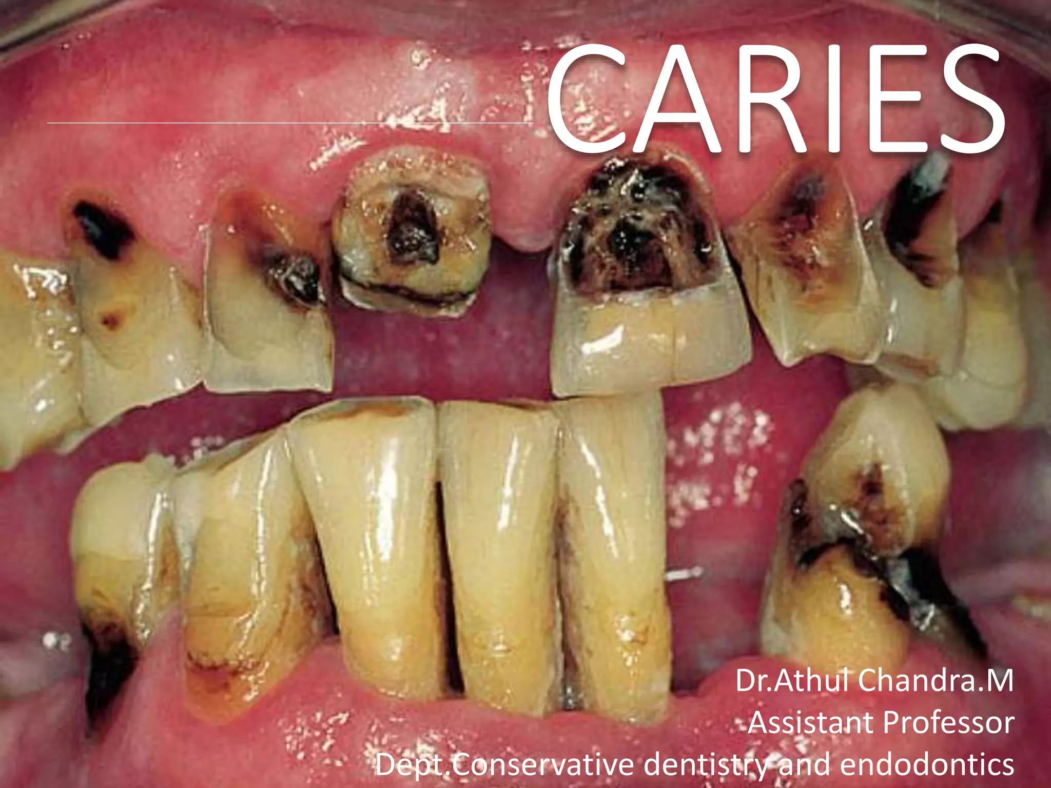
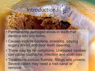
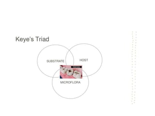
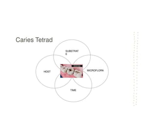
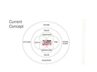
Dr.Athul Chandra.M
Assistant Professor
Dept.Conservative dentistry and endodontics
Introduction
• Permanently damaged areas in teeth that
develop into tiny holes.
• Causes include bacteria, snacking, sipping
sugary drinks and poor teeth cleaning.
• There may be no symptoms. Untreated cavities
can cause toothache, infection and tooth loss.
• Treatments include fluoride, fillings and crowns.
Severe cases may need a root canal or
removal.
Definition
Dental caries is an irreversible microbial
disease of the calcified tissues of the teeth,
characterized by demineralization of the
inorganic portion and destruction of the
organic substance of the tooth, which often
leads to cavitation
- Shafer,1993
Theories of etiology
Factors affecting the
formation of caries
Tooth Morphologic
factors
HOST
SUBSTRATE
Micro organisms
Bacteria
Time
MODIFYING FACTORS
AGE SEX OCCUPATION
RACE HEREDITARY MEDICATIONS
CLASSIFICATION OF CARIES
BASED ONANATOMICAL SITE
• OCCLUSAL (PITAND FISSURE)
• ROOT CARIES
• SMOOTH SURFACE CARIES (PROXIMALAND CERVICAL
CARIES )
• LINEAR ENAMELCARIES ( ODONTOCLASIA )
BASED ON PROGRESSION
• ACUTE CARIES
• CHRONIC CARIES
• ARRESTEDCARIES
BASED ON ORIGIN
• INITILAL / PRIMARY
• RECURRENT / SECONDARY
BASED ON THE EXTENT
• INCIPIENT CARIES
• OCCULT CARIES
• CAVITATION
BASED ON PATHWAY OF CARIES
SPREAD
• FORWARD CARIES
• BACKWARDS CARIES
BASED ON NUMBER OF SURFACES
INVOLVED
• SIMPLE
• COMPOUND
• COMPLEX
BASED ON CHRONOLOGY
• EARLY CHILDHOOD (NURSING BOTTLE & RAMPANT
CARIES )
• ADOLECENT CARIES
• ADULT CARIES
MATERNALLY DERIVED STREPROCOCCUS
MUTANS DISEASE - MDSMD
BASED ON THE REMANANT
CARIES
• RESIDUAL CARIES
CARIES which are not removed during a restorative procedure, either by accident,
neglect or intention.
Sometimes a small amount of acutely carious dentin close to the pulp is covered with
a specific capping material to stimulate dentin deposition, isolating caries from pulp.
The carious dentin can be removed at a later time.
BASED ON TEETH SURFACE TO BE
RESTORED
• OCCLUSAL
• MESIAL
• DISTAL
• FACIAL
• BUCCAL
• LINGUAL
• MOD
G V BLACKS CLASSIFICATION
WHO CLASSIFICATION
• D1 – clinically detectable enamel lesion with intact
surface
• D2 – clinically detectable cavity but limited to enamel
only
• D3 – clinically detectable cavity in dentin
• D4 – lesions extending to pulp D2 D3 D4
RADIATION CARIES
• Associated with xerostomia
Decreased salivary secretion
Increase viscosity of saliva
Lower pH
Lesion around tooth neck- loss of crown
Brown black discoloration in occlusal and incisal edges-wears away
Spot depression –spreads from any surface
BASED ON TISSUE INVOLVEMENT
• INTITIAL CARIES ( DEMINERALIZATION)
• SUPERFICIAL CARIES (CARIES SUPERFICIALIS)
• MODERATE CARIES (CARIES MEDIA)
• DEEP (CARIES PROFUNDA)
• DEEP COMPLICATED (CARIES PROFUNDA
COMPLICATA)
BY MOUNT & HUME
• Site 1 : pit and fissures
• Site 2 : proximal enamel below the contact area
• Site 3 : cervical 1/3rd
• Size 1 :minimal
• Size 2 :moderate
• Size 3 : enlarged
• Size 4 : extensive
Sit
e
Size 1 2` 3 4
1 1.1 1.2 1.3 1.4
2 2.1 2.2 2.3 2.4
3 3.1 3.2 3.3 3.4

DENTAL CARIES

  • 1.
  • 3.
    Introduction • Permanently damagedareas in teeth that develop into tiny holes. • Causes include bacteria, snacking, sipping sugary drinks and poor teeth cleaning. • There may be no symptoms. Untreated cavities can cause toothache, infection and tooth loss. • Treatments include fluoride, fillings and crowns. Severe cases may need a root canal or removal.
  • 4.
    Definition Dental caries isan irreversible microbial disease of the calcified tissues of the teeth, characterized by demineralization of the inorganic portion and destruction of the organic substance of the tooth, which often leads to cavitation - Shafer,1993
  • 5.
  • 16.
  • 20.
  • 25.
  • 33.
  • 37.
  • 42.
  • 48.
    AGE SEX OCCUPATION RACEHEREDITARY MEDICATIONS
  • 50.
  • 51.
    BASED ONANATOMICAL SITE •OCCLUSAL (PITAND FISSURE) • ROOT CARIES • SMOOTH SURFACE CARIES (PROXIMALAND CERVICAL CARIES ) • LINEAR ENAMELCARIES ( ODONTOCLASIA )
  • 52.
    BASED ON PROGRESSION •ACUTE CARIES • CHRONIC CARIES • ARRESTEDCARIES
  • 53.
    BASED ON ORIGIN •INITILAL / PRIMARY • RECURRENT / SECONDARY
  • 54.
    BASED ON THEEXTENT • INCIPIENT CARIES • OCCULT CARIES • CAVITATION
  • 55.
    BASED ON PATHWAYOF CARIES SPREAD • FORWARD CARIES • BACKWARDS CARIES
  • 56.
    BASED ON NUMBEROF SURFACES INVOLVED • SIMPLE • COMPOUND • COMPLEX
  • 57.
    BASED ON CHRONOLOGY •EARLY CHILDHOOD (NURSING BOTTLE & RAMPANT CARIES ) • ADOLECENT CARIES • ADULT CARIES MATERNALLY DERIVED STREPROCOCCUS MUTANS DISEASE - MDSMD
  • 58.
    BASED ON THEREMANANT CARIES • RESIDUAL CARIES CARIES which are not removed during a restorative procedure, either by accident, neglect or intention. Sometimes a small amount of acutely carious dentin close to the pulp is covered with a specific capping material to stimulate dentin deposition, isolating caries from pulp. The carious dentin can be removed at a later time.
  • 59.
    BASED ON TEETHSURFACE TO BE RESTORED • OCCLUSAL • MESIAL • DISTAL • FACIAL • BUCCAL • LINGUAL • MOD
  • 60.
    G V BLACKSCLASSIFICATION
  • 61.
    WHO CLASSIFICATION • D1– clinically detectable enamel lesion with intact surface • D2 – clinically detectable cavity but limited to enamel only • D3 – clinically detectable cavity in dentin • D4 – lesions extending to pulp D2 D3 D4
  • 62.
    RADIATION CARIES • Associatedwith xerostomia Decreased salivary secretion Increase viscosity of saliva Lower pH Lesion around tooth neck- loss of crown Brown black discoloration in occlusal and incisal edges-wears away Spot depression –spreads from any surface
  • 63.
    BASED ON TISSUEINVOLVEMENT • INTITIAL CARIES ( DEMINERALIZATION) • SUPERFICIAL CARIES (CARIES SUPERFICIALIS) • MODERATE CARIES (CARIES MEDIA) • DEEP (CARIES PROFUNDA) • DEEP COMPLICATED (CARIES PROFUNDA COMPLICATA)
  • 64.
    BY MOUNT &HUME • Site 1 : pit and fissures • Site 2 : proximal enamel below the contact area • Site 3 : cervical 1/3rd • Size 1 :minimal • Size 2 :moderate • Size 3 : enlarged • Size 4 : extensive
  • 65.
    Sit e Size 1 2`3 4 1 1.1 1.2 1.3 1.4 2 2.1 2.2 2.3 2.4 3 3.1 3.2 3.3 3.4