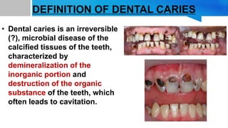
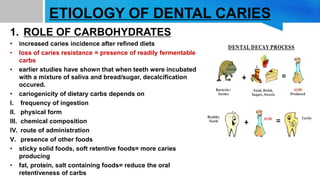
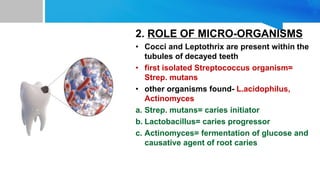
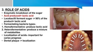
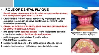
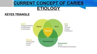
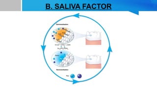
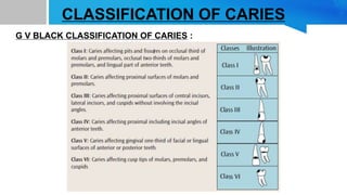
-SHRUTI ANANYA
3rd YEAR BDS
DEFINITION OF DENTAL CARIES
• Dental caries is an irreversible
(?), microbial disease of the
calcified tissues of the teeth,
characterized by
demineralization of the
inorganic portion and
destruction of the organic
substance of the teeth, which
often leads to cavitation.
FACTORS AFFECTING CARIES PREVALENCE
• RACE
Blacks> whites -unsatisfactory evidence regarding this probably due to
difference in the diet or exposure to fluoride play a major role in determining
caries rate
• AGE
Children from low socio-economic groups> children from families at higher
socioeconomic level
• SEX
Females>Males = in Girls, teeth erupt before boys of the same age
• FAMILIAL
Siblings of individuals with high caries susceptibility are generally caries active
children of parents with low caries incidence tend to have low caries, and vice
versa
ETIOLOGY OF DENTAL CARIES
1. ROLE OF CARBOHYDRATES
• increased caries incidence after refined diets
• loss of caries resistance = presence of readily fermentable
carbs
• earlier studies have shown that when teeth were incubated
with a mixture of saliva and bread/sugar, decalcification
occured.
• cariogenicity of dietary carbs depends on
I. frequency of ingestion
II. physical form
III. chemical composition
IV. route of administration
V. presence of other foods
• sticky solid foods, soft retentive foods= more caries
producing
• fat, protein, salt containing foods= reduce the oral
retentiveness of carbs
2. ROLE OF MICRO-ORGANISMS
• Cocci and Leptothrix are present within the
tubules of decayed teeth
• first isolated Streptococcus organism=
Strep. mutans
• other organisms found- L.acidophilus,
Actinomyces
a. Strep. mutans= caries initiator
b. Lactobacillus= caries progressor
c. Actinomyces= fermentation of glucose and
causative agent of root caries
3. ROLE OF ACIDS
• Enzymatic breakdown of the sugar
• Acid produced= lactic acid
• Lacobacilli ferment sugar -> 90% of the
product= lactic acid
• Fermentating bacteria types-
1. Homofermentative- produce lactic acid
2. Heterofermentative- produce a mixture
of metabolites
• Localization of acids- important for
caries progress
• Dental plaque -> localization
4. ROLE OF DENTAL PLAQUE
• Dental plaque = tenacious, thin film, that may accumulate on teeth
to a perceptible degree within 24-48 hrs
• Characteristic feature- resists removal by physiologic and oral
cleansing forces such as saliva and tongue movement but is
removable by brushing.
• DENTAL PLAQUE IS A PREREQUISITE FOR THE DEVELOPMENT
OF SMOOTH SURFACE CARIES
• imp component= acquired pellicle - forms just prior to bacterial
colonization and may facilitate plaque formation
• pellicle= glycoprotein derived from the saliva
• PLAQUES are classified as-
1. supragingival- imp role in the pathogenesis of dental caries
2. subgingival/marginal - initiation of periodontal diseases
Application of disclosing solution
for identification of plaque
LOCALIZATION OF MICROBIAL FLORA
1. PIT AND FISSURE CARIES- S. mutans
Lactobacillus
2. SMOOTH SURFACE- S. mutans
3. ROOT SURFACE- A. viscous
A. naeslundii
4. DEEP DENTINAL CARIES- Lactobacillus
A. naeslundii
other filamentous rods
CURRENT CONCEPT OF CARIES
ETIOLOGY
KEYES TRIANGLE
MODIFIED KEYES TRIANGLE
FACTORS CAUSING DENTAL CARIES
A. TOOTH FACTOR
• Surface enamel is more resistant to caries than the subsurface
enamel
• Caries develop in deep, narrow, occlusal fissures or buccal or
lingual pits {retentive areas trap food}
• Certain surfaces are more prone than others-
a. 36,46- descending order = occlusal, buccal,mesial, distal
b. 16,26- descending order = occlusal, mesial, lingual, buccal, distal
c. 12,22- lingual>labial {presence of pit}
• MANDIBULAR FIRST MOLARS ARE THE MOST SUSCEPTIBLE
PERMANENT TEETH, FOLLOWED BY MAXILLARY FIRST MOLARS,
MAX AND MAND SECOND MOLARS.
• Malaligned teeth, out of place teeth, rotated teeth are more prone to
caries {difficulty in cleansing}
B. SALIVA FACTOR
• Teeth is constantly bathed in saliva
• Calcium and phosphate in saliva - natural defense mechanism
against the dissolution of teeth
• AMYLASE {most imp oral enzyme} = responsible for degradation
of starches
• When saliva is saturated with calcium and phosphate, the pH is
known as “critical pH” below which dissolution of inorganic
material occurs
• Salivary gland aplasia and xerostomia = rampant dental caries {no
removal of bacteria and food debris from the mouth due to lack of
flow of saliva}
3. THE DIET FACTOR
X
• Physical form of the diet is responsible for difference
in caries experience
• Diet rich in roughage = cleansing the teeth of
adherent debris during mastication
• Refined diet = cling tenaciously to the teeth and are
not removed
• Increased carbohydrate content = increased caries
{due to high fermentation}
CLASSIFICATION OF CARIES
G V BLACK CLASSIFICATION OF CARIES :
CLASSIFICATION BASED ON ANATOMICAL SITE OF THE
LESION:
1. Pit and fissure caries
2. Smooth surface caries
3. Cervical caries
4. Root caries
1. PIT AND FISSURE CARIES
• Develop on the occlusal surface of molars and pre molars,
buccal and lingual surfaces of molars, and palatal surface of
maxillary incisors
• Their poor self cleansing features -> accumulation of food
debris -> caries
• Enamel directly bordering the pit and fissure may appear
bluish white as it becomes undermined
• Undermining occurs through the lateral spread of caries
2. SMOOTH SURFACE CARIES
• Develops on the proximal surfaces of the teeth
or on the gingival third of the buccal and lingual
surfaces
• Preceeded by microbial plaque - ensures the
adhesion of carbs and microbes on the tooth
surface
• Proximal caries begin just below the contact
point - in the early stage, appears as faint white
opacity of the enamel without much loss of
continuity of the enamel surface
3. CERVICAL CARIES
• Occurs on buccal, lingual, labial surfaces just
near the gingiva
• Extends laterally towards the proximal
surfaces
• When it extends below the free gingival margin
-> root caries
• CRESCENT SHAPED CAVITY
• Open cavity
• No narrow point of penetration
• Occurs on any tooth without any predilection
• Directly related to lack of oral hygiene
4. ROOT CARIES
• Aka “caries of cementum” and “senile caries”
• Defined as - soft, progressive lesion that is found
anywhere on the root surface and has lost connective
tissue attachment and is exposed to the oral
environment
• M/c found in older age groups with significant gingival
recession and exposed root
• Microbes responsible = filameous {rather than coccal}
• Teeth most frequently affected = mandibular molars,
pm, canines
• Teeth least frequently affected = mandibular incisors
CLASSIFICATION BASED ON SEVERITY AND RATE OF
PROGRESSION:
I. ACUTE DENTAL CARIES:
• Rapid clinical course
• Early pulp involvement
• Most frequent in children and young adults {because the dentinal tubules are open and
show no sclerosis}
• Process is so rapid that there is no time for deposition of sclerotic / reparative dentin
• Entrance remains small
• Diffuse involvement of dentin = large internal excavation {gives way on probing while
diagnosis}
• Dentin is usually stained light yellow
II. CHRONIC DENTAL CARIES
• Progress slowly and pulp involvement is
much later
• Most common in adults
• Entrance is large
• Slow progression = time for sclerosis of
dentin and deposition of reparative dentin
• Carious dentin is dark brown
• Shallow cavity with minimun softening of
dentin
OTHER CLINICAL VARIETIES OF DENTAL CARIES
1. RAMPANT CARIES
• Characterized by sudden, severe destruction of teeth, affecting the surfaces that are
relatively caries free
• Involves 10 or more than 10 teeth over a period of 1 year
• Observed in primary dentition of children and permanent dentition of teenagers
• BOTH THE ARCHES ARE INVOLVED
2. NURSING BOTTLE CARIES
• Aka baby bottle syndrome / bottle mouth
syndrome
• Form of rampant caries
• Due to prolonged use of nursing bottle
containing milk or milk products
• Widespread destruction of deciduous
dentition
• AFFECTS ONLY THE MAXILLARY ARCH -
MOST COMMONLY THE CENTRAL AND
LATERAL INCISORS FOLLOWED BY FIRST
MOLARS AND CUSPIDS
• Mandibular incisors are protected and
covered by the tongue
3. RECURRENT CARIES
• Occurs in the immediate vicinity of a restoration
• Due to inadequate extension of the margins of the
restoration - favours the retention of debris
• Occur beneath the restoration where carious dentin
is not removed properly prior to restoration
4. ARRESTED CARIES
• Have become static and show no signs of
progression
• Characterized by large open cavity with
lack of food retention + superficially
softened and decalcifed dentin is
gradually burnished untill it takes on a
brown stained, polished appearance and
is hard = EBURNATION OF DENTIN
• In arrested caries- sclerosis of dentinal
tubules + secondary dentin formation
occur
5. RADIATION CARIES
• Rampant caries in patients undergoing radiation
therapy in the head and neck region
• Differs from other caries by involving cusp tips,
incisal edges, and cervical areas
• Starts as a diffuse area of demineralization,
encircling the entire crown of the tooth
• Main complication of radiation = xerostomia
• Multiple doses of radiation - weaken the DEJ
• In radiation caries, enamel is destroyed with
partial disintegration of dentin and leaves the
crown reduced to an irregularly shaped,
discolored stump projecting over gingiva
6. ADOLESCENT CARIES
• Two chronological periods when acute, rapidly progressing caries is observed:
A.4-8yrs
B.11-18yrs
• The latter period is referred to as the adolescent caries {11-18yrs}
• Characteristics - small opening in the enamel with its extreme undermining
• Rapid progressing = little time for formation of reparative dentin
7. EARLY CHILDHOOD CARIES (ECC)
• Defination acc to American Academy of Pediatric Dentistry
- “the presence of one or more decayed (non
cavitated/cavitated lesions), missing (due to caries) or
filled tooth surfaces in a primary tooth in a child 71 months
of age or younger”
• Also any sign of smooth surface caries in children younger
than 3yrs is suggestive of severe ECC
• Mostly seen in lower socio economic groups
• Often associated with lack of oral hygiene and improper
feeding pattern
• Starts with maxillary anterior teeth, maxillary and
mandibular first primary molars, and sometimes the
mandibular canines
• Mandibular incisors - unaffected by the protective effect of
saliva of submandibular glands and tongue
CLASSIFICATION BASED ON SEVERITY OF DENTAL
CARIES
SEQUELAE OF DENTAL CARIES
I. ENAMEL CARIES
• Initiation- formation of dental plaque
• Stages - 1. early enamel caries
2. phase of non-bacterial destruction
3. cavity formation
4. bacterial invasion of enamel
5. undermining of enamel from below after
spread into the dentin
• Types- 1. smooth surface caries
2. pit and fissure caries
• Body of the lesion = area of greatest demineralization (5-
25% loss)
II. DENTINAL CARIES
• Begins from the DEJ
• Rapid involvement of greater dentinal tubules- each acts as a tract leading to the pulp
• Events in dentinal caries- 1. non bacterial, pre cavitation and softening of the matrix
2. widening of tubules by demineralization
3. migration of pioneer bacteria through the tubules
4. distortion of the tubules by the expanding masses of bacterial mass
5. breakdown of matrix and its progressive disintergration
III. PULPITIS
• Most common cause of dental pain and loss of teeth
• Caused by irritation or infection of pulp
• Pulpal pain is poorly localized
IV. APICAL PERIODONTITIS
• Inflammation of the periodontal ligament around the
root apex
• Common cause - 1. spread of infection following pulp
necrosis
2. occlusal trauma
3. chemical irritation (over extended
RCT medications)
V. PERIAPICAL ABSCESS AND PERIAPICAL
GRANULOMA
• PERIAPICAL ABSCESS - inflammatory reaction that
preceeds pulp necrosis
• It is the acute extension of inflammatory reaction of a
necrotic tissue
• No well defined border
• PERIAPICAL GRANULOMA - well circumscribed
radioluscent periapical lesion without a sclerotic border
• localized mass of chronic granaluation tissue at the
apex of non-vital tissue
• Less than 1cm with a well defined border
PERIAPICAL CYST
• Due to bacterial infection and necrosis of pulp, has a well defined border
• Usual successor of periapical granuloma
• Greater than 1cm
VI. OSTEOMYELITIS
• Inflammation of bone and bone
marrow
• Occurs secondary to the
inflammation of the soft tissue
components of the bone
VII. PERIOSTEITIS
• Focal gross thickening of the periosteum of the bone
with peripheral reactive bone formation
• Results from mild irritation or infection
VIII. CELLULITIS
• Diffuse inflammation of the soft
tissue
• Tends to spread through tissue
spaces and facial planes
• Caused by organisms like
Streptococci
• May become localized and result in
facial abcess.
DIAGNOSIS OF DENTAL CARIES
1. Visual assessment
2. Radiographic diagnosis
3. Infrared laser fluorescence
4. Digital imaging fiber-optic transillumination
5. Tuned aperture computed tomography (TACT)
6. Terahertz imaging
7. Caries activity tests:
a) Lactobacillus colony test
b) Colorimetric snyder test
c) Swab test
d) Salivary S.mutans level test
e) S.mutans dip-slide method
f) Buffer capacity test
g) Enamel solubility test
h) Salivary reductase test
MANAGEMENT AND TREATMENT
• Dental caries in its early stages, can be stopped and reversed
• Fluorides = helps tooth in early stage to remineralize
• Once cavity is formed -> go for a restorative treatment
• Tooth brushing twice a day {plaque control} , mouth rinsing and dental floss ->
prevention of caries
• Topical fluoride application in pedo patients
• Application of pit and fissure sealants
• Stimulation of saliva with the help of sugar-free chewing gums like Xylitol
chewing gums
THANK YOU

Dental Caries

  • 1.
  • 2.
    DEFINITION OF DENTALCARIES • Dental caries is an irreversible (?), microbial disease of the calcified tissues of the teeth, characterized by demineralization of the inorganic portion and destruction of the organic substance of the teeth, which often leads to cavitation.
  • 3.
    FACTORS AFFECTING CARIESPREVALENCE • RACE Blacks> whites -unsatisfactory evidence regarding this probably due to difference in the diet or exposure to fluoride play a major role in determining caries rate • AGE Children from low socio-economic groups> children from families at higher socioeconomic level • SEX Females>Males = in Girls, teeth erupt before boys of the same age • FAMILIAL Siblings of individuals with high caries susceptibility are generally caries active children of parents with low caries incidence tend to have low caries, and vice versa
  • 4.
    ETIOLOGY OF DENTALCARIES 1. ROLE OF CARBOHYDRATES • increased caries incidence after refined diets • loss of caries resistance = presence of readily fermentable carbs • earlier studies have shown that when teeth were incubated with a mixture of saliva and bread/sugar, decalcification occured. • cariogenicity of dietary carbs depends on I. frequency of ingestion II. physical form III. chemical composition IV. route of administration V. presence of other foods • sticky solid foods, soft retentive foods= more caries producing • fat, protein, salt containing foods= reduce the oral retentiveness of carbs
  • 5.
    2. ROLE OFMICRO-ORGANISMS • Cocci and Leptothrix are present within the tubules of decayed teeth • first isolated Streptococcus organism= Strep. mutans • other organisms found- L.acidophilus, Actinomyces a. Strep. mutans= caries initiator b. Lactobacillus= caries progressor c. Actinomyces= fermentation of glucose and causative agent of root caries
  • 6.
    3. ROLE OFACIDS • Enzymatic breakdown of the sugar • Acid produced= lactic acid • Lacobacilli ferment sugar -> 90% of the product= lactic acid • Fermentating bacteria types- 1. Homofermentative- produce lactic acid 2. Heterofermentative- produce a mixture of metabolites • Localization of acids- important for caries progress • Dental plaque -> localization
  • 7.
    4. ROLE OFDENTAL PLAQUE • Dental plaque = tenacious, thin film, that may accumulate on teeth to a perceptible degree within 24-48 hrs • Characteristic feature- resists removal by physiologic and oral cleansing forces such as saliva and tongue movement but is removable by brushing. • DENTAL PLAQUE IS A PREREQUISITE FOR THE DEVELOPMENT OF SMOOTH SURFACE CARIES • imp component= acquired pellicle - forms just prior to bacterial colonization and may facilitate plaque formation • pellicle= glycoprotein derived from the saliva • PLAQUES are classified as- 1. supragingival- imp role in the pathogenesis of dental caries 2. subgingival/marginal - initiation of periodontal diseases Application of disclosing solution for identification of plaque
  • 8.
    LOCALIZATION OF MICROBIALFLORA 1. PIT AND FISSURE CARIES- S. mutans Lactobacillus 2. SMOOTH SURFACE- S. mutans 3. ROOT SURFACE- A. viscous A. naeslundii 4. DEEP DENTINAL CARIES- Lactobacillus A. naeslundii other filamentous rods
  • 9.
    CURRENT CONCEPT OFCARIES ETIOLOGY KEYES TRIANGLE
  • 10.
  • 11.
  • 12.
    A. TOOTH FACTOR •Surface enamel is more resistant to caries than the subsurface enamel • Caries develop in deep, narrow, occlusal fissures or buccal or lingual pits {retentive areas trap food} • Certain surfaces are more prone than others- a. 36,46- descending order = occlusal, buccal,mesial, distal b. 16,26- descending order = occlusal, mesial, lingual, buccal, distal c. 12,22- lingual>labial {presence of pit} • MANDIBULAR FIRST MOLARS ARE THE MOST SUSCEPTIBLE PERMANENT TEETH, FOLLOWED BY MAXILLARY FIRST MOLARS, MAX AND MAND SECOND MOLARS. • Malaligned teeth, out of place teeth, rotated teeth are more prone to caries {difficulty in cleansing}
  • 13.
  • 14.
    • Teeth isconstantly bathed in saliva • Calcium and phosphate in saliva - natural defense mechanism against the dissolution of teeth • AMYLASE {most imp oral enzyme} = responsible for degradation of starches • When saliva is saturated with calcium and phosphate, the pH is known as “critical pH” below which dissolution of inorganic material occurs • Salivary gland aplasia and xerostomia = rampant dental caries {no removal of bacteria and food debris from the mouth due to lack of flow of saliva}
  • 15.
    3. THE DIETFACTOR X • Physical form of the diet is responsible for difference in caries experience • Diet rich in roughage = cleansing the teeth of adherent debris during mastication • Refined diet = cling tenaciously to the teeth and are not removed • Increased carbohydrate content = increased caries {due to high fermentation}
  • 16.
    CLASSIFICATION OF CARIES GV BLACK CLASSIFICATION OF CARIES :
  • 17.
    CLASSIFICATION BASED ONANATOMICAL SITE OF THE LESION: 1. Pit and fissure caries 2. Smooth surface caries 3. Cervical caries 4. Root caries
  • 18.
    1. PIT ANDFISSURE CARIES • Develop on the occlusal surface of molars and pre molars, buccal and lingual surfaces of molars, and palatal surface of maxillary incisors • Their poor self cleansing features -> accumulation of food debris -> caries • Enamel directly bordering the pit and fissure may appear bluish white as it becomes undermined • Undermining occurs through the lateral spread of caries
  • 19.
    2. SMOOTH SURFACECARIES • Develops on the proximal surfaces of the teeth or on the gingival third of the buccal and lingual surfaces • Preceeded by microbial plaque - ensures the adhesion of carbs and microbes on the tooth surface • Proximal caries begin just below the contact point - in the early stage, appears as faint white opacity of the enamel without much loss of continuity of the enamel surface
  • 20.
    3. CERVICAL CARIES •Occurs on buccal, lingual, labial surfaces just near the gingiva • Extends laterally towards the proximal surfaces • When it extends below the free gingival margin -> root caries • CRESCENT SHAPED CAVITY • Open cavity • No narrow point of penetration • Occurs on any tooth without any predilection • Directly related to lack of oral hygiene
  • 21.
    4. ROOT CARIES •Aka “caries of cementum” and “senile caries” • Defined as - soft, progressive lesion that is found anywhere on the root surface and has lost connective tissue attachment and is exposed to the oral environment • M/c found in older age groups with significant gingival recession and exposed root • Microbes responsible = filameous {rather than coccal} • Teeth most frequently affected = mandibular molars, pm, canines • Teeth least frequently affected = mandibular incisors
  • 22.
    CLASSIFICATION BASED ONSEVERITY AND RATE OF PROGRESSION: I. ACUTE DENTAL CARIES: • Rapid clinical course • Early pulp involvement • Most frequent in children and young adults {because the dentinal tubules are open and show no sclerosis} • Process is so rapid that there is no time for deposition of sclerotic / reparative dentin • Entrance remains small • Diffuse involvement of dentin = large internal excavation {gives way on probing while diagnosis} • Dentin is usually stained light yellow
  • 23.
    II. CHRONIC DENTALCARIES • Progress slowly and pulp involvement is much later • Most common in adults • Entrance is large • Slow progression = time for sclerosis of dentin and deposition of reparative dentin • Carious dentin is dark brown • Shallow cavity with minimun softening of dentin
  • 24.
    OTHER CLINICAL VARIETIESOF DENTAL CARIES 1. RAMPANT CARIES • Characterized by sudden, severe destruction of teeth, affecting the surfaces that are relatively caries free • Involves 10 or more than 10 teeth over a period of 1 year • Observed in primary dentition of children and permanent dentition of teenagers • BOTH THE ARCHES ARE INVOLVED
  • 25.
    2. NURSING BOTTLECARIES • Aka baby bottle syndrome / bottle mouth syndrome • Form of rampant caries • Due to prolonged use of nursing bottle containing milk or milk products • Widespread destruction of deciduous dentition • AFFECTS ONLY THE MAXILLARY ARCH - MOST COMMONLY THE CENTRAL AND LATERAL INCISORS FOLLOWED BY FIRST MOLARS AND CUSPIDS • Mandibular incisors are protected and covered by the tongue
  • 26.
    3. RECURRENT CARIES •Occurs in the immediate vicinity of a restoration • Due to inadequate extension of the margins of the restoration - favours the retention of debris • Occur beneath the restoration where carious dentin is not removed properly prior to restoration
  • 27.
    4. ARRESTED CARIES •Have become static and show no signs of progression • Characterized by large open cavity with lack of food retention + superficially softened and decalcifed dentin is gradually burnished untill it takes on a brown stained, polished appearance and is hard = EBURNATION OF DENTIN • In arrested caries- sclerosis of dentinal tubules + secondary dentin formation occur
  • 28.
    5. RADIATION CARIES •Rampant caries in patients undergoing radiation therapy in the head and neck region • Differs from other caries by involving cusp tips, incisal edges, and cervical areas • Starts as a diffuse area of demineralization, encircling the entire crown of the tooth • Main complication of radiation = xerostomia • Multiple doses of radiation - weaken the DEJ • In radiation caries, enamel is destroyed with partial disintegration of dentin and leaves the crown reduced to an irregularly shaped, discolored stump projecting over gingiva
  • 29.
    6. ADOLESCENT CARIES •Two chronological periods when acute, rapidly progressing caries is observed: A.4-8yrs B.11-18yrs • The latter period is referred to as the adolescent caries {11-18yrs} • Characteristics - small opening in the enamel with its extreme undermining • Rapid progressing = little time for formation of reparative dentin
  • 30.
    7. EARLY CHILDHOODCARIES (ECC) • Defination acc to American Academy of Pediatric Dentistry - “the presence of one or more decayed (non cavitated/cavitated lesions), missing (due to caries) or filled tooth surfaces in a primary tooth in a child 71 months of age or younger” • Also any sign of smooth surface caries in children younger than 3yrs is suggestive of severe ECC • Mostly seen in lower socio economic groups • Often associated with lack of oral hygiene and improper feeding pattern • Starts with maxillary anterior teeth, maxillary and mandibular first primary molars, and sometimes the mandibular canines • Mandibular incisors - unaffected by the protective effect of saliva of submandibular glands and tongue
  • 31.
    CLASSIFICATION BASED ONSEVERITY OF DENTAL CARIES
  • 32.
  • 33.
    I. ENAMEL CARIES •Initiation- formation of dental plaque • Stages - 1. early enamel caries 2. phase of non-bacterial destruction 3. cavity formation 4. bacterial invasion of enamel 5. undermining of enamel from below after spread into the dentin • Types- 1. smooth surface caries 2. pit and fissure caries • Body of the lesion = area of greatest demineralization (5- 25% loss)
  • 34.
    II. DENTINAL CARIES •Begins from the DEJ • Rapid involvement of greater dentinal tubules- each acts as a tract leading to the pulp • Events in dentinal caries- 1. non bacterial, pre cavitation and softening of the matrix 2. widening of tubules by demineralization 3. migration of pioneer bacteria through the tubules 4. distortion of the tubules by the expanding masses of bacterial mass 5. breakdown of matrix and its progressive disintergration
  • 35.
    III. PULPITIS • Mostcommon cause of dental pain and loss of teeth • Caused by irritation or infection of pulp • Pulpal pain is poorly localized
  • 37.
    IV. APICAL PERIODONTITIS •Inflammation of the periodontal ligament around the root apex • Common cause - 1. spread of infection following pulp necrosis 2. occlusal trauma 3. chemical irritation (over extended RCT medications)
  • 39.
    V. PERIAPICAL ABSCESSAND PERIAPICAL GRANULOMA • PERIAPICAL ABSCESS - inflammatory reaction that preceeds pulp necrosis • It is the acute extension of inflammatory reaction of a necrotic tissue • No well defined border • PERIAPICAL GRANULOMA - well circumscribed radioluscent periapical lesion without a sclerotic border • localized mass of chronic granaluation tissue at the apex of non-vital tissue • Less than 1cm with a well defined border
  • 40.
    PERIAPICAL CYST • Dueto bacterial infection and necrosis of pulp, has a well defined border • Usual successor of periapical granuloma • Greater than 1cm
  • 41.
    VI. OSTEOMYELITIS • Inflammationof bone and bone marrow • Occurs secondary to the inflammation of the soft tissue components of the bone
  • 42.
    VII. PERIOSTEITIS • Focalgross thickening of the periosteum of the bone with peripheral reactive bone formation • Results from mild irritation or infection
  • 43.
    VIII. CELLULITIS • Diffuseinflammation of the soft tissue • Tends to spread through tissue spaces and facial planes • Caused by organisms like Streptococci • May become localized and result in facial abcess.
  • 44.
    DIAGNOSIS OF DENTALCARIES 1. Visual assessment 2. Radiographic diagnosis 3. Infrared laser fluorescence 4. Digital imaging fiber-optic transillumination 5. Tuned aperture computed tomography (TACT) 6. Terahertz imaging 7. Caries activity tests: a) Lactobacillus colony test b) Colorimetric snyder test c) Swab test d) Salivary S.mutans level test e) S.mutans dip-slide method f) Buffer capacity test g) Enamel solubility test h) Salivary reductase test
  • 45.
    MANAGEMENT AND TREATMENT •Dental caries in its early stages, can be stopped and reversed • Fluorides = helps tooth in early stage to remineralize • Once cavity is formed -> go for a restorative treatment • Tooth brushing twice a day {plaque control} , mouth rinsing and dental floss -> prevention of caries • Topical fluoride application in pedo patients • Application of pit and fissure sealants • Stimulation of saliva with the help of sugar-free chewing gums like Xylitol chewing gums
  • 46.