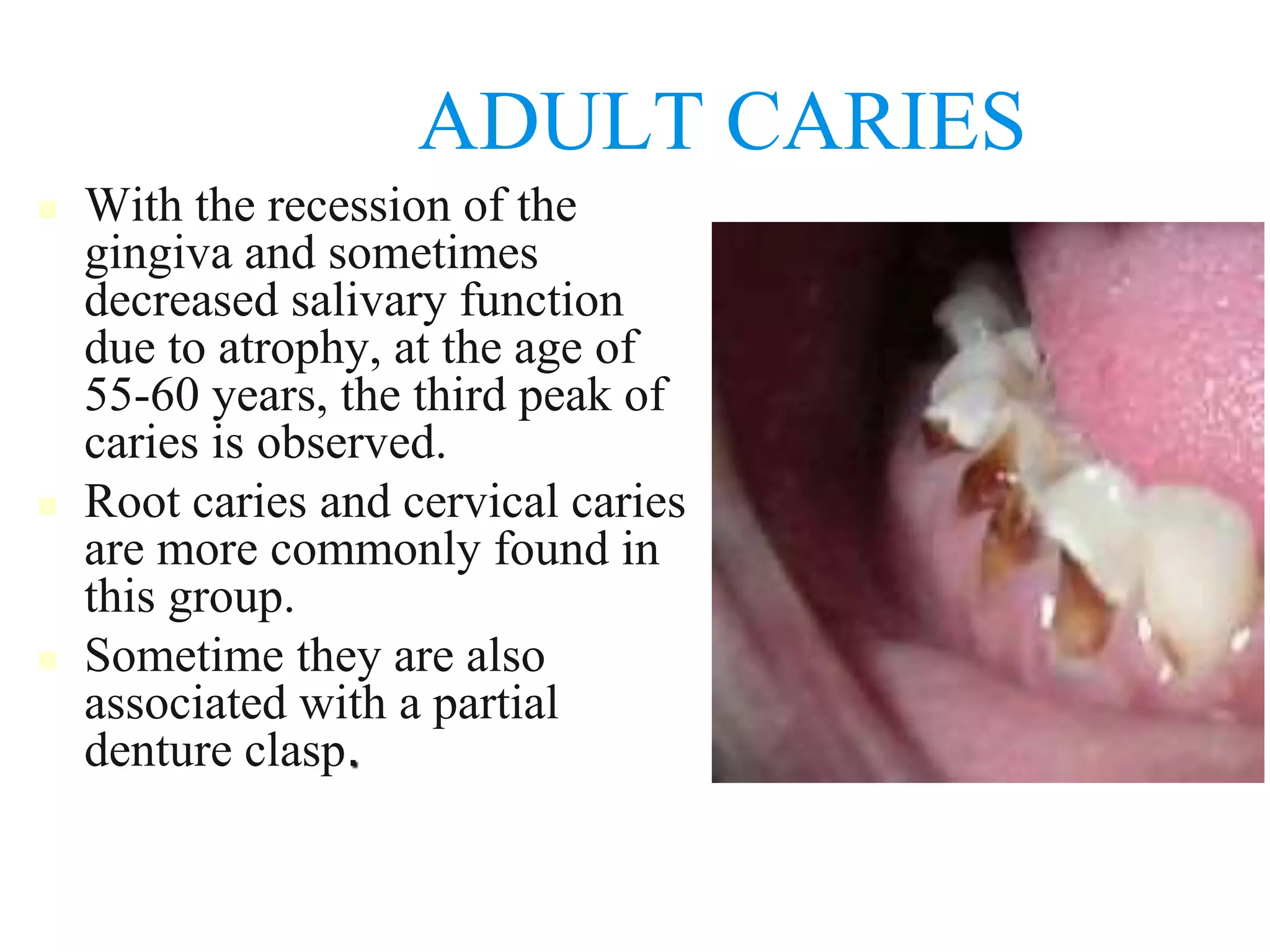
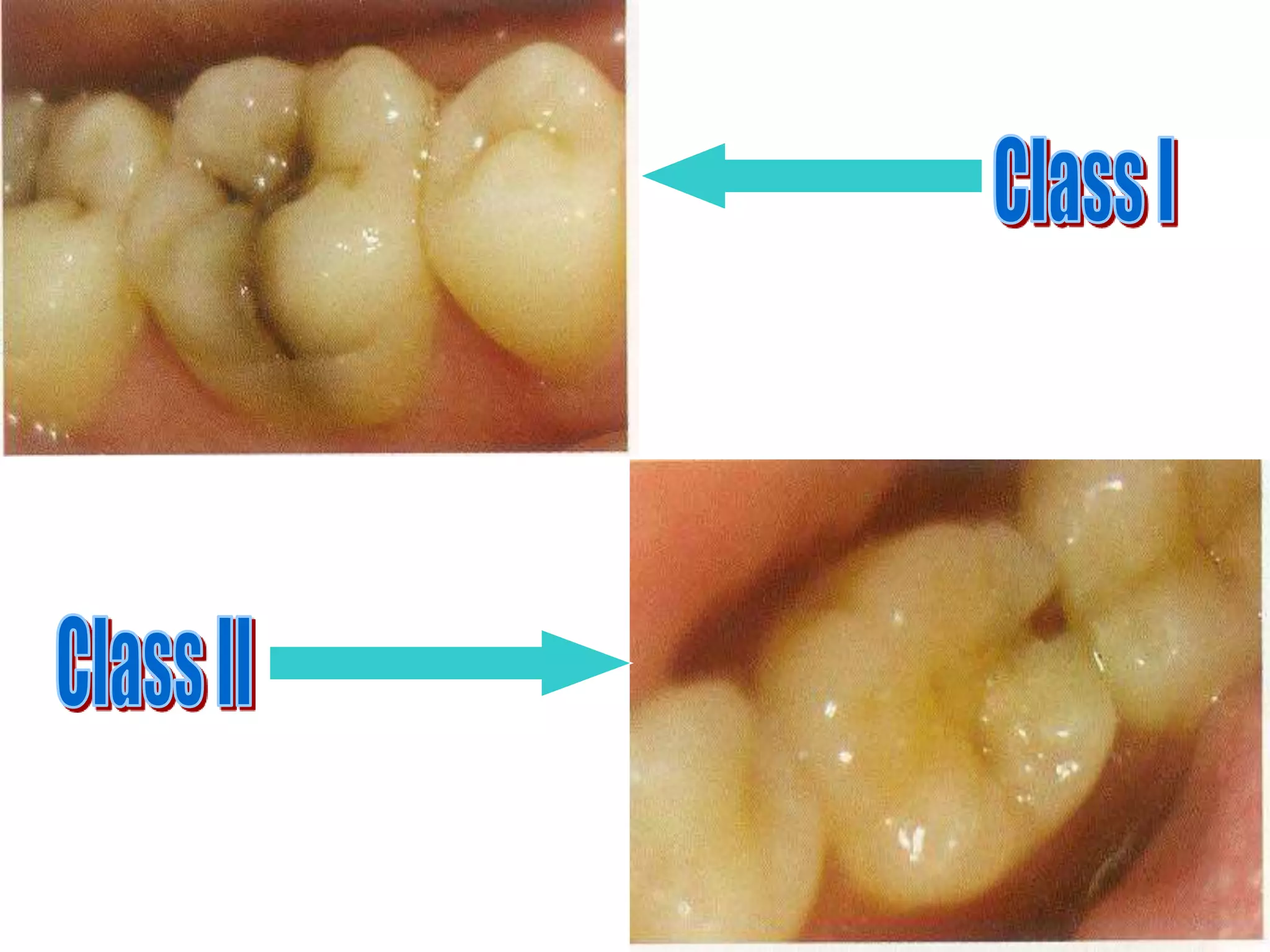
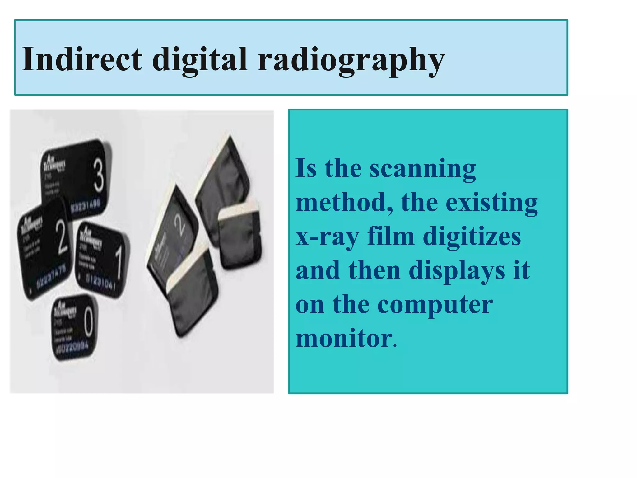
CLASSIFICATION OF
DENTAL CARIES
Dr. Mohamed Salim
(Primary prevention)
Prevent, Arrest, Reverse
(Secondary prevention)
Deep scaling Restoration
Periodontal surgery Endodontics
Exodontics
(Tertiary prevention)
Prosthodontics orthodontics
1.BASED ON ANATOMICAL SITE
OCCLUSAL
(PIT AND FISSURE)
ROOT CARIES
SMOOTH
SURFACE
CARIES
(PROXIMAL
AND CERVICAL
CARIES)
A- PIT AND FISSURE CARIES
 Highest prevalence of all caries bacteria rapidly colonize
the pits and fissures of the newly erupted teeth.
 These early colonizers form a “bacterial plug” that
remains in the site for long time, perhaps even the life of
the tooth
 Type & nature of the organisms prevalent in the oral
cavity determine the type of organisms colonizing the pit
& fissure
 The appearance of s.mutans in pits and
fissures is usually followed by caries 6 to 24
months later.
 Sealing of pits and fissures just after tooth
eruption may be the most important event in
their resistance to caries.
 Shape, morphological variation and depth of
pit and fissures contributes to their high
susceptibility to caries.
-enamel in the bottom of pit or fissure is very thin, so early
dentin involvement frequently occurs.
here the caries follows the direction of the enamel rods.
-Caries is triangular in shape with the apex facing the
surface of tooth and the base towards the DEJ.
-when reaches DEJ, greater number of dentinal tubules are
involved.
- it produces greater cavitation than the smooth surface
caries and there is more undermining of enamel.
B- Smooth surface caries
 Less favorable site for plaque attachment, usually
attaches on the smooth surface that are near the gingiva
or are under proximal contact..
 In very young patients the gingival papilla completely
fills the interproximal space under a proximal contact
 The earliest manifestation of incipient caries (early caries) of
enamel is usually seen beneath dental plaque as areas of
decalcification (white spots).
 The proximal surfaces are particularly
susceptible to caries due to extra shelter
provided to resident plaque owing to the
proximal contact area immediately occlusal
to plaque.
 Lesion have a broad area of origin and a
conical, or pointed extension towards DEJ.
 V shape with apex directed towards DEJ.
 After caries penetrate the DEJ softening of
dentin spread rapidly and pulpally
C- ROOT SURFACE CARIES
 The proximal root surface, particularly near the cervical
line, often is unaffected by the action of hygiene
procedures, such as flossing, because it may have concave
anatomic surface contours (fluting) and occasional
roughness at the termination of the enamel.
 These conditions, when coupled with exposure to the oral
environment (as a result of gingival recession), favor the
formation of mature, caries-producing plaque and proximal
root-surface caries.
 Caries originating on the root is alarming because
1. it has a comparatively rapid progression
2. it is closer to the pulp
3. it is more difficult to restore
 The root surface is softer than the enamel and
readily allows plaque formation in the absence of
good oral hygiene.
 The cementum covering the root surface is
extremely thin and provides little resistance to caries
attack.
 Root caries lesions progress more rapidly because of
the lack of protection from and enamel covering.
2.BASED ON PROGRESSION
ACUTE CARIES
CHRONIC CARIES
ARRESTED CARIES
A- ACUTE CARIES
 Active caries lesion: a progressive carious lesion.
 Acute caries is a rapid process involving a large number
of teeth.
 These lesions are lighter colored than the other types,
being light brown or grey.
 Pulp exposures and sensitive teeth are often observed in
patients with acute caries.
 It has been suggested that saliva does not easily penetrate
the small opening to the carious lesion, so there are little
opportunity for buffering or neutralizaton
B- CHRONIC CARIES
 These lesions are usually of long-standing
involvement, affect a fewer number of teeth, and are
smaller than acute caries.
 Pain is not a common feature because of protection
afforded to the pulp by secondary dentin
 The decalcified dentin is dark brown.
 Pulp prognosis is hopeful in that the deepest of
lesions usually requires only prophylactic capping
and protective bases.
C- ARRESTED CARIES:-
Arrested (inactive) carious lesion: A lesion that may have
formed years previously and then stopped further
progression.
 Caries which becomes stationary or static and does not
show any tendency for further progression
 Both deciduous and permanent affected
 With the shift in the oral conditions, even advanced
lesions may become arrested .
 Arrested caries involving dentin shows a marked brown
pigmentation
 Sclerosis of dentinal tubules and secondary dentin
formation commonly occur
3.Based on virginity of lesion
INITIAL/PRIMARY RECURRENT/SECONDARY
A- PRIMARY CARIES(INITIAL)
 lesions on unrestored tooth surface.
 A primary caries is one in which the lesion
constitutes the initial attack on the tooth
surface.
 The designation of primary is based on the
initial location of the lesion on the surface
rather than the extent of damage.
B- SECONDARY CARIES
(RECURRENT)
 This type of caries is observed around the edges and under
restorations.
 lesions that developed adjacent to a filling.
 The common locations of secondary caries are the rough
or overhanging margin and fracture place in all locations
of the mouth.
 It may be result of poor adaptation of a restoration, which
allows for a marginal leakage, or it may be due to
inadequate extension of the restoration.
 In addition caries may remain if there has not been
complete excavation of the original lesion, which later
may appear as a residual or recurrent caries.
4.Based on tissue involvement
1. Initial caries
2. Superficial caries
3. Moderate caries
4. Deep caries
5. Deep complicated caries
Dental caries can be divided into 4 or 5 stages
 Initial caries: Demineralization without
structural defect. This stage can be reversed by
fluoridation and enhanced mouth hygiene
 Superficial caries (caries superficialis):Enamel
caries. Caries has affected the enamel layer, but
has not yet penetrated the dentin.
3. Moderate caries (Caries media): Dentin caries.
Extensive structural defect. Caries has penetrated up to
the dentin and spreads two-dimensionally beneath the
enamel defect where the dentin offers little resistance.
4. Deep caries (Caries profunda): Deep structural defect.
Caries has penetrated up to the dentin layers of the
tooth close to the pulp.
5. Deep complicated caries (Caries profunda complicata)
:Caries has led to the opening of the pulp cavity (open
pulp).
5. BASED ON NUMBER OF TOOTH
SURFACE INVOLVED
Simple
Compound
Complex
A caries involving only one tooth
surface
A caries involving two surfaces
of tooth
A caries that involves more than
two surfaces of a tooth
6. BASED ON CHRONOLOGY
EARLY CHILDHOOD CARIES
ADOLESCENT CARIES
ADULT CARIES
 the number of new lesions occurring in a year,
shows three peaks-at the ages 4-8, 11-19 and 55-
65 years
Rampant caries: is the name given to multiple
active carious lesions occurring in the same patient.
This frequently involves surfaces of teeth that do not
usually experience dental caries eg, bottle or nursing
caries, baby caries, radiation caries, or drug-induced
caries.
EARLY CHILDHOOD CARIES
 Early childhood caries
would include, two
variants: Nursing caries
and rampant caries.
 The difference primarily
exist in involvement of the
teeth[ mandibular incisors ]
in the carious process in
rampant caries as opposed
to nursing caries.
TEENAGE CARIES
(ADOLESCENT CARIES)
 This type of caries is a variant of rampant
caries where the teeth generally considered
immune to decay are involved.
 The caries is also described to be of a rapidly
burrowing type, with a small enamel opening.
 The presence of a large pulp chamber causing
early pulp involvement
ADULT CARIES
 With the recession of the
gingiva and sometimes
decreased salivary function
due to atrophy, at the age of
55-60 years, the third peak of
caries is observed.
 Root caries and cervical caries
are more commonly found in
this group.
 Sometime they are also
associated with a partial
denture clasp.
7.BASED ON SURFACES TO BE
RESTORED
 Most widespread clinical utilization
O for occlusal surfaces
M for mesial surfaces
D for distal surfaces
F for facial surfaces
B for buccal surfaces
L for lingual surface
Various combinations are also possible, such as MOD –
for mesio-occluso-distal surfaces.
8. BLACK’S CLASSIFICATION
Class 1 lesions:
 Lesions that begin in the structural defects of teeth such as
pits, fissures and defective grooves.
Locations include
 Occlusal surface of molars and premolars.
 occlusal two thirds of buccal and lingual surfaces of
molars and premolars.
 Lingual surfaces of anterior tooth.
Class 2 lesions:
 They are found on the proximal surfaces of the bicuspids
and molars.
Class 3 lesions:
 Lesions found on the proximal surfaces of anterior teeth
that do not involve or necessitate the removal of the
incisal angle.
Class 4 lesions:
 Lesions found on the proximal surfaces of anterior teeth
that involve the incisal angle.
Class 5 lesions:
 Lesions that are found at the gingival third of the facial
and lingual surfaces of anterior and posterior teeth.
Class 6 (Simon’s modification):
 Lesions involving cuspal tips and incisal edges of teeth.
Diagnosis of dental caries
Diagnosis: is an art and science that results
from the synthesis of scientific knowledge, clinical
experience & common sense.
Caries diagnosis implies deciding whether a lesion is
active, progressing rapidly or slowly or whether is
already arrested.
Conventional diagnostic methods
By visually with the clinician’s eyes using direct
vision or vision assisted with a mirror and a
standard dental operatory light. In addition, a
small, rounded-end dental explorer or probe can
assist with the detection of small defects.
Use of explorer :
Explorer is useful to remove plaque and debris and
check the surface characteristics of suspected
carious lesions. gentle pressure just required to
blanch a fingernail without causing any pain or
damage, All surfaces of a tooth are cleaned of debris
and plaque, using an air syringe and examined
visually. Suspicious areas are explored to check for
the surface texture
Use of explorer is not advocated
because; Sharp tips physically damage small lesions
with intact surfaces, Probing can cause fracture &
cavitation of incipient lesion. It may spread the
organism in the mouth
Mechanical binding may be due to non-carious
reasons , Shape of fissure , Sharpness of explorer ,
Force of application , Path of explorer placement
All discolored areas should be explored using
gentle pressure. There is no need to penetrate a
suspected lesion with an explorer. If a discolored
and non-cavitated area is soft when explored, it is
recorded as non-cavitated carious pit or fissure . A
cavity is detected when there is an actual hole in the
tooth in which an explorer could easily enter the
space. An active cavity has soft walls or floors
RADIOGRAPHY
Carious lesions are detectable radiographically when there
has been enough demineralization to allow it to be
differentiate from normal. They are valuable in detecting
proximal caries which may go undetected during clinical
examination. On average they have around 50% to 70%
sensitivity in detecting carious lesions. 40% demineralization
is required for definitive decision on caries
bitewing radiography—either conventional or
digital. It has been shown that there is essentially
no difference between the diagnostic capabilities
of film and digital radiography when used for
bitewings.
Radiographic examinations include;
A- Bitewing radiographs
B- IOPA radiographs using paralleling technique
C- Dental panoramic tomograph
The two important decisions related to radiographic
examination are
(1) when to take a radiograph and
(2) how to evaluate a radiograph for presence of
signs of dental caries
Pitfalls Of Radiography
* 2 dimensional view of 3 dimensional object
* Radiographic depth of a lesion is often less than
actual depth
*Overlapping of proximal surfaces on a radiograph
*Occlusal (incipient) caries of enamel difficult to
detect
* Dental anomalies like hypoplastic pits mimic
proximal caries
Is filmless imaging
system, a method of
capturing radiographic
image using a sensor
Digital radiography
Direct digital radiography
A sensor placed into the
mouth of patient and
exposed to x-rays ,the
sensor captures the
radiographic image and
then transmits the image to
a monitor of computer
Types of system
Is the scanning
method, the existing
x-ray film digitizes
and then displays it
on the computer
monitor.
Indirect digital radiography
Advantages
1-Less radiation exposure
2-Images can be manipulated
3-Storage and archiving of patient information
4-Patient education & interaction
5-Instant image display
6-Eliminates film and processing expenses
Disadvantages
1-Expensive
2-Sensor size
3-Infection control
4-Image resolution less than conventional film
Intraoral camera
Intra-oral cameras are an
important part of
dentistry chair units contribute
to enhance improving patient
careduringthetreatments
Types of IOC
Wireless Camera
The wireless camera may be
powered by a battery or a
cable.
Wired camera
The wired camera can be
connected to the computer
using USB cable
FIBEROPTIC TRANSILLUMINATION
Different index of light transmission for decayed & sound
tooth. Decayed tooth structure has decreased index &
appears dark. The tooth is illuminated using fiberoptics.
Have a high level intra & inter-examiner variability.
Digital imaging FOTI introduced, images captured by a
camera & fed into the computer for image analysis. DIFOTI
can detect caries on all types of teeth & also detect incipient
& recurrent caries before their visibility on radiographs
ELECTRIC MEASUREMENTS FOR CARIES
Tooth demineralization due to caries process causes
increased porosity of tooth structure. This porosity
contains fluid containing ions. This leads increased
electrical conductivity, conversely, leads to
decreased electrical resistance or impedance
Factors affecting electrical measurements
* Porosity
* Surface area
* Thickness of the tissues
* Hydration of enamel
* Temperature
* Concentrations of ions in the dental tissue fluids
Diagnodent
is a laser-based instrument which uses
fluorescent properties of the carious lesion
to produce a quantitative reading of
infected carious tissue, particularly dentine
caries. Diagnodent should be used with
care. it can produce false positives due to
stain or dental materials.

Dental caries classification.ppt

  • 1.
  • 2.
    (Primary prevention) Prevent, Arrest,Reverse (Secondary prevention) Deep scaling Restoration Periodontal surgery Endodontics Exodontics (Tertiary prevention) Prosthodontics orthodontics
  • 3.
    1.BASED ON ANATOMICALSITE OCCLUSAL (PIT AND FISSURE) ROOT CARIES SMOOTH SURFACE CARIES (PROXIMAL AND CERVICAL CARIES)
  • 4.
    A- PIT ANDFISSURE CARIES  Highest prevalence of all caries bacteria rapidly colonize the pits and fissures of the newly erupted teeth.  These early colonizers form a “bacterial plug” that remains in the site for long time, perhaps even the life of the tooth  Type & nature of the organisms prevalent in the oral cavity determine the type of organisms colonizing the pit & fissure
  • 5.
     The appearanceof s.mutans in pits and fissures is usually followed by caries 6 to 24 months later.  Sealing of pits and fissures just after tooth eruption may be the most important event in their resistance to caries.  Shape, morphological variation and depth of pit and fissures contributes to their high susceptibility to caries.
  • 6.
    -enamel in thebottom of pit or fissure is very thin, so early dentin involvement frequently occurs. here the caries follows the direction of the enamel rods. -Caries is triangular in shape with the apex facing the surface of tooth and the base towards the DEJ. -when reaches DEJ, greater number of dentinal tubules are involved. - it produces greater cavitation than the smooth surface caries and there is more undermining of enamel.
  • 7.
    B- Smooth surfacecaries  Less favorable site for plaque attachment, usually attaches on the smooth surface that are near the gingiva or are under proximal contact..  In very young patients the gingival papilla completely fills the interproximal space under a proximal contact  The earliest manifestation of incipient caries (early caries) of enamel is usually seen beneath dental plaque as areas of decalcification (white spots).
  • 8.
     The proximalsurfaces are particularly susceptible to caries due to extra shelter provided to resident plaque owing to the proximal contact area immediately occlusal to plaque.  Lesion have a broad area of origin and a conical, or pointed extension towards DEJ.  V shape with apex directed towards DEJ.  After caries penetrate the DEJ softening of dentin spread rapidly and pulpally
  • 9.
    C- ROOT SURFACECARIES  The proximal root surface, particularly near the cervical line, often is unaffected by the action of hygiene procedures, such as flossing, because it may have concave anatomic surface contours (fluting) and occasional roughness at the termination of the enamel.  These conditions, when coupled with exposure to the oral environment (as a result of gingival recession), favor the formation of mature, caries-producing plaque and proximal root-surface caries.  Caries originating on the root is alarming because 1. it has a comparatively rapid progression 2. it is closer to the pulp 3. it is more difficult to restore
  • 10.
     The rootsurface is softer than the enamel and readily allows plaque formation in the absence of good oral hygiene.  The cementum covering the root surface is extremely thin and provides little resistance to caries attack.  Root caries lesions progress more rapidly because of the lack of protection from and enamel covering.
  • 12.
    2.BASED ON PROGRESSION ACUTECARIES CHRONIC CARIES ARRESTED CARIES
  • 13.
    A- ACUTE CARIES Active caries lesion: a progressive carious lesion.  Acute caries is a rapid process involving a large number of teeth.  These lesions are lighter colored than the other types, being light brown or grey.  Pulp exposures and sensitive teeth are often observed in patients with acute caries.  It has been suggested that saliva does not easily penetrate the small opening to the carious lesion, so there are little opportunity for buffering or neutralizaton
  • 14.
    B- CHRONIC CARIES These lesions are usually of long-standing involvement, affect a fewer number of teeth, and are smaller than acute caries.  Pain is not a common feature because of protection afforded to the pulp by secondary dentin  The decalcified dentin is dark brown.  Pulp prognosis is hopeful in that the deepest of lesions usually requires only prophylactic capping and protective bases.
  • 15.
    C- ARRESTED CARIES:- Arrested(inactive) carious lesion: A lesion that may have formed years previously and then stopped further progression.  Caries which becomes stationary or static and does not show any tendency for further progression  Both deciduous and permanent affected  With the shift in the oral conditions, even advanced lesions may become arrested .  Arrested caries involving dentin shows a marked brown pigmentation  Sclerosis of dentinal tubules and secondary dentin formation commonly occur
  • 16.
    3.Based on virginityof lesion INITIAL/PRIMARY RECURRENT/SECONDARY
  • 17.
    A- PRIMARY CARIES(INITIAL) lesions on unrestored tooth surface.  A primary caries is one in which the lesion constitutes the initial attack on the tooth surface.  The designation of primary is based on the initial location of the lesion on the surface rather than the extent of damage.
  • 18.
    B- SECONDARY CARIES (RECURRENT) This type of caries is observed around the edges and under restorations.  lesions that developed adjacent to a filling.  The common locations of secondary caries are the rough or overhanging margin and fracture place in all locations of the mouth.  It may be result of poor adaptation of a restoration, which allows for a marginal leakage, or it may be due to inadequate extension of the restoration.  In addition caries may remain if there has not been complete excavation of the original lesion, which later may appear as a residual or recurrent caries.
  • 20.
    4.Based on tissueinvolvement 1. Initial caries 2. Superficial caries 3. Moderate caries 4. Deep caries 5. Deep complicated caries
  • 21.
    Dental caries canbe divided into 4 or 5 stages  Initial caries: Demineralization without structural defect. This stage can be reversed by fluoridation and enhanced mouth hygiene  Superficial caries (caries superficialis):Enamel caries. Caries has affected the enamel layer, but has not yet penetrated the dentin.
  • 22.
    3. Moderate caries(Caries media): Dentin caries. Extensive structural defect. Caries has penetrated up to the dentin and spreads two-dimensionally beneath the enamel defect where the dentin offers little resistance. 4. Deep caries (Caries profunda): Deep structural defect. Caries has penetrated up to the dentin layers of the tooth close to the pulp. 5. Deep complicated caries (Caries profunda complicata) :Caries has led to the opening of the pulp cavity (open pulp).
  • 23.
    5. BASED ONNUMBER OF TOOTH SURFACE INVOLVED Simple Compound Complex A caries involving only one tooth surface A caries involving two surfaces of tooth A caries that involves more than two surfaces of a tooth
  • 24.
    6. BASED ONCHRONOLOGY EARLY CHILDHOOD CARIES ADOLESCENT CARIES ADULT CARIES
  • 25.
     the numberof new lesions occurring in a year, shows three peaks-at the ages 4-8, 11-19 and 55- 65 years Rampant caries: is the name given to multiple active carious lesions occurring in the same patient. This frequently involves surfaces of teeth that do not usually experience dental caries eg, bottle or nursing caries, baby caries, radiation caries, or drug-induced caries.
  • 26.
    EARLY CHILDHOOD CARIES Early childhood caries would include, two variants: Nursing caries and rampant caries.  The difference primarily exist in involvement of the teeth[ mandibular incisors ] in the carious process in rampant caries as opposed to nursing caries.
  • 27.
    TEENAGE CARIES (ADOLESCENT CARIES) This type of caries is a variant of rampant caries where the teeth generally considered immune to decay are involved.  The caries is also described to be of a rapidly burrowing type, with a small enamel opening.  The presence of a large pulp chamber causing early pulp involvement
  • 28.
    ADULT CARIES  Withthe recession of the gingiva and sometimes decreased salivary function due to atrophy, at the age of 55-60 years, the third peak of caries is observed.  Root caries and cervical caries are more commonly found in this group.  Sometime they are also associated with a partial denture clasp.
  • 29.
    7.BASED ON SURFACESTO BE RESTORED  Most widespread clinical utilization O for occlusal surfaces M for mesial surfaces D for distal surfaces F for facial surfaces B for buccal surfaces L for lingual surface Various combinations are also possible, such as MOD – for mesio-occluso-distal surfaces.
  • 30.
    8. BLACK’S CLASSIFICATION Class1 lesions:  Lesions that begin in the structural defects of teeth such as pits, fissures and defective grooves. Locations include  Occlusal surface of molars and premolars.  occlusal two thirds of buccal and lingual surfaces of molars and premolars.  Lingual surfaces of anterior tooth. Class 2 lesions:  They are found on the proximal surfaces of the bicuspids and molars.
  • 31.
    Class 3 lesions: Lesions found on the proximal surfaces of anterior teeth that do not involve or necessitate the removal of the incisal angle. Class 4 lesions:  Lesions found on the proximal surfaces of anterior teeth that involve the incisal angle. Class 5 lesions:  Lesions that are found at the gingival third of the facial and lingual surfaces of anterior and posterior teeth. Class 6 (Simon’s modification):  Lesions involving cuspal tips and incisal edges of teeth.
  • 35.
  • 36.
    Diagnosis: is anart and science that results from the synthesis of scientific knowledge, clinical experience & common sense. Caries diagnosis implies deciding whether a lesion is active, progressing rapidly or slowly or whether is already arrested.
  • 37.
    Conventional diagnostic methods Byvisually with the clinician’s eyes using direct vision or vision assisted with a mirror and a standard dental operatory light. In addition, a small, rounded-end dental explorer or probe can assist with the detection of small defects.
  • 38.
    Use of explorer: Explorer is useful to remove plaque and debris and check the surface characteristics of suspected carious lesions. gentle pressure just required to blanch a fingernail without causing any pain or damage, All surfaces of a tooth are cleaned of debris and plaque, using an air syringe and examined visually. Suspicious areas are explored to check for the surface texture
  • 39.
    Use of exploreris not advocated because; Sharp tips physically damage small lesions with intact surfaces, Probing can cause fracture & cavitation of incipient lesion. It may spread the organism in the mouth Mechanical binding may be due to non-carious reasons , Shape of fissure , Sharpness of explorer , Force of application , Path of explorer placement
  • 40.
    All discolored areasshould be explored using gentle pressure. There is no need to penetrate a suspected lesion with an explorer. If a discolored and non-cavitated area is soft when explored, it is recorded as non-cavitated carious pit or fissure . A cavity is detected when there is an actual hole in the tooth in which an explorer could easily enter the space. An active cavity has soft walls or floors
  • 41.
    RADIOGRAPHY Carious lesions aredetectable radiographically when there has been enough demineralization to allow it to be differentiate from normal. They are valuable in detecting proximal caries which may go undetected during clinical examination. On average they have around 50% to 70% sensitivity in detecting carious lesions. 40% demineralization is required for definitive decision on caries
  • 42.
    bitewing radiography—either conventionalor digital. It has been shown that there is essentially no difference between the diagnostic capabilities of film and digital radiography when used for bitewings.
  • 43.
    Radiographic examinations include; A-Bitewing radiographs B- IOPA radiographs using paralleling technique C- Dental panoramic tomograph The two important decisions related to radiographic examination are (1) when to take a radiograph and (2) how to evaluate a radiograph for presence of signs of dental caries
  • 44.
    Pitfalls Of Radiography *2 dimensional view of 3 dimensional object * Radiographic depth of a lesion is often less than actual depth *Overlapping of proximal surfaces on a radiograph *Occlusal (incipient) caries of enamel difficult to detect * Dental anomalies like hypoplastic pits mimic proximal caries
  • 45.
    Is filmless imaging system,a method of capturing radiographic image using a sensor Digital radiography
  • 46.
    Direct digital radiography Asensor placed into the mouth of patient and exposed to x-rays ,the sensor captures the radiographic image and then transmits the image to a monitor of computer Types of system
  • 47.
    Is the scanning method,the existing x-ray film digitizes and then displays it on the computer monitor. Indirect digital radiography
  • 48.
    Advantages 1-Less radiation exposure 2-Imagescan be manipulated 3-Storage and archiving of patient information 4-Patient education & interaction 5-Instant image display 6-Eliminates film and processing expenses
  • 49.
  • 50.
    Intraoral camera Intra-oral camerasare an important part of dentistry chair units contribute to enhance improving patient careduringthetreatments
  • 51.
    Types of IOC WirelessCamera The wireless camera may be powered by a battery or a cable. Wired camera The wired camera can be connected to the computer using USB cable
  • 52.
    FIBEROPTIC TRANSILLUMINATION Different indexof light transmission for decayed & sound tooth. Decayed tooth structure has decreased index & appears dark. The tooth is illuminated using fiberoptics. Have a high level intra & inter-examiner variability. Digital imaging FOTI introduced, images captured by a camera & fed into the computer for image analysis. DIFOTI can detect caries on all types of teeth & also detect incipient & recurrent caries before their visibility on radiographs
  • 53.
    ELECTRIC MEASUREMENTS FORCARIES Tooth demineralization due to caries process causes increased porosity of tooth structure. This porosity contains fluid containing ions. This leads increased electrical conductivity, conversely, leads to decreased electrical resistance or impedance
  • 54.
    Factors affecting electricalmeasurements * Porosity * Surface area * Thickness of the tissues * Hydration of enamel * Temperature * Concentrations of ions in the dental tissue fluids
  • 55.
    Diagnodent is a laser-basedinstrument which uses fluorescent properties of the carious lesion to produce a quantitative reading of infected carious tissue, particularly dentine caries. Diagnodent should be used with care. it can produce false positives due to stain or dental materials.