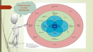
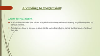
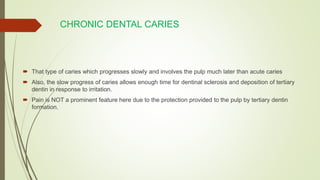
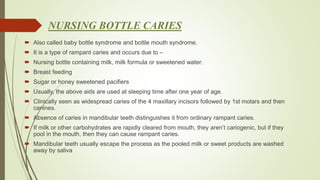
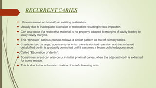
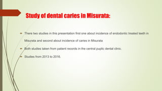
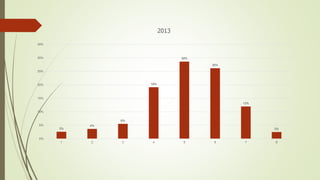
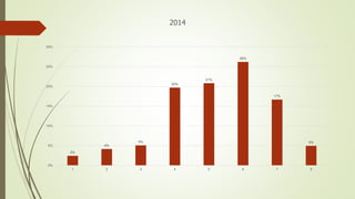
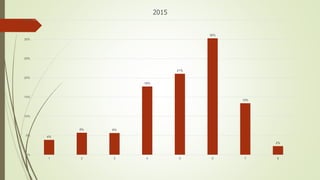
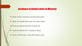
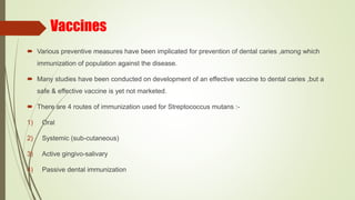
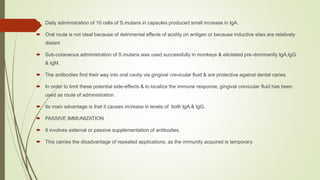
Presented by:
Dr. Nihal Mohamed
What is dental caries?
Dental caries is defined as an “irreversible disease of calcified tissues of
teeth, characterized by demineralization of the inorganic portion and
destruction of the organic substance of the tooth, which often leads to
cavitation”.
Classification of dental caries
According to anatomic site:
 Pit and fissure caries
 Smooth surface caries
 Root surface caries
According to extent:
 Incipient caries (reversible)
 Cavitated (irreversible)
According to progression:
 Acute caries
 Chronic caries
 Arrested caries
 Recurrent caries
 Radiation caries
 Nursing bottle caries
According to anatomic site
 Pit and fissure caries:
the pit and fissures of tooth provide a location for carries formulation.
 As the decay progresses, caries in enamel nearest the surface of the tooth spreads gradually
deeper.
 Once the caries reaches the dentin at the dentino-enamel junction, the decay quickly spreads
laterally.
 Within the dentin, the decay follows a triangle pattern that points to the tooth’s pulp.
 This pattern of decay is typically described as tow triangles (one triangle in enamel, and another in
dentin) with their bases conjoined to each other at the dentino-enamel junction (DEJ).
 This base to base pattern is typical of pit and caries.
• SMOOTH SURFACE CARIES
 Develops on proximal surfaces of all teeth or on gingival 1/3rd of buccal and lingual surfaces.
 Here, the caries is preceded by formation of dental plaque, quite unlike the pit and fissure caries.
 Presence of plaque ensures retention of carbohydrate and bacteria on tooth surfaces, leading to
subsequent acid production and demineralization of enamel.
• Smooth surface caries
 Three types of smooth surface caries:
 Proximal caries: also called interproximal caries, form on the smooth surfaces between adjacent
teeth.
 Root caries: form on the root surfaces of teeth.
 The Third type of smooth-surface caries occur on any other smooth tooth surface.
• Interproximal caries
 Proximal caries are the most difficult type to detect.
 Cannot be detected visually or manually with a dental explorer.
 Proximal caries form cervically (toward the roots of a tooth) just under the contact between two
teeth.
 Radiographs are needed for early discovery.
 The disintegration in the enamel in smooth surface caries is also pictures as cone.
 But the base of cone is enamel surface & apex directed towards apex.
• Root surface caries
 usually occur when the root surfaces have been exposed due to gingival recession.
 The root surface is more vulnerable to the demineralization process than enamel because
cementum begins to demineralize at 6.7 PH, which is higher than enamel’s critical PH.
 Regardless, it is easier to arrest the progression of root caries than enamel caries because roots
have a greater reuptake of fluoride than enamel.
According to progression:
ACUTE DENTAL CARIES
 It is that form of caries that follows a rapid clinical course and results in early pulpal involvement by
carious process.
 Pain is more likely to be seen in acute dental caries than chronic caries, but this is not a hard and
fast rule
CHRONIC DENTAL CARIES
 That type of caries which progresses slowly and involves the pulp much later than acute caries
 Also, the slow progress of caries allows enough time for dentinal sclerosis and deposition of tertiary
dentin in response to irritation.
 Pain is NOT a prominent feature here due to the protection provided to the pulp by tertiary dentin
formation.
NURSING BOTTLE CARIES
 Also called baby bottle syndrome and bottle mouth syndrome.
 It is a type of rampant caries and occurs due to –
 Nursing bottle containing milk, milk formula or sweetened water.
 Breast feeding
 Sugar or honey sweetened pacifiers
 Usually, the above aids are used at sleeping time after one year of age.
 Clinically seen as widespread caries of the 4 maxillary incisors followed by 1st molars and then
canines.
 Absence of caries in mandibular teeth distinguishes it from ordinary rampant caries.
 If milk or other carbohydrates are rapidly cleared from mouth, they aren’t cariogenic, but if they
pool in the mouth, then they can cause rampant caries.
 Mandibular teeth usually escape the process as the pooled milk or sweet products are washed
away by saliva
RECURRENT CARIES
 Occurs around or beneath an existing restoration.
 Usually due to inadequate extension of restoration resulting in food impaction
 Can also occur if a restorative material is not properly adapted to margins of cavity leading to
leaky cavity margins.
 This “renewed” carious process follows a similar pattern as that of primary caries.
 Characterized by large, open cavity in which there is no food retention and the softened
decalcified dentin is gradually burnished until it assumes a brown polished appearance.
 Called “Eburnation of dentin”.
 Sometimes arrest can also occur in initial proximal caries, when the adjacent tooth is extracted
for some reason.
 This is due to the automatic creation of a self cleansing area
RADIATION CARIES
 Rampant caries occurring in patients receiving radiotherapy in head & neck region is called as
RADIATION CARIES.
 Xerostomia is a major complication of radiotherapy of head & neck region. This, coupled with
increase in viscosity and low pH of saliva results in decreased anticariogenic actions of saliva.
ARRESTED CARIES
 It is that caries which becomes static or stationary and does not progress any further.
 Can occur in both deciduous and permanent dentition.
 Occurs almost exclusively on occlusal surface caries.
Etiological theories
 (1) Acidogenic Theory
 (2) Proteolytic Theory
 (3) Proteolysis-chelation Theory
Acidogenic Theory
 According to this theory dental decay is a chemo parasitic process
 It is a two stage process there is decalcification of the enamel which also results in the
destruction of the dentin. in the second stage there is dissolution of the softened residue of the
enamel and dentin
 In the first stage there is destruction is done by the acid attack where as the dissolution of the
residue is carried by the proteolytic action of the bacteria
 This whole process is supported by the presence of carbohydrates microorganisms and dental
plaque
. PROTEOLYTIC THEORY
 The evidence given by the acidogenic theory was considerable but it was not conclusive, and
alternative explanation was given in the form of proteolytic theory.
 There has been evidence that the organic portion of the tooth plays an important role in the
development of dental caries
 There are some enamel structure which are made of the organic material such enamel
lamellae and enamel rods
 These structure prove to be the path ways for the advancing microorganisms
 It has been established that enamel contains 0.56 % of organic matter of which 0.18% is
keratin and 0.17 % is a soluble protein
 Microorganisms produce proteolytic enzymes, which destroy the organic matrix of enamel,
loosening the apatite crystals….so they are eventually lost and tissue collapses
 However, even though proteolysis may not play any role in initiation of caries, their role in
progression of more advanced carious lesions cannot be ruled out.
. PROTEOLYSIS – CHELATION THEORY
Some of the minor flaws of the acidogenic and the proteolytic theory were addressed in the
proteolysis chelation theory.
CHELATION
It is a process in which there is complexion of the metal ions to form complex substance through
coordinate covalent bond which results in:
poorly dissociated or weakly ionized compound
 Chelation is independent of the PH of the medium.
 The bacterial attack on the surface of the enamel results in the breakdown of the protein ,chiefly keratin
and results in the formation of soluble chelates with calcium which decalcify enamel even at neutral
PH..
 Mucopolysaccarides may also act as secondary chelators.
 However, although chelation may not be actually responsible for initiating caries, it may still have some
role to play in advanced carious lesion where the pH levels return to neutral
Histopathology of dental caries
 Histopathology of caries in enamel:
Several zones can be distinguished beginning from the dental side of the lesion
 Zone 1: translucent zone
 Zone 2: dark zone
 Zone 3: body of the lesion
 Zone 4: surface zone
 Histopathology of caries in dentin
Zones of dental caries
 Zone 1: normal dentin
 Zone 2: sub transparent dentin
 Zone 3: transparent dentin
 Zone 4: turbid dentin
 Zone 5: infected dentin
CONTRIBUTING FACTORS – SYSTEMIC FACTORS
1. HEREDITY: -
2. 2. PREGNANCY & LACTATION: -
Signs and symptoms
 Caries initially involves only the enamel and produces no symptoms. a cavity that invades the
dentin causes pain, first when hot, cold, or sweet foods or beverages contact the involved tooth,
and later with chewing or percussion. Pain can be intense and persistent when the pulp is
severely involved
Spread from maxillary teeth
 may cause purulent sinusitis,
 meningitis,
 brain abscess,
 orbital cellulitis,
 and cavernous sinus thrombosis
Spread from mandibular teeth
 Spread from mandibular teeth may cause
 Ludwig's angina,
 parapharyngeal abscess,
 mediastinitis,
 pericarditis,
 emphysema, and jugular thrombophlebitis.
Diagnosis
 The traditional method of detecting caries signs is by visual inspection of dental surfaces, with the aid of a bright light
and dental mirror.
 Reflecting light onto the mouth mirror also can be done to search for dark shadows that could indicate dentin lesions.
 While the use of a dental probe continues to be controversial, it is extremely helpful when used correctly and
judiciously.
 During a visual–tactile examination, the dentist will also use a syringe or drying tool to blast air on to the tooth, which
makes it easier to see some lesions.
 Other tools used in visual–tactile examination may include magnifying devices to look at teeth, or orthodontic elastic
separators to separate teeth over the course of 2 to 3 days for a closer look between teeth prone to caries lesions.
 Fiber-optic transillumination is also sometimes used. This is a method by which visible light is emitted through the
tooth using an intense light source. If the transmitted light reveals a shadow, this may indicate a carious lesion.
Radiographic Methods
Radiographic examinations include;
 Bitewing radiographs
 IOPA radiographs using paralleling technique
 Dental panoramic tomography
CARIES ACTIVITY TESTS
 Lactobacillus colony count test
 Snyder test
 Alban’s test
 Swab test
 Reductase test
 Enamel solubility test
 Saliva Flow test
Study of dental caries in Misurata:
 There two studies in this presentation first one about incidence of endodontic treated teeth in
Misurata and second about incidence of caries in Misurata
 Both studies taken from patient records in the central puplic dental clinic.
 Studies from 2013 to 2016.
Incidence of endodontic treated teeth in Misurata
 The study include 4710 cases.
 The study does not include age and sex
 The study does not include deciduous teeth.
 The study not care if patient complete treatment or not.
keys for pictures
 1: central incisors
 2: lateral incisors
 3: canines
 4: first premolars
 5: second premolars
 6: first molars
 7: second molars
 8: third molars
3%
4%
6%
19%
29%
26%
12%
2%
0%
5%
10%
15%
20%
25%
30%
35%
1 2 3 4 5 6 7 8
2013
2%
4%
5%
20%
21%
26%
17%
5%
0%
5%
10%
15%
20%
25%
30%
1 2 3 4 5 6 7 8
2014
4%
6% 6%
18%
21%
30%
13%
2%
0%
5%
10%
15%
20%
25%
30%
35%
1 2 3 4 5 6 7 8
2015
5%
6% 6%
16%
17%
34%
13%
3%
0%
5%
10%
15%
20%
25%
30%
35%
40%
1 2 3 4 5 6 7 8
2016
Incidence of dental caries in Misurata
 Study include deciduous and permanent teeth
 Study not include third molar, non carious lesion
 Pedo are patients less than 12 years old
 Adult are patients from 13 years to above.
 Study of 1000 cases , about 250 cases in year.
0%
10%
20%
30%
40%
50%
60%
pedo adult
1%
44%
0%
55%
2013
RCT SC
0%
10%
20%
30%
40%
50%
60%
pedo adult
0%
54%
3%
42%
2014
RCT SC
0%
10%
20%
30%
40%
50%
60%
pedo adult
3%
56%
4%
36%
2015
RCT SC
0%
10%
20%
30%
40%
50%
60%
pedo adult
1%
52%
2%
44%
2016
RCT SC
Vaccines
 Various preventive measures have been implicated for prevention of dental caries ,among which
immunization of population against the disease.
 Many studies have been conducted on development of an effective vaccine to dental caries ,but a
safe & effective vaccine is yet not marketed.
 There are 4 routes of immunization used for Streptococcus mutans :-
1) Oral
2) Systemic (sub-cutaneous)
3) Active gingivo-salivary
4) Passive dental immunization
 Daily administration of 10 cells of S.mutans in capsules produced small increase in IgA.
 Oral route is not ideal because of detrimental effects of acidity on antigen or because inductive sites are relatively
distant
 Sub-cutaneous administration of S.mutans was used successfully in monkeys & elicitated pre-dominantly IgA,IgG
& IgM.
 The antibodies find their way into oral cavity via gingival crevicular fluid & are protective against dental caries.
 In order to limit these potential side-effects & to localize the immune response, gingival crevicular fluid has been
used as route of administration.
 Its main advantage is that it causes increase in levels of both IgA & IgG.
 PASSIVE IMMUNIZATION
 It involves external or passive supplementation of antibodies.
 This carries the disadvantage of repeated applications, as the immunity acquired is temporary.
 Fluoride treatment used abroad has successfully limited caries progression ,but not sufficient to
control this disease even when used together with professional tooth cleaning & dietary
counseling in populations highly exposed to these cariogenic bacteria.
Conclusion
 The caries process can be understood in very simple terms as being the result of acids
generated by dental biofilm from dietary fermentable carbohydrates causing demineralization
of tooth mineral and ultimately leading to a caries lesion.
 However , the complex and dynamic environment created by various contributing factors must
be taken into account to fully understand the caries disease process.
Cariology
Cariology

Cariology

  • 1.
  • 2.
    What is dentalcaries? Dental caries is defined as an “irreversible disease of calcified tissues of teeth, characterized by demineralization of the inorganic portion and destruction of the organic substance of the tooth, which often leads to cavitation”.
  • 4.
    Classification of dentalcaries According to anatomic site:  Pit and fissure caries  Smooth surface caries  Root surface caries According to extent:  Incipient caries (reversible)  Cavitated (irreversible) According to progression:  Acute caries  Chronic caries
  • 5.
     Arrested caries Recurrent caries  Radiation caries  Nursing bottle caries
  • 6.
    According to anatomicsite  Pit and fissure caries: the pit and fissures of tooth provide a location for carries formulation.  As the decay progresses, caries in enamel nearest the surface of the tooth spreads gradually deeper.  Once the caries reaches the dentin at the dentino-enamel junction, the decay quickly spreads laterally.  Within the dentin, the decay follows a triangle pattern that points to the tooth’s pulp.
  • 7.
     This patternof decay is typically described as tow triangles (one triangle in enamel, and another in dentin) with their bases conjoined to each other at the dentino-enamel junction (DEJ).  This base to base pattern is typical of pit and caries.
  • 8.
    • SMOOTH SURFACECARIES  Develops on proximal surfaces of all teeth or on gingival 1/3rd of buccal and lingual surfaces.  Here, the caries is preceded by formation of dental plaque, quite unlike the pit and fissure caries.  Presence of plaque ensures retention of carbohydrate and bacteria on tooth surfaces, leading to subsequent acid production and demineralization of enamel.
  • 9.
    • Smooth surfacecaries  Three types of smooth surface caries:  Proximal caries: also called interproximal caries, form on the smooth surfaces between adjacent teeth.  Root caries: form on the root surfaces of teeth.  The Third type of smooth-surface caries occur on any other smooth tooth surface.
  • 10.
    • Interproximal caries Proximal caries are the most difficult type to detect.  Cannot be detected visually or manually with a dental explorer.  Proximal caries form cervically (toward the roots of a tooth) just under the contact between two teeth.  Radiographs are needed for early discovery.  The disintegration in the enamel in smooth surface caries is also pictures as cone.  But the base of cone is enamel surface & apex directed towards apex.
  • 11.
    • Root surfacecaries  usually occur when the root surfaces have been exposed due to gingival recession.  The root surface is more vulnerable to the demineralization process than enamel because cementum begins to demineralize at 6.7 PH, which is higher than enamel’s critical PH.  Regardless, it is easier to arrest the progression of root caries than enamel caries because roots have a greater reuptake of fluoride than enamel.
  • 12.
    According to progression: ACUTEDENTAL CARIES  It is that form of caries that follows a rapid clinical course and results in early pulpal involvement by carious process.  Pain is more likely to be seen in acute dental caries than chronic caries, but this is not a hard and fast rule
  • 13.
    CHRONIC DENTAL CARIES That type of caries which progresses slowly and involves the pulp much later than acute caries  Also, the slow progress of caries allows enough time for dentinal sclerosis and deposition of tertiary dentin in response to irritation.  Pain is NOT a prominent feature here due to the protection provided to the pulp by tertiary dentin formation.
  • 14.
    NURSING BOTTLE CARIES Also called baby bottle syndrome and bottle mouth syndrome.  It is a type of rampant caries and occurs due to –  Nursing bottle containing milk, milk formula or sweetened water.  Breast feeding  Sugar or honey sweetened pacifiers  Usually, the above aids are used at sleeping time after one year of age.  Clinically seen as widespread caries of the 4 maxillary incisors followed by 1st molars and then canines.  Absence of caries in mandibular teeth distinguishes it from ordinary rampant caries.  If milk or other carbohydrates are rapidly cleared from mouth, they aren’t cariogenic, but if they pool in the mouth, then they can cause rampant caries.  Mandibular teeth usually escape the process as the pooled milk or sweet products are washed away by saliva
  • 15.
    RECURRENT CARIES  Occursaround or beneath an existing restoration.  Usually due to inadequate extension of restoration resulting in food impaction  Can also occur if a restorative material is not properly adapted to margins of cavity leading to leaky cavity margins.  This “renewed” carious process follows a similar pattern as that of primary caries.  Characterized by large, open cavity in which there is no food retention and the softened decalcified dentin is gradually burnished until it assumes a brown polished appearance.  Called “Eburnation of dentin”.  Sometimes arrest can also occur in initial proximal caries, when the adjacent tooth is extracted for some reason.  This is due to the automatic creation of a self cleansing area
  • 16.
    RADIATION CARIES  Rampantcaries occurring in patients receiving radiotherapy in head & neck region is called as RADIATION CARIES.  Xerostomia is a major complication of radiotherapy of head & neck region. This, coupled with increase in viscosity and low pH of saliva results in decreased anticariogenic actions of saliva.
  • 17.
    ARRESTED CARIES  Itis that caries which becomes static or stationary and does not progress any further.  Can occur in both deciduous and permanent dentition.  Occurs almost exclusively on occlusal surface caries.
  • 18.
    Etiological theories  (1)Acidogenic Theory  (2) Proteolytic Theory  (3) Proteolysis-chelation Theory
  • 19.
    Acidogenic Theory  Accordingto this theory dental decay is a chemo parasitic process  It is a two stage process there is decalcification of the enamel which also results in the destruction of the dentin. in the second stage there is dissolution of the softened residue of the enamel and dentin  In the first stage there is destruction is done by the acid attack where as the dissolution of the residue is carried by the proteolytic action of the bacteria  This whole process is supported by the presence of carbohydrates microorganisms and dental plaque
  • 20.
    . PROTEOLYTIC THEORY The evidence given by the acidogenic theory was considerable but it was not conclusive, and alternative explanation was given in the form of proteolytic theory.  There has been evidence that the organic portion of the tooth plays an important role in the development of dental caries  There are some enamel structure which are made of the organic material such enamel lamellae and enamel rods  These structure prove to be the path ways for the advancing microorganisms
  • 21.
     It hasbeen established that enamel contains 0.56 % of organic matter of which 0.18% is keratin and 0.17 % is a soluble protein  Microorganisms produce proteolytic enzymes, which destroy the organic matrix of enamel, loosening the apatite crystals….so they are eventually lost and tissue collapses  However, even though proteolysis may not play any role in initiation of caries, their role in progression of more advanced carious lesions cannot be ruled out.
  • 22.
    . PROTEOLYSIS –CHELATION THEORY Some of the minor flaws of the acidogenic and the proteolytic theory were addressed in the proteolysis chelation theory. CHELATION It is a process in which there is complexion of the metal ions to form complex substance through coordinate covalent bond which results in: poorly dissociated or weakly ionized compound
  • 23.
     Chelation isindependent of the PH of the medium.  The bacterial attack on the surface of the enamel results in the breakdown of the protein ,chiefly keratin and results in the formation of soluble chelates with calcium which decalcify enamel even at neutral PH..  Mucopolysaccarides may also act as secondary chelators.  However, although chelation may not be actually responsible for initiating caries, it may still have some role to play in advanced carious lesion where the pH levels return to neutral
  • 24.
    Histopathology of dentalcaries  Histopathology of caries in enamel: Several zones can be distinguished beginning from the dental side of the lesion  Zone 1: translucent zone  Zone 2: dark zone  Zone 3: body of the lesion  Zone 4: surface zone
  • 25.
     Histopathology ofcaries in dentin Zones of dental caries  Zone 1: normal dentin  Zone 2: sub transparent dentin  Zone 3: transparent dentin  Zone 4: turbid dentin  Zone 5: infected dentin
  • 26.
    CONTRIBUTING FACTORS –SYSTEMIC FACTORS 1. HEREDITY: - 2. 2. PREGNANCY & LACTATION: -
  • 27.
    Signs and symptoms Caries initially involves only the enamel and produces no symptoms. a cavity that invades the dentin causes pain, first when hot, cold, or sweet foods or beverages contact the involved tooth, and later with chewing or percussion. Pain can be intense and persistent when the pulp is severely involved
  • 28.
    Spread from maxillaryteeth  may cause purulent sinusitis,  meningitis,  brain abscess,  orbital cellulitis,  and cavernous sinus thrombosis
  • 29.
    Spread from mandibularteeth  Spread from mandibular teeth may cause  Ludwig's angina,  parapharyngeal abscess,  mediastinitis,  pericarditis,  emphysema, and jugular thrombophlebitis.
  • 30.
    Diagnosis  The traditionalmethod of detecting caries signs is by visual inspection of dental surfaces, with the aid of a bright light and dental mirror.  Reflecting light onto the mouth mirror also can be done to search for dark shadows that could indicate dentin lesions.  While the use of a dental probe continues to be controversial, it is extremely helpful when used correctly and judiciously.  During a visual–tactile examination, the dentist will also use a syringe or drying tool to blast air on to the tooth, which makes it easier to see some lesions.  Other tools used in visual–tactile examination may include magnifying devices to look at teeth, or orthodontic elastic separators to separate teeth over the course of 2 to 3 days for a closer look between teeth prone to caries lesions.  Fiber-optic transillumination is also sometimes used. This is a method by which visible light is emitted through the tooth using an intense light source. If the transmitted light reveals a shadow, this may indicate a carious lesion.
  • 31.
    Radiographic Methods Radiographic examinationsinclude;  Bitewing radiographs  IOPA radiographs using paralleling technique  Dental panoramic tomography
  • 32.
    CARIES ACTIVITY TESTS Lactobacillus colony count test  Snyder test  Alban’s test  Swab test  Reductase test  Enamel solubility test  Saliva Flow test
  • 33.
    Study of dentalcaries in Misurata:  There two studies in this presentation first one about incidence of endodontic treated teeth in Misurata and second about incidence of caries in Misurata  Both studies taken from patient records in the central puplic dental clinic.  Studies from 2013 to 2016.
  • 34.
    Incidence of endodontictreated teeth in Misurata  The study include 4710 cases.  The study does not include age and sex  The study does not include deciduous teeth.  The study not care if patient complete treatment or not.
  • 35.
    keys for pictures 1: central incisors  2: lateral incisors  3: canines  4: first premolars  5: second premolars  6: first molars  7: second molars  8: third molars
  • 36.
  • 37.
  • 38.
  • 39.
  • 40.
    Incidence of dentalcaries in Misurata  Study include deciduous and permanent teeth  Study not include third molar, non carious lesion  Pedo are patients less than 12 years old  Adult are patients from 13 years to above.  Study of 1000 cases , about 250 cases in year.
  • 41.
  • 42.
  • 43.
  • 44.
  • 45.
    Vaccines  Various preventivemeasures have been implicated for prevention of dental caries ,among which immunization of population against the disease.  Many studies have been conducted on development of an effective vaccine to dental caries ,but a safe & effective vaccine is yet not marketed.  There are 4 routes of immunization used for Streptococcus mutans :- 1) Oral 2) Systemic (sub-cutaneous) 3) Active gingivo-salivary 4) Passive dental immunization
  • 46.
     Daily administrationof 10 cells of S.mutans in capsules produced small increase in IgA.  Oral route is not ideal because of detrimental effects of acidity on antigen or because inductive sites are relatively distant  Sub-cutaneous administration of S.mutans was used successfully in monkeys & elicitated pre-dominantly IgA,IgG & IgM.  The antibodies find their way into oral cavity via gingival crevicular fluid & are protective against dental caries.  In order to limit these potential side-effects & to localize the immune response, gingival crevicular fluid has been used as route of administration.  Its main advantage is that it causes increase in levels of both IgA & IgG.  PASSIVE IMMUNIZATION  It involves external or passive supplementation of antibodies.  This carries the disadvantage of repeated applications, as the immunity acquired is temporary.
  • 47.
     Fluoride treatmentused abroad has successfully limited caries progression ,but not sufficient to control this disease even when used together with professional tooth cleaning & dietary counseling in populations highly exposed to these cariogenic bacteria.
  • 48.
    Conclusion  The cariesprocess can be understood in very simple terms as being the result of acids generated by dental biofilm from dietary fermentable carbohydrates causing demineralization of tooth mineral and ultimately leading to a caries lesion.  However , the complex and dynamic environment created by various contributing factors must be taken into account to fully understand the caries disease process.