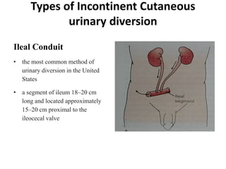
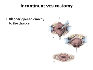
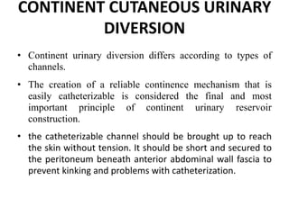
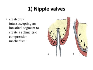
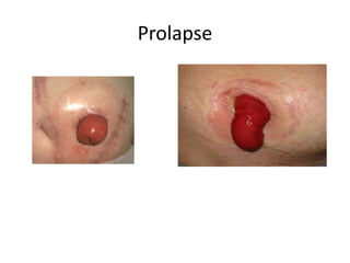
Cutaneous urinary diversion
By
Mohamed Elgendy
Assistant lecturer of urology
Assuit University
Definition
• Artifictial orifice for urinary diversion.
Indication of urinary diversion
• When the bladder has to be removed either due to malignant
tumour or post tramatic.
• When the sphincters of the bladder & the detrusor muscle
have been damaged or have lost their normal neurological
control.
• When there is irremovable obstruction in the bladder &
distal to that.
• neglected Ectopic vesicae or failure of repair
• Incurable vesico- vagina fistula.
Types/Classification
(1) According to elevation from surrounding
surface: Two types of stomas may be made on
the anterior abdominal wall:
• The flush stoma is preferable for the continent
type of diversion.
• The stoma that protrudes is preferable for
incontinent type of diversion.
Types/Classification Cont.
2) According to Shape:
1. Nipple Stoma:
“Rosebud”
• the stoma is grasped and
brought out for a
distance of 5 to 6 cm
through the abdominal
wall.
• it is preferred in the
incontinent stoma
2) Loop End Ileostomy
• It is preferred in obese
patients that have a
thick abdominal wall
and often a thick, short
ileal mesentery
• the distal end of the
loop is closed and the
bowel is drawn through
the rent in the
abdominal wall.
3) Flush Stoma
 It is preferred in the continent
types of urinary diversion.
 It can be done by appendix or
tapered ileum.
 It can be placed in umbilicus or
away from umbilicus.
 To avoid stomal stenosis a skin
flap should be incorporated in the
stoma.
 It has different shape V shaped
and VQZ stomaplasty.
Flush Stoma
The site of the stoma should be
selected preoperatively
 done by marking the stomal site with the patient in the
sitting position, as well as in the supine position.
 at least 5 cm away from the planned incision line.
 well away from skin creases, scars, belt lines, and bone
prominences.
• placed through the belly of the rectus muscle.
 be brought through a circular incision made at the
predetermined site.
Types of Incontinent Cutaneous
urinary diversion
Ileal Conduit
• the most common method of
urinary diversion in the United
States
• a segment of ileum 18–20 cm
long and located approximately
15–20 cm proximal to the
ileocecal valve
Ureterocutaneous shunt
• Ureter is implantened
direct to the skin
• Common in developing
country
 Loop ureterostomy
• Anterior part of the
ureter is implanted to
skin with maintenance
of posterior wall
continuity.
Incontinent vesicostomy
• Bladder opened directly
to the the skin
CONTINENT CUTANEOUS URINARY
DIVERSION
• Continent urinary diversion differs according to types of
channels.
• The creation of a reliable continence mechanism that is
easily catheterizable is considered the final and most
important principle of continent urinary reservoir
construction.
• the catheterizable channel should be brought up to reach
the skin without tension. It should be short and secured to
the peritoneum beneath anterior abdominal wall fascia to
prevent kinking and problems with catheterization.
1) Nipple valves
• created by
intussuscepting an
intestinal segment to
create a sphincteric
compression
mechanism.
2) Mitrofanoff
• using the vermiform appendix
• Its opened distal end was implanted submucosally, and
the base was brought to the abdominal wall.
• His principle was that any supple tube implanted
submucosally with sufficient muscle backing acts as a
flap valve and results in a reliable continent
cutaneous catheterizable channel.
• As the reservoir fills, the rise in intravesical pressure is
transmitted through the epithelium and to the
implanted conduit, clothing its lumen.
Mitrofanoff
3) Yang-Monti technique
• In this procedure, a 2
cm segment of small
bowel is opened
longitudinally along the
antimesenteric border
and then closed
transversely.
• its problem is the
relatively short length of
the channel.
4) Casale
• modification of Yang-
Monti technique
• Increase length of the
tube
• Two segment open
opposite each other
Paramesentric
• But its blood supply is
irreliable.
5) In situ ileocecal valve
• used as continence
mechanism in
reservoirs formed by
the cecum
• A short segment of
terminal ileum, whether
tailored or not, is used
as catheterizable
channel.
6) Continent Vesicostomy
• Parallel incisions 3 cm
apart are made into the
anterior bladder and used
to form a long rectangular
flap.
• The full-thickness strip is
tubularized down to the
bladder.
• The bladder mucosa from
either side of the tube is
then mobilized and closed
over the mucosal tube to
create a flap valve.
Complication of cutaneous urinary
stoma
Early complications
–Ischemia
–hemorrhage
–stenosis
–fistula
–retraction.
Late complications
– Prolapse
– obstruction
– para stomal
hernia
– skin irritation
Stoma Ischemia/Necrosis
Causes
–Aggressive
stripping of
mesentery
–Stenotic fascia
defect
–Extensive tension
Hemorrhage
• Mild hemorrhage common and self limiting.
– Usually mucosal.
– Apply pressure
• Active bleeding
– Implies failure to ligate a mesenteric vessel
– Identify and ligate.
Stomal Stenosis/Stricture
• Could manifest early or
late
• Ischemia is usual
underlying factor
• May leads to upper-tract
obstruction.
• Other causes: -Infection
and retraction
• Treat initially with dilation
• Definitive Stoma revision
Edema
• TTT mainly medical.
Mucocutaneous Separation
• Separation along
mucocutaneous border
• Caused by underlying
tension or separation of
sutures
• Usually treated
medically.
• Could lead to stricture.
Peristomal abscess and fistula
• Caused mainly by
infected
hematoma
• May lead to fistula
Stoma Retraction
• Causes
– Tension
– Obesity
– Steroids use. Poor
wound healing
• Can lead to leakage and
severe skin problem.
• Most eventually need
revision
Prolapse
Parastomal Hernia
• Predisposing factors
– Stoma placement lateral to rectus
(common)
– Large stoma
– Obesity
– Prior abdominal incisions
– Malnutrition
– Wound infection
• May present with
intestinal obstruction
• Minor cases ttt by
Abdominal binder
• Major cases – Repair
with mesh.
Peristomal Skin Complication
causes
• Due to prolonged contact of the skin with
urine.
• Pressure trauma from belt
• Allergic reaction in sensitive patient.
Skin Complications
• Fungal infections
• Ttt by Antifungal
powder
(Nystatin) or
systemic therapy
with fluconozole
Skin Complications
(Pyoderma Gangrenosum)
• Treatment conflicting
–Wound
debridement
–Steroids
injection
–Systemic
therapy
Skin Complications
• Allergic contact
dermatitis
• Ttt by Identify irritant
(skin patch test) and
discontinue products.
• Apply hydrocortisone
cream for short period
• Topical antihistamines
Skin Complications(Granulomas)
May be due to
– Granulation
tissue (poor
wound healing
and infection)
–Bowel
metaplasia
Skin Complications(Stoma warts)
• Warty raised
macerated
lesions around
the stoma.
• May cause
bleeding and
pain.
Pyelonephritis & Renal Deterioration
• Pyelonephritis occurs in approximately 10% of
patients who have undergone urinary diversion.
• Obstruction and stasis of urine within the
reconstructed urinary tract are risk factors for the
development of infection
• The presence of hydronephrosis, particularly in
patients with a conduit diversion, may indicate
the presence of ureteric reflux or obstruction at
the ureterovesical junction.
finally
• “care and expertise are important in
creating urinary stomas because
some patients must live with the
technical result for the rest of their
lives”
Thank you

Cutaneous urinary Stoma

  • 1.
    Cutaneous urinary diversion By MohamedElgendy Assistant lecturer of urology Assuit University
  • 2.
    Definition • Artifictial orificefor urinary diversion.
  • 3.
    Indication of urinarydiversion • When the bladder has to be removed either due to malignant tumour or post tramatic. • When the sphincters of the bladder & the detrusor muscle have been damaged or have lost their normal neurological control. • When there is irremovable obstruction in the bladder & distal to that. • neglected Ectopic vesicae or failure of repair • Incurable vesico- vagina fistula.
  • 4.
    Types/Classification (1) According toelevation from surrounding surface: Two types of stomas may be made on the anterior abdominal wall: • The flush stoma is preferable for the continent type of diversion. • The stoma that protrudes is preferable for incontinent type of diversion.
  • 5.
    Types/Classification Cont. 2) Accordingto Shape: 1. Nipple Stoma: “Rosebud” • the stoma is grasped and brought out for a distance of 5 to 6 cm through the abdominal wall. • it is preferred in the incontinent stoma
  • 6.
    2) Loop EndIleostomy • It is preferred in obese patients that have a thick abdominal wall and often a thick, short ileal mesentery • the distal end of the loop is closed and the bowel is drawn through the rent in the abdominal wall.
  • 7.
    3) Flush Stoma It is preferred in the continent types of urinary diversion.  It can be done by appendix or tapered ileum.  It can be placed in umbilicus or away from umbilicus.  To avoid stomal stenosis a skin flap should be incorporated in the stoma.  It has different shape V shaped and VQZ stomaplasty.
  • 8.
  • 9.
    The site ofthe stoma should be selected preoperatively  done by marking the stomal site with the patient in the sitting position, as well as in the supine position.  at least 5 cm away from the planned incision line.  well away from skin creases, scars, belt lines, and bone prominences. • placed through the belly of the rectus muscle.  be brought through a circular incision made at the predetermined site.
  • 10.
    Types of IncontinentCutaneous urinary diversion Ileal Conduit • the most common method of urinary diversion in the United States • a segment of ileum 18–20 cm long and located approximately 15–20 cm proximal to the ileocecal valve
  • 11.
    Ureterocutaneous shunt • Ureteris implantened direct to the skin • Common in developing country
  • 12.
     Loop ureterostomy •Anterior part of the ureter is implanted to skin with maintenance of posterior wall continuity.
  • 13.
    Incontinent vesicostomy • Bladderopened directly to the the skin
  • 14.
    CONTINENT CUTANEOUS URINARY DIVERSION •Continent urinary diversion differs according to types of channels. • The creation of a reliable continence mechanism that is easily catheterizable is considered the final and most important principle of continent urinary reservoir construction. • the catheterizable channel should be brought up to reach the skin without tension. It should be short and secured to the peritoneum beneath anterior abdominal wall fascia to prevent kinking and problems with catheterization.
  • 15.
    1) Nipple valves •created by intussuscepting an intestinal segment to create a sphincteric compression mechanism.
  • 16.
    2) Mitrofanoff • usingthe vermiform appendix • Its opened distal end was implanted submucosally, and the base was brought to the abdominal wall. • His principle was that any supple tube implanted submucosally with sufficient muscle backing acts as a flap valve and results in a reliable continent cutaneous catheterizable channel. • As the reservoir fills, the rise in intravesical pressure is transmitted through the epithelium and to the implanted conduit, clothing its lumen.
  • 17.
  • 18.
    3) Yang-Monti technique •In this procedure, a 2 cm segment of small bowel is opened longitudinally along the antimesenteric border and then closed transversely. • its problem is the relatively short length of the channel.
  • 19.
    4) Casale • modificationof Yang- Monti technique • Increase length of the tube • Two segment open opposite each other Paramesentric • But its blood supply is irreliable.
  • 20.
    5) In situileocecal valve • used as continence mechanism in reservoirs formed by the cecum • A short segment of terminal ileum, whether tailored or not, is used as catheterizable channel.
  • 21.
    6) Continent Vesicostomy •Parallel incisions 3 cm apart are made into the anterior bladder and used to form a long rectangular flap. • The full-thickness strip is tubularized down to the bladder. • The bladder mucosa from either side of the tube is then mobilized and closed over the mucosal tube to create a flap valve.
  • 22.
    Complication of cutaneousurinary stoma Early complications –Ischemia –hemorrhage –stenosis –fistula –retraction. Late complications – Prolapse – obstruction – para stomal hernia – skin irritation
  • 23.
  • 24.
    Hemorrhage • Mild hemorrhagecommon and self limiting. – Usually mucosal. – Apply pressure • Active bleeding – Implies failure to ligate a mesenteric vessel – Identify and ligate.
  • 25.
    Stomal Stenosis/Stricture • Couldmanifest early or late • Ischemia is usual underlying factor • May leads to upper-tract obstruction. • Other causes: -Infection and retraction • Treat initially with dilation • Definitive Stoma revision
  • 26.
  • 27.
    Mucocutaneous Separation • Separationalong mucocutaneous border • Caused by underlying tension or separation of sutures • Usually treated medically. • Could lead to stricture.
  • 28.
    Peristomal abscess andfistula • Caused mainly by infected hematoma • May lead to fistula
  • 29.
    Stoma Retraction • Causes –Tension – Obesity – Steroids use. Poor wound healing • Can lead to leakage and severe skin problem. • Most eventually need revision
  • 30.
  • 31.
    Parastomal Hernia • Predisposingfactors – Stoma placement lateral to rectus (common) – Large stoma – Obesity – Prior abdominal incisions – Malnutrition – Wound infection • May present with intestinal obstruction • Minor cases ttt by Abdominal binder • Major cases – Repair with mesh.
  • 32.
    Peristomal Skin Complication causes •Due to prolonged contact of the skin with urine. • Pressure trauma from belt • Allergic reaction in sensitive patient.
  • 33.
    Skin Complications • Fungalinfections • Ttt by Antifungal powder (Nystatin) or systemic therapy with fluconozole
  • 34.
    Skin Complications (Pyoderma Gangrenosum) •Treatment conflicting –Wound debridement –Steroids injection –Systemic therapy
  • 35.
    Skin Complications • Allergiccontact dermatitis • Ttt by Identify irritant (skin patch test) and discontinue products. • Apply hydrocortisone cream for short period • Topical antihistamines
  • 36.
    Skin Complications(Granulomas) May bedue to – Granulation tissue (poor wound healing and infection) –Bowel metaplasia
  • 37.
    Skin Complications(Stoma warts) •Warty raised macerated lesions around the stoma. • May cause bleeding and pain.
  • 38.
    Pyelonephritis & RenalDeterioration • Pyelonephritis occurs in approximately 10% of patients who have undergone urinary diversion. • Obstruction and stasis of urine within the reconstructed urinary tract are risk factors for the development of infection • The presence of hydronephrosis, particularly in patients with a conduit diversion, may indicate the presence of ureteric reflux or obstruction at the ureterovesical junction.
  • 39.
    finally • “care andexpertise are important in creating urinary stomas because some patients must live with the technical result for the rest of their lives”
  • 40.