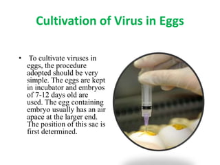
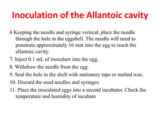
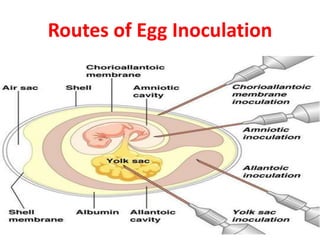
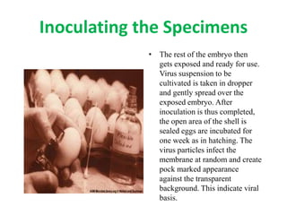
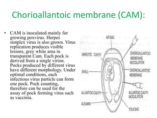
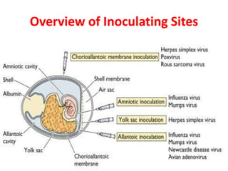
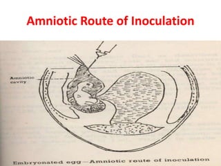
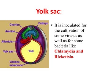
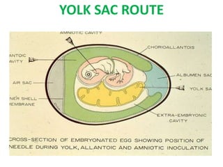
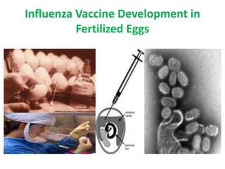
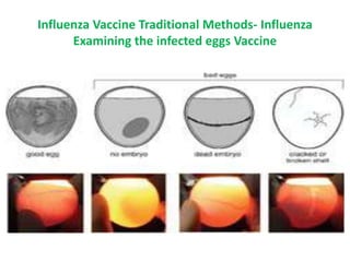
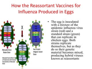
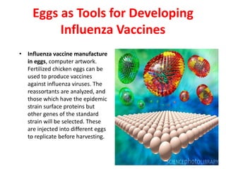
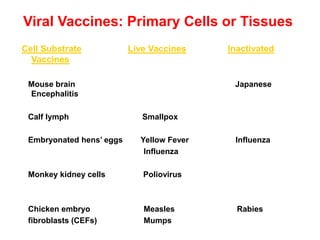
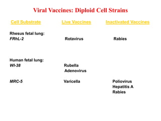
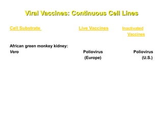
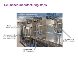
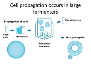
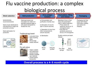
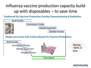
SlideShare a Scribd company logo
1 of 62
Viruses are Different From Other Microbes
• Viruses are obligate intracellular parasites. They depend
totally on their host cells for their existence. Their total host
dependence makes it, extremely difficult to get good insight
of them natural conditions, because the internal
characteristics of the host cells are likely to interfere with the
observations. Due to these reasons, it has been found
desirable that viruses are cultivated or grown in the
laboratory itself.
Isolation of Virus
• Laboratory animals
• Fertilized Hen’s Egg
– Chorioallantoic membrane
– Allantoic cavity
– Amniotic cavity
– Yolk sac
• Organ/Tissue/Cell Culture
• Growth identified by serological method like neutralization.
Virus Culture
Embryonated Egg Chorioallantioc membrane (CAM)
Allantoic cavity
Amniotic cavity
Yolk Sac
Cell Lines/ Tissue cultures Primary
Diploid/ Secondary
Continuous
Animal inoculation Suckling
Embryonated Hen’s Egg
Cultivation of Viruses and Bacteria
• Chorioallantoic membrane (CAM) – visible lesions called pocks. Each
infectious virus particle forms one pock. e.g. Variola, Vaccinia virus
• Allantoic cavity – Influenza virus (vaccine production) & paramyxoviruses
• Amniotic cavity – primary isolation of Influenza virus
• Yolk sac – Chlamydia, Rickettsia & some viruses
Embryonated eggs:
• The Embryonated hen’s egg was first used for cultivation of viruses by
Good Pasteur and Burnet (1931). Cultivation of viruses in organized
tissues like chick embryo necessitates a different type of approach.. For all
practical purposes they all themselves behave as tissue cultures. The
process of cultivation of viruses in embryonated eggs depends on the type
of egg which is used. The egg used for cultivation must be sterile and the
shell should be intact and healthy.
Burnet as Director of the Hall Institute, 1944-1965
F.M. Burnet in the laboratory
in the early 1950's,
was experimenting
on influenza virus genetics,
using the developing hen's egg
Only Embryonated Eggs Are Suitable
for Growing Virus
Inoculated eggs are candled
daily to see the chicken
embryos inside.
Eggs are Used for Mass Vaccine Production
in Influenza
 Animals and chick embryo
were the first method that was used
to cultivate virus. This method is rarely
used as it is not convenient.
However, when preparing for bulk virus,
(e.g. antigen or vaccine production)
the usage of chick embryo is useful.
Advantages of Fertile Eggs
 Fertile chicken eggs provide
a convenient, space-saving incubator
for many kinds of animal viruses.
Different viruses can be injected
into an egg at different sites and
the egg can be easily observed
for viral replication throughout
the development of the chicken embryo.
Advantages of Using Embryonated Eggs
• Isolation and cultivation of many avian and few mammalian
viruses
• Ideal receptacle for virus to grow
• Sterile & wide range of tissues and fluids
• Cost- much less
• Maintenance-easier
• Less labor
• Readily available
Advantages of Fertilized Eggs are
 Free from bacteria and many latent viruses.
 Free from specific and
non specific factors of defense.
Structure and Utility of Fertilized Egg
Routes of Injecting the Fertilized Eggs
Cultivation of Virus in Eggs
• To cultivate viruses in
eggs, the procedure
adopted should be very
simple. The eggs are kept
in incubator and embryos
of 7-12 days old are
used. The egg containing
embryo usually has an air
apace at the larger end.
The position of this sac is
first determined.
Begin you Exercise with
Candling Eggs
• Candling is the process of
holding a strong light above
or below the egg to observe
the embryo. A candling lamp
consists of a strong electric
bulb covered by a plastic or
aluminum container that has
a handle and an aperture.
The egg is placed against
this aperture and illuminated
by the light. If you do not
have a candling lamp,
improvise. Try using a torch.
Marking the inoculation site:
1. Hold the blunt end of the egg
against the aperture of the
candling lamp and note the
position of the head of the
embryo.
2. Turn the egg a quarter turn away
from the head.
3. Draw a line on the shell marking
the edge of the air sac.
4. Draw an X approximately 2 mm
above this line.
5. The X marks the inoculation
site.
• Eggs: 9-day old or 10-day old embryonated eggs. Candle the eggs
and mark the inoculation sites as described in Section 5. Eggs
should be placed in an egg rack with the inoculation site uppermost.
• Egg shell punch.
• Cotton wool.
• A 70 percent alcohol solution in water.
• Syringe 1 mL.
• Needles preferably 25 gauge, 16 mm.
• Stationery tape (also called cello or sticky tape) or melted wax to
seal the inoculation site.
• Inoculum. This must be free of microbial contamination.
• Discard tray.
Materials Needed for Egg
Inoculation
1. Use cotton wool and 70 percent alcohol to swab the end of the
eggs to be inoculated. Allow the alcohol to evaporate.
2. Swab the eggshell punch with 70 percent alcohol solution.
Place used cotton wool in discard tray.
3. Pierce a hole in the end of the egg at the marked inoculation
site.
4. Attach needle to 1 mL syringe.
5. Draw inoculum into 1 mL syringe.
Inoculation of the Allantoic
cavity
6 Keeping the needle and syringe vertical, place the needle
through the hole in the eggshell. The needle will need to
penetrate approximately 16 mm into the egg to reach the
allantoic cavity.
7. Inject 0.1 mL of inoculum into the egg.
8. Withdraw the needle from the egg.
9. Seal the hole in the shell with stationery tape or melted wax.
10. Discard the used needles and syringes.
11. Place the inoculated eggs into a second incubator. Check the
temperature and humidity of incubate
Inoculation of the Allantoic cavity
Piercing a hole in the egg shell
• A dental drill can be
used if it is available.
In most laboratories a
tool called an eggshell
punch can be
improvised using
materials that are
cheap and easy to
procure.
Routes of Egg Inoculation
Inoculating the Specimens
• The rest of the embryo then
gets exposed and ready for use.
Virus suspension to be
cultivated is taken in dropper
and gently spread over the
exposed embryo. After
inoculation is thus completed,
the open area of the shell is
sealed eggs are incubated for
one week as in hatching. The
virus particles infect the
membrane at random and create
pock marked appearance
against the transparent
background. This indicate viral
basis.
Chorioallantoic membrane (CAM):
• CAM is inoculated mainly for
growing poxvirus. Herpes
simplex virus is also grown. Virus
replication produces visible
lesions, grey white area in
transparent Cam. Each pock is
derived from a single virion.
Pocks produced by different virus
have different morphology. Under
optimal conditions, each
infectious virus particle can form
one pock. Pock counting,
therefore can be used for the
assay of pock forming virus such
as vaccinia.
Piercing the Chorioallantoic Membrane
• Little holes are drilled
through the egg shell for
infection of the chorio-
allantoic membrane
Can be used in few Fungal Infection
• They provide a complex
environment, including
phagocytic cells, to study
fungal host-pathogen
interaction, but are of a lower
developmental stage than
adult mice.
Piercing the Shell with Needle
Injecting Infective Material with
Needle
Overview of Inoculating Sites
• Inoculation into the allantoic cavity provides a rich
yield of influenza and some paramyxoviruses.
Allantoic inoculation is employed for growing the
influenza virus for vaccine production. Other
allantoic vaccines include Yellow fever (17D strain),
and rabies vaccines. Duck eggs are bigger and have a
longer incubation period then hen’s egg. They
therefore provide a better yield of rabies virus and
were used for the preparation of the inactivated non-
neural rabies vaccines.
Allantoic cavity:
ALLANTOIC ROUTE –
INOCULATION SITE DETERMINATION
Amniotic cavity:
• The amniotic sac is
mainly inoculated
for primary
isolation of
influenza a virus
and the mumps
virus.
Amniotic Route of Inoculation
Yolk sac:
• It is inoculated for
the cultivation of
some viruses as
well as for some
bacteria like
Chlamydia and
Rickettsia.
YOLK SAC ROUTE
Influenza Vaccine Development in
Fertilized Eggs
Influenza Vaccine Traditional Methods- Influenza
Examining the infected eggs Vaccine
How Vaccines are Produced in Eggs
• In egg culture, flu viruses are
injected into chicken egg
embryos, where they multiply.
After several days of incubation
a machine opens the egg and
harvests the virus, which is then
purified and chemically killed.
On average it takes one or two
eggs to produce a single dose of
annual flu vaccine.
In cell culture, the virus is
grown in animal or human
cells, which are available in
unlimited supply.
How the Reassortant Vaccines for
Influenza Produced in Eggs
• The egg is inoculated
with a mixture of the
epidemic influenza virus
strain (red) and a
standard strain (green)
that can replicate in
chicken eggs. Both
strains replicate
themselves, but as they
do so their genetic
material becomes mixed,
producing hybrid viruses
known as reassortants
Eggs as Tools for Developing
Influenza Vaccines
• Influenza vaccine manufacture
in eggs, computer artwork.
Fertilized chicken eggs can be
used to produce vaccines
against influenza viruses. The
reassortants are analyzed, and
those which have the epidemic
strain surface proteins but
other genes of the standard
strain will be selected. These
are injected into different eggs
to replicate before harvesting.
Eggs are Used in Mass Scale
Development of Vaccines
Egg Allergies and Vaccines
• No suitable cell culture
system exists and egg
inoculation is the method
of choice. Influenza virus
vaccines are still
cultivated in eggs, and
hence people with egg
allergies cannot tolerate
the influenza vaccines.
Follow all the Biosafety
Considerations
• All procedures involving
the manipulation of
infectious materials are
conducted within
biological safety
cabinets, specially
designed hoods, or other
physical containment
devices, or by personnel
wearing appropriate
personal protective
clothing and equipment.
EGG-BASED PRODUCTION
Advantages:
 Well established and cost-effective
 Lower cost
•Disadvantages:
• Extensive planning: long timeline for million eggs procurement
• Limited flexibility in case of exponentially increasing demand
(pandemic not contained & defeated):
•production takes too long
•eggs don’t grow on demand
• Potential impurities in eggs (antibiotics, other viruses) may
cause sterility problems
• Risk of allergies against egg albumin
• Growth of epidemic viruses in eggs result in variants that are
antigenically distinct from the original viruses
• Emerging endemic viruses sometimes do not grow at all in eggs
Cell Culture Vaccine Production
Potential Advantages
In response to the urgent and growing need for alternative means for influenza vaccine
production, a number of vaccine manufacturers have considered new approaches.
Of particularly interest has been the potential use of tissue culture cell lines to be
used either in additional to, or in lieu of, egg-based production. The chief
advantages of such a system are summarized below:
 Enables utilization of the same basic and clinically proven approach used in egg-
based production systems – i.e., production of whole-viruses, multiple disrupted
(“split”), more highly purified influenza virus antigens, or live attenuated vaccines
– while at the same time eliminating the long lead times and supply-chain
vulnerabilities required for egg-based production systems .
 Enables a more robust, consistent and reproducible means of vaccine production,
utilizing a scalable and a closed (or largely closed) bioreactor process, which may
be initiated at any time and extended for a prolonged period if needed.
 Enabling the capability of producing influenza vaccines with avian strains, which
generally cannot grow in eggs without genetic modification
Ideal characteristics of cell lines that could be utilized for production
 Replication of influenza strains to sufficient titer. Aside from embryonated eggs,
chicken embryo fibroblasts, and other avian cells ,replication of influenza
viruses has otherwise been documented in hamster cells (BHK21-F and
HKCC), MDBK cells, PER.C6 cells, Vero cells, and MDCK cells. A
prerequisite for a successful infection is the addition of proteases to the
medium, preferably trypsin or similar serine proteases, as these proteases
extracellularly cleave the precursor protein of hemagglutinin (HA0) into active
hemagglutinin (HA1 and HA2). Only cleaved hemagglutinin leads to the
adsorption of the influenza viruses on cells with subsequent virus assimilation
into the cell, which leads to further replication. Of the various continuous
mammalian cell lines that have been adequately tested – including diploid cell
lines (MRC-5, WI-38, and FRhl-2) and continuous cell lines (PER.C6, NIH-
3T3, BHK, CHO, Vero and MDCK) – only Vero, PER.C6 and MDCK have
consistently yielded influenza viruses titers that are sufficiently high enough to
be considered commercially viable.
 Established Regulatory Precedent: Immortalized (continuous) cell
lines are the only mammalian cell lines that have been documented to
support sufficient replication of influenza viruses. Of the three leading
candidates (Vero, PER.C6 and MDCK) Vero cells have been the only ones
to have been used in a widely used, commercially available vaccine. The
main regulatory concern related to these cell lines is that each of them has
been documented to be tumorigenic in animals at some point during their
passage history. Of the three, MDCK cells are the most tumorigenic, the
mechanism for which remains unknown. This and other regulatory aspects
related to cell line characterization have posed significant challenges for the
approval of mammalian cell-derived influenza vaccines
 Ability to Propagate Cells in a Chemically Defined Medium Under Serum-
free Conditions. In order to develop a process suitable for large-scale
commercial production, cells can be propagated and maintained as either
anchorage-dependent (adherent) or non-anchorage dependent. In the case
of adherent cells, after the initial proliferation phase, the nutrient medium is
removed and fresh medium is added to the cells, with infection of the cells
with influenza viruses taking place simultaneously or shortly thereafter.
After a specified time post infection, a protease (most often trypsin) is
added in order to obtain an optimum virus replication. The initial and
subsequent additions of trypsin later on in the typical process are generally
labor-intensive, with an increased potential for contamination of the cell
culture by adventitious agents. A more cost-effective alternative is cell
proliferation in fermenter systems utilizing cells growing adherently on
microcarriers under serum-free conditions, but such a process also requires
opening of the culture vessels several times and thus brings with it an
increased risk of contamination. The most desirable system would be the
propagation of cells in suspension. This not only eliminates the need for
trypsin, but also reduces cost, labor and the risk of contamination and also
allows for the formation of microvilli on the entire cell surface, thus
improving process yield and efficiency.
Types of Cell Substrates Used in Viral
Vaccines
 Primary Cells or Tissues: used without passage in
tissue culture
 Diploid Cells: cells with a finite lifespan and passage in
tissue culture
 Continuous Cell Lines: immortal, neoplastic cells with
unrestricted passage in tissue culture
 Non-tumorigenic
Cell substrates used for vaccine production
• Primary cells & Tissues (1954)
– Calf lymph for smallpox vaccines
– AGMK cells for polio vaccines
– Embryonated hens’ eggs for influenza, yellow fever vaccines
– Chicken embryo cell culture for measles, mumps vaccines
– Mouse brain for inactivated JEV vaccine
• Human diploid cells (introduced in 1960s)
– MRC-5, WI-38 for rubella, varicella vaccines
• CHO cells for highly purified, subunit investigational vaccines (1980s)
• Vero cells at non-tumorigenic passages for highly purified, inactivated
vaccine (IPV) (1980s)
• Vero cells at non-tumorigenic passages for live-attenuated vaccines (late
1990s)
• In vitro transformed human cells (e.g., 293, PER.C6) for defective vaccines
(early 2000s)
Viral Vaccines: Primary Cells or Tissues
Cell Substrate Live Vaccines Inactivated
Vaccines
Mouse brain Japanese
Encephalitis
Calf lymph Smallpox
Embryonated hens’ eggs Yellow Fever Influenza
Influenza
Monkey kidney cells Poliovirus
Chicken embryo Measles Rabies
fibroblasts (CEFs) Mumps
Viral Vaccines: Diploid Cell Strains
Cell Substrate Live Vaccines Inactivated Vaccines
Rhesus fetal lung:
FRhL-2 Rotavirus Rabies
Human fetal lung:
WI-38 Rubella
Adenovirus
MRC-5 Varicella Poliovirus
Hepatitis A
Rabies
Viral Vaccines: Continuous Cell Lines
Cell Substrate Live Vaccines Inactivated
Vaccines
African green monkey kidney:
Vero Poliovirus Poliovirus
(Europe) (U.S.)
Cell-based manufacturing steps
Cell propagation occurs in large
fermenters
Propagation of cells
Seed
cells Pre-culture
Production
fermenter
Virus infection
Virus propagation
Flu vaccine production: a complex
biological process
Optimized strains are
inoculated into millions of
specially prepared eggs or
cell culture bioreactors to
amplify the virus
Traditional egg based
OR
Flu cell culture
• Long lead times
• Open handling
steps
• Secure egg-
supply required
• Highly scalable
and responsive,
closed process
• Readily available
raw materials
• Closed system
bioreactors
WHO/CDC/EA
recommend strains
Seed viruses distributed to
manufacturers to begin the
production process
Seed optimization for
production
a) Concentration and
purification of whole virus
b) Disruption of virus for
split or subunit vaccines
c) Separation of virus from
other components for
subunit vaccines
d) Sterilization
Blending of virus in final
dose and presentation
(with or without adjuvant)
•Three strains for
seasonal vaccine at 15
micrograms each
•One strain for
pandemic vaccine as
low as 3.75 micrograms
Quality controls and batch
release
Multi
Dose Vial
Pre- filled
syringe
Packaging into final
boxes with Patient
Information Leaflets
and cartons for
shipping
Strain selection Bulk production
Virus antigen
isolation
Formulation, filling
and release
Packaging
Whole
virus
Subunits
purified
X month X month X month X month X month
Overall process is a 4- 6 month cycle
Or
The vaccine landscape is changing and is
moving to new manufacturing methods
Egg-based
production
technology
Cell Culture-based
production
technology
Cell Free
production
technology
System Eggs Mammalian Cells Insect and Bacterial Cells Recomb Cells
Lead Times 6-9 months ~3-6 months ~3 months Days?
Maturity On the market New on the market In 2-4 years time -
Facility design options
Disposable Bioreactors
Disposable DSP
Traditional Fixed Manufacturing
• Reduced capital investment
• Faster set up/ project throughput
• Easily moved
• Opportunity to ‘stockpile’ capacity
• Capital intensive manufacturing
• Long set up time
• Significant validation costs
• Highly localized
500 liter Wave Bioreactor
(GE Healthcare)
Influenza vaccine production capacity build
up with disposables – to save time
Traditional Flu Vaccine Production Facility Commissioning & Validation:
Build Facility
Validate Equipment
Commission Facility
Validate Process
Build Facility
Validate Equipment
Commission Facility
Validate Process
Time Saved
Saving
~60% in
time
Single-use Insect Cell Culture-Based Flu Vaccine Production:

More Related Content

What's hot

Monoclonal antibodies & hybridoma technology
Monoclonal antibodies & hybridoma technologyMonoclonal antibodies & hybridoma technology
Monoclonal antibodies & hybridoma technology
Ajay Dominic
 
Antimicrobial susceptibility test and assay bls 206
Antimicrobial susceptibility test and assay bls 206Antimicrobial susceptibility test and assay bls 206
Antimicrobial susceptibility test and assay bls 206
Bruno Mmassy
 
Antimicrobial susceptibility testing – disk diffusion methods
Antimicrobial susceptibility testing – disk diffusion methodsAntimicrobial susceptibility testing – disk diffusion methods
Antimicrobial susceptibility testing – disk diffusion methods
Ann Sam
 

What's hot (20)

Vaccine production
Vaccine productionVaccine production
Vaccine production
 
Vaccine
Vaccine Vaccine
Vaccine
 
Recent advances in microbiology
Recent advances in microbiologyRecent advances in microbiology
Recent advances in microbiology
 
Vaccines
VaccinesVaccines
Vaccines
 
MIC Testing
MIC TestingMIC Testing
MIC Testing
 
NANOPARTICLES IN VACCINE DELIVERY SYSTEM
NANOPARTICLES IN VACCINE DELIVERY SYSTEMNANOPARTICLES IN VACCINE DELIVERY SYSTEM
NANOPARTICLES IN VACCINE DELIVERY SYSTEM
 
Monoclonal antibodies & hybridoma technology
Monoclonal antibodies & hybridoma technologyMonoclonal antibodies & hybridoma technology
Monoclonal antibodies & hybridoma technology
 
Cultivation of viruses
Cultivation of virusesCultivation of viruses
Cultivation of viruses
 
Antibody engineering by R.S.Priyengha
Antibody engineering by R.S.PriyenghaAntibody engineering by R.S.Priyengha
Antibody engineering by R.S.Priyengha
 
bacteriophage
bacteriophage bacteriophage
bacteriophage
 
MIC.pptx
MIC.pptxMIC.pptx
MIC.pptx
 
Vaccine preparation part 2
Vaccine preparation part 2Vaccine preparation part 2
Vaccine preparation part 2
 
Tissue culture and virology (cpe, plaque assay)
Tissue culture and virology (cpe, plaque assay)Tissue culture and virology (cpe, plaque assay)
Tissue culture and virology (cpe, plaque assay)
 
Routes for inocculation of virus in an emryonated egg
Routes for inocculation of virus in an emryonated eggRoutes for inocculation of virus in an emryonated egg
Routes for inocculation of virus in an emryonated egg
 
Medical Microbiology Laboratory (Bacillus spp.)
Medical Microbiology Laboratory (Bacillus spp.)Medical Microbiology Laboratory (Bacillus spp.)
Medical Microbiology Laboratory (Bacillus spp.)
 
Vaccine production techniques.pptx
Vaccine production techniques.pptxVaccine production techniques.pptx
Vaccine production techniques.pptx
 
CULTIVATION OF VIRUS : Embryonated eggs
CULTIVATION OF VIRUS : Embryonated eggsCULTIVATION OF VIRUS : Embryonated eggs
CULTIVATION OF VIRUS : Embryonated eggs
 
Bacteriophages
Bacteriophages Bacteriophages
Bacteriophages
 
Antimicrobial susceptibility test and assay bls 206
Antimicrobial susceptibility test and assay bls 206Antimicrobial susceptibility test and assay bls 206
Antimicrobial susceptibility test and assay bls 206
 
Antimicrobial susceptibility testing – disk diffusion methods
Antimicrobial susceptibility testing – disk diffusion methodsAntimicrobial susceptibility testing – disk diffusion methods
Antimicrobial susceptibility testing – disk diffusion methods
 

Similar to egg n cell vaccine production.ppt

Similar to egg n cell vaccine production.ppt (20)

Egg inoculation new1
Egg inoculation new1Egg inoculation new1
Egg inoculation new1
 
new Virus culture techniques
new Virus culture techniques new Virus culture techniques
new Virus culture techniques
 
Viral vaccines
Viral vaccinesViral vaccines
Viral vaccines
 
Viral Cell Culture by Arijit Pani
Viral Cell Culture by Arijit PaniViral Cell Culture by Arijit Pani
Viral Cell Culture by Arijit Pani
 
cultivation of viruses
cultivation of virusescultivation of viruses
cultivation of viruses
 
APPLIED VIROLOGY Embryo and CPE.sk.pptx
APPLIED VIROLOGY Embryo and CPE.sk.pptxAPPLIED VIROLOGY Embryo and CPE.sk.pptx
APPLIED VIROLOGY Embryo and CPE.sk.pptx
 
Embryonated chicken egg.pdf
Embryonated chicken egg.pdfEmbryonated chicken egg.pdf
Embryonated chicken egg.pdf
 
Cultivation of viruses
Cultivation of virusesCultivation of viruses
Cultivation of viruses
 
rgdrgdr rhdr44 fefs fes fefsef esf grsgs.pptx
rgdrgdr rhdr44 fefs fes  fefsef esf grsgs.pptxrgdrgdr rhdr44 fefs fes  fefsef esf grsgs.pptx
rgdrgdr rhdr44 fefs fes fefsef esf grsgs.pptx
 
Viral innoculation methods.pdf
Viral innoculation methods.pdfViral innoculation methods.pdf
Viral innoculation methods.pdf
 
Cultivation of virus
Cultivation of virus Cultivation of virus
Cultivation of virus
 
Virus Cultivation - Purposes and Methods
Virus Cultivation - Purposes and MethodsVirus Cultivation - Purposes and Methods
Virus Cultivation - Purposes and Methods
 
VIROLOGY - CULTIVATION AND DETECTION OF VIRUS
VIROLOGY - CULTIVATION AND DETECTION OF VIRUSVIROLOGY - CULTIVATION AND DETECTION OF VIRUS
VIROLOGY - CULTIVATION AND DETECTION OF VIRUS
 
Presentation on Advanced Virology in virus cultivation.pptx
Presentation on Advanced Virology in virus cultivation.pptxPresentation on Advanced Virology in virus cultivation.pptx
Presentation on Advanced Virology in virus cultivation.pptx
 
Virus isolation & cultivation
Virus isolation & cultivationVirus isolation & cultivation
Virus isolation & cultivation
 
cultivation of virus
cultivation of viruscultivation of virus
cultivation of virus
 
Cultivation of viruses uhf copy - copy
Cultivation of  viruses uhf   copy - copyCultivation of  viruses uhf   copy - copy
Cultivation of viruses uhf copy - copy
 
Virology.pptx
Virology.pptxVirology.pptx
Virology.pptx
 
Egg inoculation by Chinithung ngullie (2)
Egg inoculation by Chinithung ngullie (2)Egg inoculation by Chinithung ngullie (2)
Egg inoculation by Chinithung ngullie (2)
 
CULTIVATION OF VIRUS.pptx
CULTIVATION OF VIRUS.pptxCULTIVATION OF VIRUS.pptx
CULTIVATION OF VIRUS.pptx
 

Recently uploaded

Spellings Wk 4 and Wk 5 for Grade 4 at CAPS
Spellings Wk 4 and Wk 5 for Grade 4 at CAPSSpellings Wk 4 and Wk 5 for Grade 4 at CAPS
Spellings Wk 4 and Wk 5 for Grade 4 at CAPS
AnaAcapella
 

Recently uploaded (20)

Details on CBSE Compartment Exam.pptx1111
Details on CBSE Compartment Exam.pptx1111Details on CBSE Compartment Exam.pptx1111
Details on CBSE Compartment Exam.pptx1111
 
Wellbeing inclusion and digital dystopias.pptx
Wellbeing inclusion and digital dystopias.pptxWellbeing inclusion and digital dystopias.pptx
Wellbeing inclusion and digital dystopias.pptx
 
80 ĐỀ THI THỬ TUYỂN SINH TIẾNG ANH VÀO 10 SỞ GD – ĐT THÀNH PHỐ HỒ CHÍ MINH NĂ...
80 ĐỀ THI THỬ TUYỂN SINH TIẾNG ANH VÀO 10 SỞ GD – ĐT THÀNH PHỐ HỒ CHÍ MINH NĂ...80 ĐỀ THI THỬ TUYỂN SINH TIẾNG ANH VÀO 10 SỞ GD – ĐT THÀNH PHỐ HỒ CHÍ MINH NĂ...
80 ĐỀ THI THỬ TUYỂN SINH TIẾNG ANH VÀO 10 SỞ GD – ĐT THÀNH PHỐ HỒ CHÍ MINH NĂ...
 
dusjagr & nano talk on open tools for agriculture research and learning
dusjagr & nano talk on open tools for agriculture research and learningdusjagr & nano talk on open tools for agriculture research and learning
dusjagr & nano talk on open tools for agriculture research and learning
 
Philosophy of china and it's charactistics
Philosophy of china and it's charactisticsPhilosophy of china and it's charactistics
Philosophy of china and it's charactistics
 
COMMUNICATING NEGATIVE NEWS - APPROACHES .pptx
COMMUNICATING NEGATIVE NEWS - APPROACHES .pptxCOMMUNICATING NEGATIVE NEWS - APPROACHES .pptx
COMMUNICATING NEGATIVE NEWS - APPROACHES .pptx
 
Spellings Wk 4 and Wk 5 for Grade 4 at CAPS
Spellings Wk 4 and Wk 5 for Grade 4 at CAPSSpellings Wk 4 and Wk 5 for Grade 4 at CAPS
Spellings Wk 4 and Wk 5 for Grade 4 at CAPS
 
Understanding Accommodations and Modifications
Understanding  Accommodations and ModificationsUnderstanding  Accommodations and Modifications
Understanding Accommodations and Modifications
 
General Principles of Intellectual Property: Concepts of Intellectual Proper...
General Principles of Intellectual Property: Concepts of Intellectual  Proper...General Principles of Intellectual Property: Concepts of Intellectual  Proper...
General Principles of Intellectual Property: Concepts of Intellectual Proper...
 
HMCS Max Bernays Pre-Deployment Brief (May 2024).pptx
HMCS Max Bernays Pre-Deployment Brief (May 2024).pptxHMCS Max Bernays Pre-Deployment Brief (May 2024).pptx
HMCS Max Bernays Pre-Deployment Brief (May 2024).pptx
 
This PowerPoint helps students to consider the concept of infinity.
This PowerPoint helps students to consider the concept of infinity.This PowerPoint helps students to consider the concept of infinity.
This PowerPoint helps students to consider the concept of infinity.
 
Sensory_Experience_and_Emotional_Resonance_in_Gabriel_Okaras_The_Piano_and_Th...
Sensory_Experience_and_Emotional_Resonance_in_Gabriel_Okaras_The_Piano_and_Th...Sensory_Experience_and_Emotional_Resonance_in_Gabriel_Okaras_The_Piano_and_Th...
Sensory_Experience_and_Emotional_Resonance_in_Gabriel_Okaras_The_Piano_and_Th...
 
Towards a code of practice for AI in AT.pptx
Towards a code of practice for AI in AT.pptxTowards a code of practice for AI in AT.pptx
Towards a code of practice for AI in AT.pptx
 
Tatlong Kwento ni Lola basyang-1.pdf arts
Tatlong Kwento ni Lola basyang-1.pdf artsTatlong Kwento ni Lola basyang-1.pdf arts
Tatlong Kwento ni Lola basyang-1.pdf arts
 
Food safety_Challenges food safety laboratories_.pdf
Food safety_Challenges food safety laboratories_.pdfFood safety_Challenges food safety laboratories_.pdf
Food safety_Challenges food safety laboratories_.pdf
 
How to setup Pycharm environment for Odoo 17.pptx
How to setup Pycharm environment for Odoo 17.pptxHow to setup Pycharm environment for Odoo 17.pptx
How to setup Pycharm environment for Odoo 17.pptx
 
FICTIONAL SALESMAN/SALESMAN SNSW 2024.pdf
FICTIONAL SALESMAN/SALESMAN SNSW 2024.pdfFICTIONAL SALESMAN/SALESMAN SNSW 2024.pdf
FICTIONAL SALESMAN/SALESMAN SNSW 2024.pdf
 
AIM of Education-Teachers Training-2024.ppt
AIM of Education-Teachers Training-2024.pptAIM of Education-Teachers Training-2024.ppt
AIM of Education-Teachers Training-2024.ppt
 
Jamworks pilot and AI at Jisc (20/03/2024)
Jamworks pilot and AI at Jisc (20/03/2024)Jamworks pilot and AI at Jisc (20/03/2024)
Jamworks pilot and AI at Jisc (20/03/2024)
 
Mehran University Newsletter Vol-X, Issue-I, 2024
Mehran University Newsletter Vol-X, Issue-I, 2024Mehran University Newsletter Vol-X, Issue-I, 2024
Mehran University Newsletter Vol-X, Issue-I, 2024
 

egg n cell vaccine production.ppt

  • 1.
  • 2. Viruses are Different From Other Microbes • Viruses are obligate intracellular parasites. They depend totally on their host cells for their existence. Their total host dependence makes it, extremely difficult to get good insight of them natural conditions, because the internal characteristics of the host cells are likely to interfere with the observations. Due to these reasons, it has been found desirable that viruses are cultivated or grown in the laboratory itself.
  • 3. Isolation of Virus • Laboratory animals • Fertilized Hen’s Egg – Chorioallantoic membrane – Allantoic cavity – Amniotic cavity – Yolk sac • Organ/Tissue/Cell Culture • Growth identified by serological method like neutralization.
  • 4. Virus Culture Embryonated Egg Chorioallantioc membrane (CAM) Allantoic cavity Amniotic cavity Yolk Sac Cell Lines/ Tissue cultures Primary Diploid/ Secondary Continuous Animal inoculation Suckling
  • 5. Embryonated Hen’s Egg Cultivation of Viruses and Bacteria • Chorioallantoic membrane (CAM) – visible lesions called pocks. Each infectious virus particle forms one pock. e.g. Variola, Vaccinia virus • Allantoic cavity – Influenza virus (vaccine production) & paramyxoviruses • Amniotic cavity – primary isolation of Influenza virus • Yolk sac – Chlamydia, Rickettsia & some viruses
  • 6. Embryonated eggs: • The Embryonated hen’s egg was first used for cultivation of viruses by Good Pasteur and Burnet (1931). Cultivation of viruses in organized tissues like chick embryo necessitates a different type of approach.. For all practical purposes they all themselves behave as tissue cultures. The process of cultivation of viruses in embryonated eggs depends on the type of egg which is used. The egg used for cultivation must be sterile and the shell should be intact and healthy.
  • 7. Burnet as Director of the Hall Institute, 1944-1965 F.M. Burnet in the laboratory in the early 1950's, was experimenting on influenza virus genetics, using the developing hen's egg
  • 8. Only Embryonated Eggs Are Suitable for Growing Virus Inoculated eggs are candled daily to see the chicken embryos inside.
  • 9. Eggs are Used for Mass Vaccine Production in Influenza  Animals and chick embryo were the first method that was used to cultivate virus. This method is rarely used as it is not convenient. However, when preparing for bulk virus, (e.g. antigen or vaccine production) the usage of chick embryo is useful.
  • 10. Advantages of Fertile Eggs  Fertile chicken eggs provide a convenient, space-saving incubator for many kinds of animal viruses. Different viruses can be injected into an egg at different sites and the egg can be easily observed for viral replication throughout the development of the chicken embryo.
  • 11. Advantages of Using Embryonated Eggs • Isolation and cultivation of many avian and few mammalian viruses • Ideal receptacle for virus to grow • Sterile & wide range of tissues and fluids • Cost- much less • Maintenance-easier • Less labor • Readily available
  • 12. Advantages of Fertilized Eggs are  Free from bacteria and many latent viruses.  Free from specific and non specific factors of defense.
  • 13. Structure and Utility of Fertilized Egg
  • 14. Routes of Injecting the Fertilized Eggs
  • 15.
  • 16. Cultivation of Virus in Eggs • To cultivate viruses in eggs, the procedure adopted should be very simple. The eggs are kept in incubator and embryos of 7-12 days old are used. The egg containing embryo usually has an air apace at the larger end. The position of this sac is first determined.
  • 17. Begin you Exercise with Candling Eggs • Candling is the process of holding a strong light above or below the egg to observe the embryo. A candling lamp consists of a strong electric bulb covered by a plastic or aluminum container that has a handle and an aperture. The egg is placed against this aperture and illuminated by the light. If you do not have a candling lamp, improvise. Try using a torch.
  • 18. Marking the inoculation site: 1. Hold the blunt end of the egg against the aperture of the candling lamp and note the position of the head of the embryo. 2. Turn the egg a quarter turn away from the head. 3. Draw a line on the shell marking the edge of the air sac. 4. Draw an X approximately 2 mm above this line. 5. The X marks the inoculation site.
  • 19. • Eggs: 9-day old or 10-day old embryonated eggs. Candle the eggs and mark the inoculation sites as described in Section 5. Eggs should be placed in an egg rack with the inoculation site uppermost. • Egg shell punch. • Cotton wool. • A 70 percent alcohol solution in water. • Syringe 1 mL. • Needles preferably 25 gauge, 16 mm. • Stationery tape (also called cello or sticky tape) or melted wax to seal the inoculation site. • Inoculum. This must be free of microbial contamination. • Discard tray. Materials Needed for Egg Inoculation
  • 20. 1. Use cotton wool and 70 percent alcohol to swab the end of the eggs to be inoculated. Allow the alcohol to evaporate. 2. Swab the eggshell punch with 70 percent alcohol solution. Place used cotton wool in discard tray. 3. Pierce a hole in the end of the egg at the marked inoculation site. 4. Attach needle to 1 mL syringe. 5. Draw inoculum into 1 mL syringe. Inoculation of the Allantoic cavity
  • 21. 6 Keeping the needle and syringe vertical, place the needle through the hole in the eggshell. The needle will need to penetrate approximately 16 mm into the egg to reach the allantoic cavity. 7. Inject 0.1 mL of inoculum into the egg. 8. Withdraw the needle from the egg. 9. Seal the hole in the shell with stationery tape or melted wax. 10. Discard the used needles and syringes. 11. Place the inoculated eggs into a second incubator. Check the temperature and humidity of incubate Inoculation of the Allantoic cavity
  • 22. Piercing a hole in the egg shell • A dental drill can be used if it is available. In most laboratories a tool called an eggshell punch can be improvised using materials that are cheap and easy to procure.
  • 23. Routes of Egg Inoculation
  • 24. Inoculating the Specimens • The rest of the embryo then gets exposed and ready for use. Virus suspension to be cultivated is taken in dropper and gently spread over the exposed embryo. After inoculation is thus completed, the open area of the shell is sealed eggs are incubated for one week as in hatching. The virus particles infect the membrane at random and create pock marked appearance against the transparent background. This indicate viral basis.
  • 25. Chorioallantoic membrane (CAM): • CAM is inoculated mainly for growing poxvirus. Herpes simplex virus is also grown. Virus replication produces visible lesions, grey white area in transparent Cam. Each pock is derived from a single virion. Pocks produced by different virus have different morphology. Under optimal conditions, each infectious virus particle can form one pock. Pock counting, therefore can be used for the assay of pock forming virus such as vaccinia.
  • 26. Piercing the Chorioallantoic Membrane • Little holes are drilled through the egg shell for infection of the chorio- allantoic membrane
  • 27. Can be used in few Fungal Infection • They provide a complex environment, including phagocytic cells, to study fungal host-pathogen interaction, but are of a lower developmental stage than adult mice.
  • 28.
  • 29. Piercing the Shell with Needle
  • 32. • Inoculation into the allantoic cavity provides a rich yield of influenza and some paramyxoviruses. Allantoic inoculation is employed for growing the influenza virus for vaccine production. Other allantoic vaccines include Yellow fever (17D strain), and rabies vaccines. Duck eggs are bigger and have a longer incubation period then hen’s egg. They therefore provide a better yield of rabies virus and were used for the preparation of the inactivated non- neural rabies vaccines. Allantoic cavity:
  • 33. ALLANTOIC ROUTE – INOCULATION SITE DETERMINATION
  • 34. Amniotic cavity: • The amniotic sac is mainly inoculated for primary isolation of influenza a virus and the mumps virus.
  • 35. Amniotic Route of Inoculation
  • 36. Yolk sac: • It is inoculated for the cultivation of some viruses as well as for some bacteria like Chlamydia and Rickettsia.
  • 38. Influenza Vaccine Development in Fertilized Eggs
  • 39. Influenza Vaccine Traditional Methods- Influenza Examining the infected eggs Vaccine
  • 40. How Vaccines are Produced in Eggs • In egg culture, flu viruses are injected into chicken egg embryos, where they multiply. After several days of incubation a machine opens the egg and harvests the virus, which is then purified and chemically killed. On average it takes one or two eggs to produce a single dose of annual flu vaccine. In cell culture, the virus is grown in animal or human cells, which are available in unlimited supply.
  • 41. How the Reassortant Vaccines for Influenza Produced in Eggs • The egg is inoculated with a mixture of the epidemic influenza virus strain (red) and a standard strain (green) that can replicate in chicken eggs. Both strains replicate themselves, but as they do so their genetic material becomes mixed, producing hybrid viruses known as reassortants
  • 42. Eggs as Tools for Developing Influenza Vaccines • Influenza vaccine manufacture in eggs, computer artwork. Fertilized chicken eggs can be used to produce vaccines against influenza viruses. The reassortants are analyzed, and those which have the epidemic strain surface proteins but other genes of the standard strain will be selected. These are injected into different eggs to replicate before harvesting.
  • 43. Eggs are Used in Mass Scale Development of Vaccines
  • 44. Egg Allergies and Vaccines • No suitable cell culture system exists and egg inoculation is the method of choice. Influenza virus vaccines are still cultivated in eggs, and hence people with egg allergies cannot tolerate the influenza vaccines.
  • 45. Follow all the Biosafety Considerations • All procedures involving the manipulation of infectious materials are conducted within biological safety cabinets, specially designed hoods, or other physical containment devices, or by personnel wearing appropriate personal protective clothing and equipment.
  • 46. EGG-BASED PRODUCTION Advantages:  Well established and cost-effective  Lower cost •Disadvantages: • Extensive planning: long timeline for million eggs procurement • Limited flexibility in case of exponentially increasing demand (pandemic not contained & defeated): •production takes too long •eggs don’t grow on demand • Potential impurities in eggs (antibiotics, other viruses) may cause sterility problems • Risk of allergies against egg albumin • Growth of epidemic viruses in eggs result in variants that are antigenically distinct from the original viruses • Emerging endemic viruses sometimes do not grow at all in eggs
  • 47. Cell Culture Vaccine Production
  • 48. Potential Advantages In response to the urgent and growing need for alternative means for influenza vaccine production, a number of vaccine manufacturers have considered new approaches. Of particularly interest has been the potential use of tissue culture cell lines to be used either in additional to, or in lieu of, egg-based production. The chief advantages of such a system are summarized below:  Enables utilization of the same basic and clinically proven approach used in egg- based production systems – i.e., production of whole-viruses, multiple disrupted (“split”), more highly purified influenza virus antigens, or live attenuated vaccines – while at the same time eliminating the long lead times and supply-chain vulnerabilities required for egg-based production systems .  Enables a more robust, consistent and reproducible means of vaccine production, utilizing a scalable and a closed (or largely closed) bioreactor process, which may be initiated at any time and extended for a prolonged period if needed.  Enabling the capability of producing influenza vaccines with avian strains, which generally cannot grow in eggs without genetic modification
  • 49. Ideal characteristics of cell lines that could be utilized for production  Replication of influenza strains to sufficient titer. Aside from embryonated eggs, chicken embryo fibroblasts, and other avian cells ,replication of influenza viruses has otherwise been documented in hamster cells (BHK21-F and HKCC), MDBK cells, PER.C6 cells, Vero cells, and MDCK cells. A prerequisite for a successful infection is the addition of proteases to the medium, preferably trypsin or similar serine proteases, as these proteases extracellularly cleave the precursor protein of hemagglutinin (HA0) into active hemagglutinin (HA1 and HA2). Only cleaved hemagglutinin leads to the adsorption of the influenza viruses on cells with subsequent virus assimilation into the cell, which leads to further replication. Of the various continuous mammalian cell lines that have been adequately tested – including diploid cell lines (MRC-5, WI-38, and FRhl-2) and continuous cell lines (PER.C6, NIH- 3T3, BHK, CHO, Vero and MDCK) – only Vero, PER.C6 and MDCK have consistently yielded influenza viruses titers that are sufficiently high enough to be considered commercially viable.
  • 50.  Established Regulatory Precedent: Immortalized (continuous) cell lines are the only mammalian cell lines that have been documented to support sufficient replication of influenza viruses. Of the three leading candidates (Vero, PER.C6 and MDCK) Vero cells have been the only ones to have been used in a widely used, commercially available vaccine. The main regulatory concern related to these cell lines is that each of them has been documented to be tumorigenic in animals at some point during their passage history. Of the three, MDCK cells are the most tumorigenic, the mechanism for which remains unknown. This and other regulatory aspects related to cell line characterization have posed significant challenges for the approval of mammalian cell-derived influenza vaccines
  • 51.  Ability to Propagate Cells in a Chemically Defined Medium Under Serum- free Conditions. In order to develop a process suitable for large-scale commercial production, cells can be propagated and maintained as either anchorage-dependent (adherent) or non-anchorage dependent. In the case of adherent cells, after the initial proliferation phase, the nutrient medium is removed and fresh medium is added to the cells, with infection of the cells with influenza viruses taking place simultaneously or shortly thereafter. After a specified time post infection, a protease (most often trypsin) is added in order to obtain an optimum virus replication. The initial and subsequent additions of trypsin later on in the typical process are generally labor-intensive, with an increased potential for contamination of the cell culture by adventitious agents. A more cost-effective alternative is cell proliferation in fermenter systems utilizing cells growing adherently on microcarriers under serum-free conditions, but such a process also requires opening of the culture vessels several times and thus brings with it an increased risk of contamination. The most desirable system would be the propagation of cells in suspension. This not only eliminates the need for trypsin, but also reduces cost, labor and the risk of contamination and also allows for the formation of microvilli on the entire cell surface, thus improving process yield and efficiency.
  • 52. Types of Cell Substrates Used in Viral Vaccines  Primary Cells or Tissues: used without passage in tissue culture  Diploid Cells: cells with a finite lifespan and passage in tissue culture  Continuous Cell Lines: immortal, neoplastic cells with unrestricted passage in tissue culture  Non-tumorigenic
  • 53. Cell substrates used for vaccine production • Primary cells & Tissues (1954) – Calf lymph for smallpox vaccines – AGMK cells for polio vaccines – Embryonated hens’ eggs for influenza, yellow fever vaccines – Chicken embryo cell culture for measles, mumps vaccines – Mouse brain for inactivated JEV vaccine • Human diploid cells (introduced in 1960s) – MRC-5, WI-38 for rubella, varicella vaccines • CHO cells for highly purified, subunit investigational vaccines (1980s) • Vero cells at non-tumorigenic passages for highly purified, inactivated vaccine (IPV) (1980s) • Vero cells at non-tumorigenic passages for live-attenuated vaccines (late 1990s) • In vitro transformed human cells (e.g., 293, PER.C6) for defective vaccines (early 2000s)
  • 54. Viral Vaccines: Primary Cells or Tissues Cell Substrate Live Vaccines Inactivated Vaccines Mouse brain Japanese Encephalitis Calf lymph Smallpox Embryonated hens’ eggs Yellow Fever Influenza Influenza Monkey kidney cells Poliovirus Chicken embryo Measles Rabies fibroblasts (CEFs) Mumps
  • 55. Viral Vaccines: Diploid Cell Strains Cell Substrate Live Vaccines Inactivated Vaccines Rhesus fetal lung: FRhL-2 Rotavirus Rabies Human fetal lung: WI-38 Rubella Adenovirus MRC-5 Varicella Poliovirus Hepatitis A Rabies
  • 56. Viral Vaccines: Continuous Cell Lines Cell Substrate Live Vaccines Inactivated Vaccines African green monkey kidney: Vero Poliovirus Poliovirus (Europe) (U.S.)
  • 58. Cell propagation occurs in large fermenters Propagation of cells Seed cells Pre-culture Production fermenter Virus infection Virus propagation
  • 59. Flu vaccine production: a complex biological process Optimized strains are inoculated into millions of specially prepared eggs or cell culture bioreactors to amplify the virus Traditional egg based OR Flu cell culture • Long lead times • Open handling steps • Secure egg- supply required • Highly scalable and responsive, closed process • Readily available raw materials • Closed system bioreactors WHO/CDC/EA recommend strains Seed viruses distributed to manufacturers to begin the production process Seed optimization for production a) Concentration and purification of whole virus b) Disruption of virus for split or subunit vaccines c) Separation of virus from other components for subunit vaccines d) Sterilization Blending of virus in final dose and presentation (with or without adjuvant) •Three strains for seasonal vaccine at 15 micrograms each •One strain for pandemic vaccine as low as 3.75 micrograms Quality controls and batch release Multi Dose Vial Pre- filled syringe Packaging into final boxes with Patient Information Leaflets and cartons for shipping Strain selection Bulk production Virus antigen isolation Formulation, filling and release Packaging Whole virus Subunits purified X month X month X month X month X month Overall process is a 4- 6 month cycle Or
  • 60. The vaccine landscape is changing and is moving to new manufacturing methods Egg-based production technology Cell Culture-based production technology Cell Free production technology System Eggs Mammalian Cells Insect and Bacterial Cells Recomb Cells Lead Times 6-9 months ~3-6 months ~3 months Days? Maturity On the market New on the market In 2-4 years time -
  • 61. Facility design options Disposable Bioreactors Disposable DSP Traditional Fixed Manufacturing • Reduced capital investment • Faster set up/ project throughput • Easily moved • Opportunity to ‘stockpile’ capacity • Capital intensive manufacturing • Long set up time • Significant validation costs • Highly localized 500 liter Wave Bioreactor (GE Healthcare)
  • 62. Influenza vaccine production capacity build up with disposables – to save time Traditional Flu Vaccine Production Facility Commissioning & Validation: Build Facility Validate Equipment Commission Facility Validate Process Build Facility Validate Equipment Commission Facility Validate Process Time Saved Saving ~60% in time Single-use Insect Cell Culture-Based Flu Vaccine Production: