Download to read offline

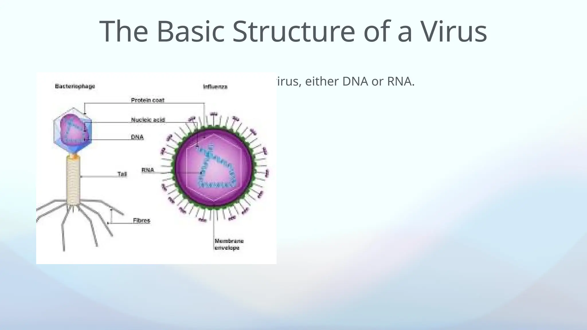


The document provides an overview of virus structure, classification, and methods for cultivation, highlighting key features such as nucleic acid types, capsid structures, and the Baltimore classification system. It details different methods for viral cultivation including animal inoculation, embryonated eggs, and tissue culture, each with its own advantages and disadvantages. Additionally, it explains cellular responses to viral infection, emphasizing the importance of virology in diagnostics and vaccine development.