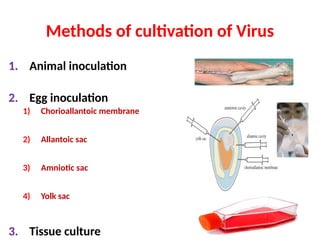
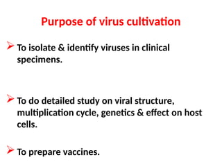
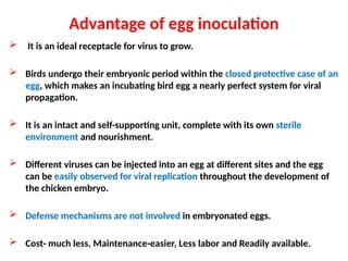
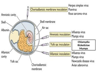
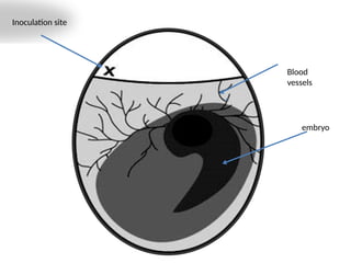
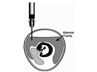
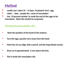
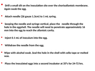
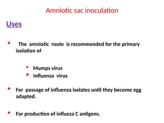
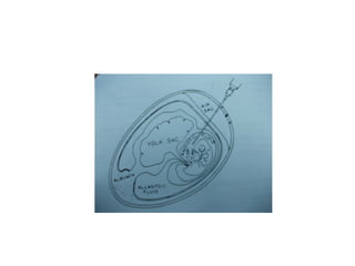
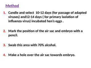
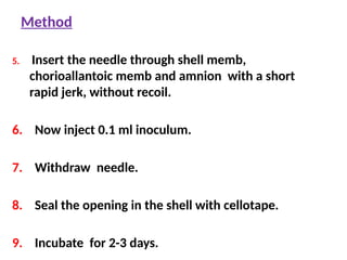
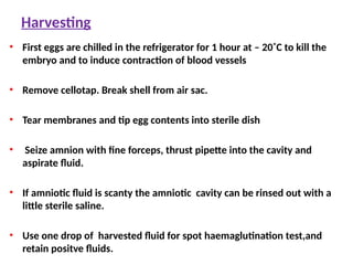
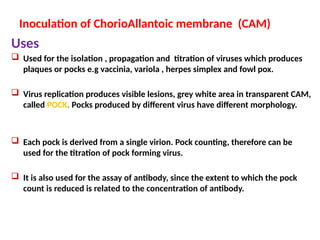
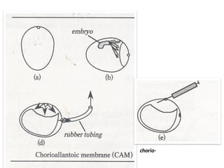
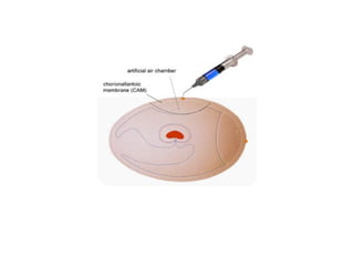
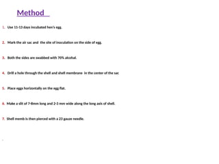
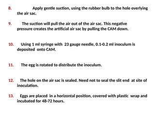
EGG INOCULATION
Principles and Technique
Dr. Jairam Choudhary
Methods of cultivation of Virus
1. Animal inoculation
2. Egg inoculation
1) Chorioallantoic membrane
2) Allantoic sac
3) Amniotic sac
4) Yolk sac
3. Tissue culture
Purpose of virus cultivation
To isolate & identify viruses in clinical
specimens.
To do detailed study on viral structure,
multiplication cycle, genetics & effect on host
cells.
To prepare vaccines.
Advantage of egg inoculation
 It is an ideal receptacle for virus to grow.
 Birds undergo their embryonic period within the closed protective case of an
egg, which makes an incubating bird egg a nearly perfect system for viral
propagation.
 It is an intact and self-supporting unit, complete with its own sterile
environment and nourishment.
 Different viruses can be injected into an egg at different sites and the egg
can be easily observed for viral replication throughout the development of
the chicken embryo.
 Defense mechanisms are not involved in embryonated eggs.
 Cost- much less, Maintenance-easier, Less labor and Readily available.
History
Goodpasture and Burnet in 1931
first used the embryonated hen’s egg
for the cultivation of virus.
Parts of Embryonated Egg
CAM
Shell and shell membrane
The membrane is closely attached to the shell. Together they
function as an exchange system and gaseous and liquid
molecules pass in both directions. This is why eggs must be
incubated in humid conditions. Eggs incubated in low humidity
will lose moisture and eventually the embryo will die.
Air sac
Eggs have one rounded blunt and other pointed end. The air sac is the
space at the rounded end and has a function in respiration and
pressure adjustments.
Chorioallantoic membrane and allantoic cavity
The membrane is attached to the embryo and functions to remove
soluble, insoluble and gaseous waste products. The sac contains
allantoic fluid. .
Yolk sac
This is attached to the embryo and contains the nutrient-rich
yolk. As the embryo develops, the yolk sac decreases in size.
Amniotic Sac
This sac surrounds the embryo. It is filled with liquid and serves
to protect the embryo against physical damage as well as
functioning as an area of exchange of molecules. As the embryo
develops, the membrane stretches and the amniotic sac is
barely visible in the fully developed embryo.
Routes of Egg Inoculation
1. Chorioallantoic membrane
2. Allantoic sac
3. Amniotic sac
4. Yolk sac
The chosen route of inoculation are
determined by selectivity of virus
1. For a certain membrane.
2. Developmental stage of the embryo
Chlamydia
Rickettsiae
Mumps
Choice of Eggs
Hen, duck, and turkey eggs are the most
common choices for inoculation .
The egg shell should be intact and healthy.
Discard cracked shell eggs.
 Specific pathogen free (SPF) eggs.
Materials Needed for Egg Inoculation
 Candle box
 Incubator
 Egg shell punch / dental drill
 Cotton swab
 70 percent alcohol
 Syringe 1 mL
 Needles preferably 25 gauge, 16 mm.
 Stationery tape (also called cello or sticky tape) or melted wax to seal the inoculation
site.
 Inoculum
Incubation
Incubation of eggs before inoculation
 Incubation temperature = 38°C to 39°C (100.4 F to102.2F)
 Humidity should be maintained at 60 to 65 percent. A tray filled with water and placed in
the bottom of the incubator is usually sufficient to maintain this level of humidity.
 Place the eggs in the incubator with the air sac on top.
 Eggs should be turned three times a day.
 Duration depends on requirement of embryonated egg ‘s age.
Incubation of eggs after inoculation
Inoculated eggs contain virus and should be placed in a different
incubator.
Eggs inoculated with virulent strains of virus should not be
incubated in the same incubator as used for eggs inoculated with
the avirulent strain of virus
Inoculated eggs are incubated under the same conditions as
uninoculated eggs
DO NOT turn the eggs.
CANDLING
 Candling is the process of holding a
strong light above or below the egg to
observe the embryo
 Candling is done in a darkened room
or in an area shielded by curtains.
 A candling lamp consists of a strong
electric bulb covered by a plastic or
aluminum container that has a handle
and an aperture. If we do not have a
candling lamp, we can try a torch.
Candling
1. Signs of viability
2. Marking positions of air sac, embryo,and
blood vessels.
3. Age of embryo.
4. Signs of death of embryo
1.Signs of viability
1. Clear well demarcated air sac.
2. Intact blood vessels.
3. Intact embryonic membrane.
4. Motility of embryo. Healthy embryos will respond to the light by moving.
2. Marking position of air sac, embryo and blood
vessels
1. Air space present at the rounded blunt end.
2. Embryo- appears as a dark shadow with
the head as a dark spot.
3. Marking of blood vessels
3.Age of embryo
 Embryo is recognized only with difficulty during the first
few days, but at 4- or 5-days incubation it may be readily
detected by candling.
 Swallowing movements are made from the 9th day
onward.
 From the 10th day the embryo rapidly increases in size and
feathers appear.
 Development of the respiratory tract occurs between the
12th and 15th day.
4. Signs of death of Embryo
 Loss of air sac.
 Disappearance or damage of blood vessels.
 Detachment of membranes from the rim of air sac.
 Diminished movement of embryo at beginning
followed by no movement .
 Falling of embryo and its membranes into the lower
part of egg.
Yolk sac inoculation
Uses
This method is used for the cultivation of Mumps virus
and bacteria like Chlamydiae and Rickettsiae.
Immune interference mechanism can occur in most of
avian viruses. So not suitable for cultivation of avian
viruses.
Method
1. Candle and select 6 -8 days incubated hen’s eggs.
2. Label -- date, sample No., route of inoculation.
3. Mark the air sac.
4. Use 70 percent alcohol to swab the end of the eggs to be
inoculated. Allow the alcohol to evaporate.
5. Drill a hole over the air sac through the shell.
6. Use a 25-27 gauge, 1 1/2-in. length needle.
7. Insert the needle verticle down through this slit just beyond the
center of egg. Correct positioning may be checked by aspirating a
little yolk into the syringe.
8. After confirming position inject 0.1 ml of inoculum.
9. Seal the slit with celotape or melted wax.
10. Incubate the eggs for 3- 8 days .
Harvesting
1. First eggs are chilled in the refrigerator for 1 hour at – 20˚c to kill the
embryo and to induce contraction of blood vessels .
2. Swab shell with spirit.
3. Break shell from air sac.
4. Tear membranes and tip egg contents into sterile dish
5. Tear yolk sac with forceps ,allow contents to escape.
6. Pick up empty sac with forceps. With fine scissors, snip off small piece from
neck to make smears on two marked slides and put sac in bottle.
7. Retain yolk sacs showing ‘elementary bodies’ in smears stained by Giemsa/
Macchivello stains
Inoculation of the Allantoic cavity
Uses
 Viruses which grow well in the allantoic endoderm are fowl plague, Newcastle disease,
influenza, mumps, and Eastern, Western, and Venezuelan encephalitis.
 Used for production of haemagglutinin and antigens from adapted strains of myxoviruses.
 This route has the advantage of :
• Simplicity of inoculation.
• when large quantities of virus-infected fluid are to be obtained for use in chemical analysis,
vaccine production and preparation of antigen for serologic tests.
Inoculation site
embryo
Blood
vessels
Method
 candle and select 10 – 12 days incubated hen’s egg.
 Label -- date, sample No., route of inoculation
 Use 70 percent alcohol to swab the end of the eggs to be
inoculated. Allow the alcohol to evaporate.
Marking the inoculation site :
 Note the position of the head of the embryo.
 Turn the egg a quarter turn away from the head.
 Mark the air-sac edge with a pencil, and the large blood vessels.
 Draw an X approximately 2 mm above this line.
 The X marks the inoculation site.
 Drill a small slit on the inoculation site over the chorioallantoic membrane.
Again swab the egg.
 Attach needle (26 gauze 1.3cm) to 1 mL syring.
 keeping the needle and syringe vertical, place the needle through the
hole in the eggshell. The needle will need to penetrate approximately 16
mm into the egg to reach the allantoic cavity.
 Inject 0.1 mL of inoculum into the egg.
 Withdraw the needle from the egg.
 Wipe with alcohol swab .Seal the hole in the shell with cello tape or melted
wax.
 Place the inoculated eggs into a second incubator at 35°c for 24-72 hrs.
Harvesting
1. First eggs are chilled in the refrigerator for 1 hour at – 20˚C to kill
the embryo and to induce contraction of blood vessels .
2. Swab shell with spirit.
3. Break shell from air sac.
4. Rupture membranes with broad tipped pipette and aspirate fluid.
5. Use 1 drop for spot haemagglutination test and put rest of the
fluid in a marked bottle.
6. Retained fluid giving positive haemagglutination.
Amniotic sac inoculation
Uses
 The amniotic route is recommended for the primary
isolation of
 Mumps virus
 Influenza virus
 For passage of influenza isolates until they become egg
adapted.
 For production of influeza C antigens.
Method
1. Candle and select 10-12 days (for passage of adapted
viruses) and12-14 days ( for primary isolation of
influenza virus) incubated hen’s eggs .
2. Mark the position of the air sac and embryo with a
pencil.
3. Swab this area with 70% alcohal.
4. Make a hole over the air sac towards embryo.
Method
5. Insert the needle through shell memb,
chorioallantoic memb and amnion with a short
rapid jerk, without recoil.
6. Now inject 0.1 ml inoculum.
7. Withdraw needle.
8. Seal the opening in the shell with cellotape.
9. Incubate for 2-3 days.
Harvesting
• First eggs are chilled in the refrigerator for 1 hour at – 20˚C to kill the
embryo and to induce contraction of blood vessels
• Remove cellotap. Break shell from air sac.
• Tear membranes and tip egg contents into sterile dish
• Seize amnion with fine forceps, thrust pipette into the cavity and
aspirate fluid.
• If amniotic fluid is scanty the amniotic cavity can be rinsed out with a
little sterile saline.
• Use one drop of harvested fluid for spot haemaglutination test,and
retain positve fluids.
Inoculation of ChorioAllantoic membrane (CAM)
Uses
 Used for the isolation , propagation and titration of viruses which produces
plaques or pocks e.g vaccinia, variola , herpes simplex and fowl pox.
 Virus replication produces visible lesions, grey white area in transparent CAM,
called POCK. Pocks produced by different virus have different morphology.
 Each pock is derived from a single virion. Pock counting, therefore can be
used for the titration of pock forming virus.
 It is also used for the assay of antibody, since the extent to which the pock
count is reduced is related to the concentration of antibody.
: Schematic diagram of the of chorio-
allantoic membrane
Method
1. Use 11-13 days incubated hen’s egg.
2. Mark the air sac and the site of inoculation on the side of egg.
3. Both the sides are swabbed with 70% alcohal.
4. Drill a hole through the shell and shell membrane in the center of the sac
5. Place eggs horizontally on the egg flat.
6. Make a slit of 7-8mm long and 2-3 mm wide along the long axis of shell.
7. Shell memb is then pierced with a 23 gauze needle.
.
 .

8. Apply gentle suction, using the rubber bulb to the hole overlying
the air sac.
9. The suction will pull the air out of the air sac. This negative
pressure creates the artificial air sac by pulling the CAM down.
10. Using 1 ml syringe with 23 gauge needle, 0.1-0.2 ml inoculum is
deposited onto CAM.
11. The egg is rotated to distribute the inoculum.
12. The hole on the air sac is sealed. Need not to seal the slit end at site of
inoculation.
13. Eggs are placed in a horizontal position, covered with plastic wrap and
incubated for 48-72 hours.
Harvesting
• First eggs are chilled in the refrigerator for 1 hour at – 20˚c.
• The shell over the false air sac is swabbed with
70%alcohal.
• Shell and shell membrane are broken to the edge of false
air sac to expose the CAM to the fullest extent with a blunt
forceps.
• If the membrane shows lesions it is cut out with sterile
scissors and placed in a sterile petri dish for further
examination.
Morphology characteristics of pocks
(after 72 hours of incubation)
Vaccinia virus pocks
3-4 mm flattened with central necrosis and ulceration.
Variola virus pocks
1 mm in diameter, greyish white, opaque, convex, round,
smooth surface and not haemorragic.
Herpex simplex virus
pin point not raised, irregular shaped showing“ lattice work
arrangement’’
vavirus pocks on the chorio-allantoic membrane
Vaccinia virus pocks on chorioallantoic membrane
Detection of viral growth
• Viruses multiplying in embryos may or may not cause effects
visible to the naked eye.
• The signs of viral growth include:
 Death of the embryo.
 Defects in embryonic development
 Localized areas of damage in the membranes, resulting in discrete
opaque spots called pocks
 Curling and dwarfing of embryos .
 Congestion and heamorrhagic changes on embryos
Replication of a virus in embryos
Determined by several methods such as
(1) Sampling of the virus in the extraembryonic fluids and
membranes or in the embryo proper for quantitative assay of
infectivity.
(2) Pathologic alterations
(3) Serologic tests
(4) Hemagglutination
(5) Antigenicity
(6) Immunogenicity
References
1.www.amrita.vlab.in
2.www.slideshare.net
3.www.fao.org
4.www.virology.ws
5.www.atcc.cutshelp.com
6.www.biology.life.org
7.dr.R. Kalamathi egg inoculation teqniques
Thank you

egg exercise.ppt........................................

  • 1.
    EGG INOCULATION Principles andTechnique Dr. Jairam Choudhary
  • 2.
    Methods of cultivationof Virus 1. Animal inoculation 2. Egg inoculation 1) Chorioallantoic membrane 2) Allantoic sac 3) Amniotic sac 4) Yolk sac 3. Tissue culture
  • 3.
    Purpose of viruscultivation To isolate & identify viruses in clinical specimens. To do detailed study on viral structure, multiplication cycle, genetics & effect on host cells. To prepare vaccines.
  • 4.
    Advantage of egginoculation  It is an ideal receptacle for virus to grow.  Birds undergo their embryonic period within the closed protective case of an egg, which makes an incubating bird egg a nearly perfect system for viral propagation.  It is an intact and self-supporting unit, complete with its own sterile environment and nourishment.  Different viruses can be injected into an egg at different sites and the egg can be easily observed for viral replication throughout the development of the chicken embryo.  Defense mechanisms are not involved in embryonated eggs.  Cost- much less, Maintenance-easier, Less labor and Readily available.
  • 5.
    History Goodpasture and Burnetin 1931 first used the embryonated hen’s egg for the cultivation of virus.
  • 6.
  • 7.
    Shell and shellmembrane The membrane is closely attached to the shell. Together they function as an exchange system and gaseous and liquid molecules pass in both directions. This is why eggs must be incubated in humid conditions. Eggs incubated in low humidity will lose moisture and eventually the embryo will die. Air sac Eggs have one rounded blunt and other pointed end. The air sac is the space at the rounded end and has a function in respiration and pressure adjustments. Chorioallantoic membrane and allantoic cavity The membrane is attached to the embryo and functions to remove soluble, insoluble and gaseous waste products. The sac contains allantoic fluid. .
  • 8.
    Yolk sac This isattached to the embryo and contains the nutrient-rich yolk. As the embryo develops, the yolk sac decreases in size. Amniotic Sac This sac surrounds the embryo. It is filled with liquid and serves to protect the embryo against physical damage as well as functioning as an area of exchange of molecules. As the embryo develops, the membrane stretches and the amniotic sac is barely visible in the fully developed embryo.
  • 9.
    Routes of EggInoculation 1. Chorioallantoic membrane 2. Allantoic sac 3. Amniotic sac 4. Yolk sac
  • 10.
    The chosen routeof inoculation are determined by selectivity of virus 1. For a certain membrane. 2. Developmental stage of the embryo
  • 11.
  • 12.
    Choice of Eggs Hen,duck, and turkey eggs are the most common choices for inoculation . The egg shell should be intact and healthy. Discard cracked shell eggs.  Specific pathogen free (SPF) eggs.
  • 13.
    Materials Needed forEgg Inoculation  Candle box  Incubator  Egg shell punch / dental drill  Cotton swab  70 percent alcohol  Syringe 1 mL  Needles preferably 25 gauge, 16 mm.  Stationery tape (also called cello or sticky tape) or melted wax to seal the inoculation site.  Inoculum
  • 14.
    Incubation Incubation of eggsbefore inoculation  Incubation temperature = 38°C to 39°C (100.4 F to102.2F)  Humidity should be maintained at 60 to 65 percent. A tray filled with water and placed in the bottom of the incubator is usually sufficient to maintain this level of humidity.  Place the eggs in the incubator with the air sac on top.  Eggs should be turned three times a day.  Duration depends on requirement of embryonated egg ‘s age.
  • 15.
    Incubation of eggsafter inoculation Inoculated eggs contain virus and should be placed in a different incubator. Eggs inoculated with virulent strains of virus should not be incubated in the same incubator as used for eggs inoculated with the avirulent strain of virus Inoculated eggs are incubated under the same conditions as uninoculated eggs DO NOT turn the eggs.
  • 16.
    CANDLING  Candling isthe process of holding a strong light above or below the egg to observe the embryo  Candling is done in a darkened room or in an area shielded by curtains.  A candling lamp consists of a strong electric bulb covered by a plastic or aluminum container that has a handle and an aperture. If we do not have a candling lamp, we can try a torch.
  • 18.
    Candling 1. Signs ofviability 2. Marking positions of air sac, embryo,and blood vessels. 3. Age of embryo. 4. Signs of death of embryo
  • 19.
    1.Signs of viability 1.Clear well demarcated air sac. 2. Intact blood vessels. 3. Intact embryonic membrane. 4. Motility of embryo. Healthy embryos will respond to the light by moving.
  • 21.
    2. Marking positionof air sac, embryo and blood vessels 1. Air space present at the rounded blunt end. 2. Embryo- appears as a dark shadow with the head as a dark spot. 3. Marking of blood vessels
  • 23.
    3.Age of embryo Embryo is recognized only with difficulty during the first few days, but at 4- or 5-days incubation it may be readily detected by candling.  Swallowing movements are made from the 9th day onward.  From the 10th day the embryo rapidly increases in size and feathers appear.  Development of the respiratory tract occurs between the 12th and 15th day.
  • 25.
    4. Signs ofdeath of Embryo  Loss of air sac.  Disappearance or damage of blood vessels.  Detachment of membranes from the rim of air sac.  Diminished movement of embryo at beginning followed by no movement .  Falling of embryo and its membranes into the lower part of egg.
  • 26.
    Yolk sac inoculation Uses Thismethod is used for the cultivation of Mumps virus and bacteria like Chlamydiae and Rickettsiae. Immune interference mechanism can occur in most of avian viruses. So not suitable for cultivation of avian viruses.
  • 28.
    Method 1. Candle andselect 6 -8 days incubated hen’s eggs. 2. Label -- date, sample No., route of inoculation. 3. Mark the air sac. 4. Use 70 percent alcohol to swab the end of the eggs to be inoculated. Allow the alcohol to evaporate. 5. Drill a hole over the air sac through the shell.
  • 29.
    6. Use a25-27 gauge, 1 1/2-in. length needle. 7. Insert the needle verticle down through this slit just beyond the center of egg. Correct positioning may be checked by aspirating a little yolk into the syringe. 8. After confirming position inject 0.1 ml of inoculum. 9. Seal the slit with celotape or melted wax. 10. Incubate the eggs for 3- 8 days .
  • 30.
    Harvesting 1. First eggsare chilled in the refrigerator for 1 hour at – 20˚c to kill the embryo and to induce contraction of blood vessels . 2. Swab shell with spirit. 3. Break shell from air sac. 4. Tear membranes and tip egg contents into sterile dish 5. Tear yolk sac with forceps ,allow contents to escape. 6. Pick up empty sac with forceps. With fine scissors, snip off small piece from neck to make smears on two marked slides and put sac in bottle. 7. Retain yolk sacs showing ‘elementary bodies’ in smears stained by Giemsa/ Macchivello stains
  • 31.
    Inoculation of theAllantoic cavity Uses  Viruses which grow well in the allantoic endoderm are fowl plague, Newcastle disease, influenza, mumps, and Eastern, Western, and Venezuelan encephalitis.  Used for production of haemagglutinin and antigens from adapted strains of myxoviruses.  This route has the advantage of : • Simplicity of inoculation. • when large quantities of virus-infected fluid are to be obtained for use in chemical analysis, vaccine production and preparation of antigen for serologic tests.
  • 32.
  • 34.
    Method  candle andselect 10 – 12 days incubated hen’s egg.  Label -- date, sample No., route of inoculation  Use 70 percent alcohol to swab the end of the eggs to be inoculated. Allow the alcohol to evaporate. Marking the inoculation site :  Note the position of the head of the embryo.  Turn the egg a quarter turn away from the head.  Mark the air-sac edge with a pencil, and the large blood vessels.  Draw an X approximately 2 mm above this line.  The X marks the inoculation site.
  • 35.
     Drill asmall slit on the inoculation site over the chorioallantoic membrane. Again swab the egg.  Attach needle (26 gauze 1.3cm) to 1 mL syring.  keeping the needle and syringe vertical, place the needle through the hole in the eggshell. The needle will need to penetrate approximately 16 mm into the egg to reach the allantoic cavity.  Inject 0.1 mL of inoculum into the egg.  Withdraw the needle from the egg.  Wipe with alcohol swab .Seal the hole in the shell with cello tape or melted wax.  Place the inoculated eggs into a second incubator at 35°c for 24-72 hrs.
  • 36.
    Harvesting 1. First eggsare chilled in the refrigerator for 1 hour at – 20˚C to kill the embryo and to induce contraction of blood vessels . 2. Swab shell with spirit. 3. Break shell from air sac. 4. Rupture membranes with broad tipped pipette and aspirate fluid. 5. Use 1 drop for spot haemagglutination test and put rest of the fluid in a marked bottle. 6. Retained fluid giving positive haemagglutination.
  • 37.
    Amniotic sac inoculation Uses The amniotic route is recommended for the primary isolation of  Mumps virus  Influenza virus  For passage of influenza isolates until they become egg adapted.  For production of influeza C antigens.
  • 39.
    Method 1. Candle andselect 10-12 days (for passage of adapted viruses) and12-14 days ( for primary isolation of influenza virus) incubated hen’s eggs . 2. Mark the position of the air sac and embryo with a pencil. 3. Swab this area with 70% alcohal. 4. Make a hole over the air sac towards embryo.
  • 40.
    Method 5. Insert theneedle through shell memb, chorioallantoic memb and amnion with a short rapid jerk, without recoil. 6. Now inject 0.1 ml inoculum. 7. Withdraw needle. 8. Seal the opening in the shell with cellotape. 9. Incubate for 2-3 days.
  • 41.
    Harvesting • First eggsare chilled in the refrigerator for 1 hour at – 20˚C to kill the embryo and to induce contraction of blood vessels • Remove cellotap. Break shell from air sac. • Tear membranes and tip egg contents into sterile dish • Seize amnion with fine forceps, thrust pipette into the cavity and aspirate fluid. • If amniotic fluid is scanty the amniotic cavity can be rinsed out with a little sterile saline. • Use one drop of harvested fluid for spot haemaglutination test,and retain positve fluids.
  • 42.
    Inoculation of ChorioAllantoicmembrane (CAM) Uses  Used for the isolation , propagation and titration of viruses which produces plaques or pocks e.g vaccinia, variola , herpes simplex and fowl pox.  Virus replication produces visible lesions, grey white area in transparent CAM, called POCK. Pocks produced by different virus have different morphology.  Each pock is derived from a single virion. Pock counting, therefore can be used for the titration of pock forming virus.  It is also used for the assay of antibody, since the extent to which the pock count is reduced is related to the concentration of antibody.
  • 43.
    : Schematic diagramof the of chorio- allantoic membrane
  • 45.
    Method 1. Use 11-13days incubated hen’s egg. 2. Mark the air sac and the site of inoculation on the side of egg. 3. Both the sides are swabbed with 70% alcohal. 4. Drill a hole through the shell and shell membrane in the center of the sac 5. Place eggs horizontally on the egg flat. 6. Make a slit of 7-8mm long and 2-3 mm wide along the long axis of shell. 7. Shell memb is then pierced with a 23 gauze needle. .  . 
  • 46.
    8. Apply gentlesuction, using the rubber bulb to the hole overlying the air sac. 9. The suction will pull the air out of the air sac. This negative pressure creates the artificial air sac by pulling the CAM down. 10. Using 1 ml syringe with 23 gauge needle, 0.1-0.2 ml inoculum is deposited onto CAM. 11. The egg is rotated to distribute the inoculum. 12. The hole on the air sac is sealed. Need not to seal the slit end at site of inoculation. 13. Eggs are placed in a horizontal position, covered with plastic wrap and incubated for 48-72 hours.
  • 47.
    Harvesting • First eggsare chilled in the refrigerator for 1 hour at – 20˚c. • The shell over the false air sac is swabbed with 70%alcohal. • Shell and shell membrane are broken to the edge of false air sac to expose the CAM to the fullest extent with a blunt forceps. • If the membrane shows lesions it is cut out with sterile scissors and placed in a sterile petri dish for further examination.
  • 48.
    Morphology characteristics ofpocks (after 72 hours of incubation) Vaccinia virus pocks 3-4 mm flattened with central necrosis and ulceration. Variola virus pocks 1 mm in diameter, greyish white, opaque, convex, round, smooth surface and not haemorragic. Herpex simplex virus pin point not raised, irregular shaped showing“ lattice work arrangement’’
  • 49.
    vavirus pocks onthe chorio-allantoic membrane Vaccinia virus pocks on chorioallantoic membrane
  • 50.
    Detection of viralgrowth • Viruses multiplying in embryos may or may not cause effects visible to the naked eye. • The signs of viral growth include:  Death of the embryo.  Defects in embryonic development  Localized areas of damage in the membranes, resulting in discrete opaque spots called pocks  Curling and dwarfing of embryos .  Congestion and heamorrhagic changes on embryos
  • 51.
    Replication of avirus in embryos Determined by several methods such as (1) Sampling of the virus in the extraembryonic fluids and membranes or in the embryo proper for quantitative assay of infectivity. (2) Pathologic alterations (3) Serologic tests (4) Hemagglutination (5) Antigenicity (6) Immunogenicity
  • 52.
  • 53.