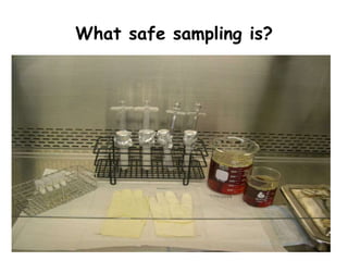
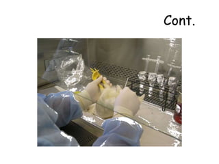
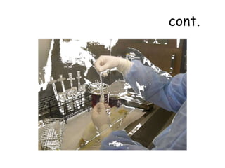
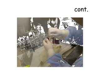
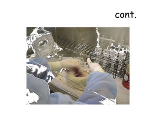
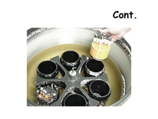
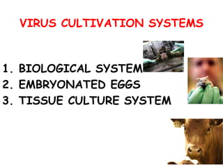
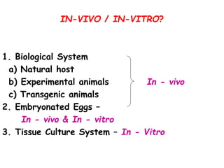
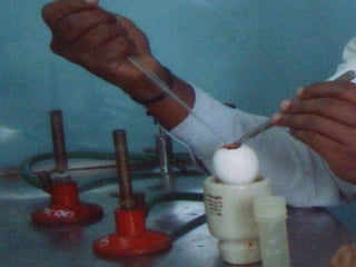
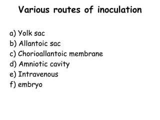
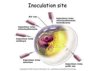
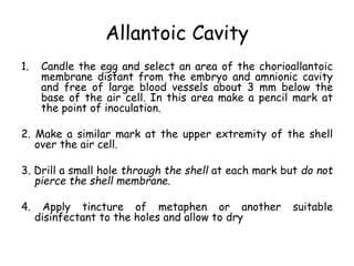
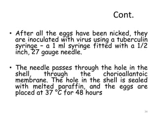
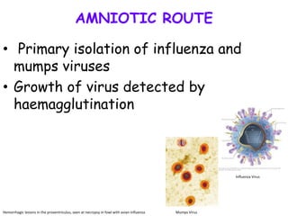
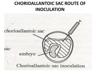
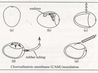
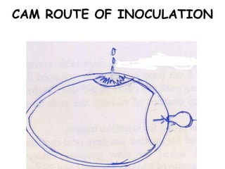
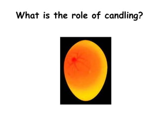
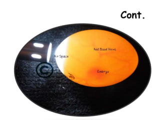
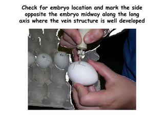
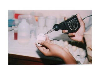
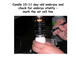
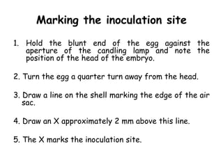
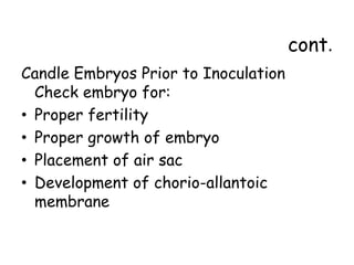
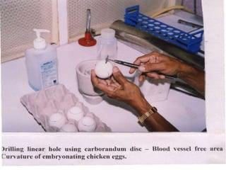
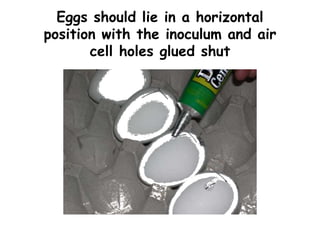
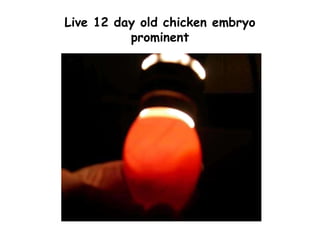
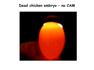
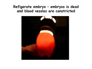
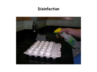
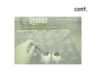
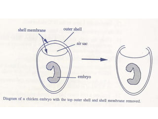
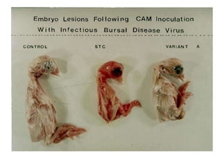
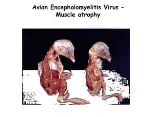
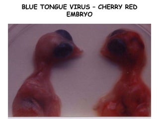
The document provides guidelines for virus isolation techniques using embryonated eggs, outlining various inoculation routes including allantoic, amniotic, chorioallantoic membrane and discussing advantages of different routes for isolating specific viruses. It describes the process of candling eggs to check embryo viability and mark inoculation sites prior to introducing viral samples via needle injection into selected areas. Proper egg storage, cleaning, and incubator maintenance are also covered.
Routes for inocculation of virus in an emryonated eggDr. Waqas Nawaz
The document describes a procedure for inoculating embryonated eggs with a virus to study viral growth. It provides details on the necessary materials, including 9-10 day old eggs, syringes, needles, inoculum, and incubators. The procedure involves candling the eggs to locate the embryo, puncturing the shell and injecting 0.1ml of viral suspension at a specific site. The eggs are then sealed and incubated. Successful inoculation is indicated by visible pocks on the eggs or embryo death. Advantages of this method include its wide use for viral isolation and growth, low cost, and sterile environment free from bacterial and viral contamination.
This document provides information on Brucella, the causative agent of brucellosis. It discusses the classification of Brucella as an alpha Proteobacteria. The main Brucella species that infect humans are B. melitensis, B. abortus, and B. suis, which are transmitted through contact with infected animals. The document outlines the morphology, culture characteristics, pathogenicity, diagnosis, treatment and prevention of Brucella infections. Brucellosis is a widespread zoonotic disease that can cause acute or chronic infections in humans.
Dr. S. Meenatchisundaram discusses poliovirus and poliomyelitis. Poliovirus is a picornavirus that causes the disease poliomyelitis. There are three types of poliovirus that can cause paralysis. The virus is transmitted via the fecal-oral route. Most infections are asymptomatic, but some cause minor illness and in rare cases paralytic poliomyelitis. Paralytic polio can affect the spinal cord and bulbar regions. Diagnosis involves culturing the virus from specimens. Vaccines including both inactivated and live attenuated versions are used to prevent the disease.
This document provides guidance on sampling and diagnosing various poultry respiratory diseases. It discusses the clinical signs, pathogens, and optimal samples for diagnosing diseases like avian influenza, Newcastle disease, infectious bronchitis, and mycoplasmosis. The document includes charts detailing the organ systems affected by each disease and the preferred diagnostic methods, such as PCR, virus isolation, or serology. It aims to help practitioners differentiate between diseases with similar respiratory signs and choose the appropriate diagnostic tests.
Newcastle Disease is a contagious and fatal viral disease affecting many avian species, especially poultry. It poses a major threat to the poultry industry in Egypt. The virus has different strains and pathotypes causing variations in disease severity and symptoms, ranging from respiratory to neurological signs. Diagnosis involves virus isolation, molecular techniques, and serology. Prevention and control relies on biosecurity measures and vaccination strategies, using live attenuated or inactivated vaccines, with mass vaccination programs aiming to produce protective antibodies in as many birds as possible to control outbreaks.
Monitor and Control of Vertically Transmitted Poultry DiseasesRafael Monleon
A presentation covering the monitor and control of common vertically transmitted diseases in poultry with concentration in chickens.
Presented at various locations including BioChek Seminar in Manila, Philippines in 2014 by Dr. Rafael Monleon
Contact me in LinkedIn for any question: www.linkedin.com/rafaelmonleon
This document discusses infectious bronchitis virus (IBV), a coronavirus that causes a highly contagious respiratory disease in chickens. IBV infects the respiratory tract, kidneys, intestines, and reproductive organs of chickens. It is transmitted through the air, feces, and fomites. Clinical signs include respiratory signs like sneezing as well as decreased egg production and thin-shelled misshapen eggs. Gross lesions include caseous plugs in the bronchi and thickened bronchial mucosa. Microscopic lesions involve the tracheal, kidney, and oviduct tissues. Diagnosis involves observing clinical signs and lesions as well as virus isolation, immunodetection assays, and inoculation of embryonated
This document discusses Avian Encephalomyelitis (AE) vaccination. It describes the importance of humoral immunity against AE and the aims of vaccination, which are to protect breeder flocks and their progeny. The two main types of vaccines discussed are live vaccines, which spread within flocks, and inactivated vaccines. Choosing the right vaccine involves ensuring the virus has not become adapted to growth in chicken embryos, which could cause clinical disease if administered via wing-web inoculation. Careful monitoring of inoculated embryos is important to detect and eliminate any adapted vaccine seed viruses.
This document discusses vaccination against infectious bursal disease virus (IBDV) in chickens. It covers hyperimmunizing breeder chickens to provide maternal antibodies to offspring, different types of IBD vaccines including live attenuated and inactivated vaccines. The document recommends choosing more invasive live vaccines when field viruses are very virulent and outlines factors to consider for optimal vaccination timing against IBDV. Characteristics of good live vaccines include early protection and minimal immunosuppression.
Pox diseases are caused by viruses in the family Poxviridae. Poxviruses that affect animals include sheep pox virus, goat pox virus, and vaccinia virus. Sheep pox and goat pox viruses cause significant disease in sheep and goats, characterized by fever and pox lesions on skin and mucous membranes. The diseases can spread rapidly between animals through direct contact or indirect contact with contaminated materials. Diagnosis involves identifying characteristic lesions and isolating virus from samples. Vaccines are available and used to control outbreaks.
COLLECTION AND TRANSPORT OF SPECIMEN LATEST.pptxSereneVarghese1
The document provides guidance on proper collection, transport, and storage of clinical specimens to ensure accurate microbiological testing and results. Key points include:
- Specimen quality directly impacts patient care, treatment decisions, and outcomes.
- Laboratories should reject poor quality specimens that could impact results.
- Proper collection, transport, and storage methods depend on specimen type to maintain organism viability until testing.
- Complete labeling and requisition forms are needed to properly identify and interpret specimen results.
This document provides information about a hot air oven and its uses. It discusses how hot air ovens work by providing temperatures higher than the atmosphere, typically between 50-250°C, and are used for rapid drying and sterilization. It also details how a hot air oven is used for the standard method of determining seed moisture content by precisely heating samples to eliminate water. Specifications are given for common crops dried in hot air ovens, including temperatures and drying times.
Yersinia pestis is the bacterium that causes bubonic and pneumonic plague in humans. It is typically transmitted via the bites of infected fleas. There have been three major pandemics of plague throughout history, including the infamous Black Death pandemic of the 14th century that killed approximately 50 million people in Europe. Y. pestis is a gram-negative rod that grows rapidly in blood and tissue fluids. It carries plasmids that are essential for virulence. After being bitten by an infected flea, the bacteria migrate to lymph nodes and multiply, causing swelling, necrosis, and sepsis if untreated. Diagnosis involves identifying the bacteria in smears, cultures, or antibodies. Streptomycin is the
This document provides information about gram positive bacilli (rods), including Bacillus species. It discusses their characteristics such as being aerobic or facultatively anaerobic, producing endospores, and being catalase-positive. Key pathogens mentioned are B. anthracis which causes anthrax, and B. cereus which can cause food poisoning. The document then focuses on B. anthracis and B. cereus, describing their microscopy, culture characteristics, biochemical tests for identification, and advanced detection techniques for B. anthracis.
This document discusses practical approaches for diagnosing viral diseases in poultry, including clinical diagnosis, rapid field diagnostic tests, serological diagnosis, molecular diagnosis, and isolation/characterization. Clinical diagnosis is based on case history, clinical signs, examination of live/dead birds, and gross lesions. Rapid field tests can detect viruses but require high viral titers. Serological tests detect antibodies but have delays. Molecular diagnosis using PCR technologies can sensitively and specifically detect pathogens. The document emphasizes that clinical signs alone are not confirmatory and that multiple diagnostic approaches should be used to accurately diagnose poultry viral diseases.
Mycobacterium avium subspecies paratuberculosis (MAP) is the causative agent of Johne's disease in ruminants. It is a gram-positive, acid-fast bacterium that survives in the environment and is resistant to heat and pasteurization. MAP has been detected in pasteurized milk and dairy products through contamination of raw milk from infected animals. This poses a potential risk to human health as MAP may play a role in Crohn's disease. Improved diagnostics, therapeutics, and management practices are needed to control MAP in animal populations and minimize risks to food safety.
Campylobacter is the leading cause of bacterial diarrhea in humans. It is commonly transmitted through contaminated or undercooked poultry, raw milk, or untreated water. Symptoms usually appear within 2 to 10 days and include diarrhea, fever, nausea, and abdominal pain. While most cases resolve on their own within a week, complications can occasionally occur such as Guillain-Barré syndrome. Proper food handling and cooking can help prevent Campylobacter infections.
This document discusses chronic respiratory disease (CRD) in chickens caused by Mycoplasma gallisepticum infection. CRD is a slow spreading upper and lower respiratory tract infection characterized by nasal discharge, coughing, and airsacculitis. M. gallisepticum is the causative agent, a small bacterium that transmits between birds through direct contact and fomites. Clinical signs include respiratory signs and decreased weight gain. At necropsy, lesions include excess mucus, airsacculitis, and consolidation of the lungs. Diagnosis involves isolating M. gallisepticum from tracheal samples. Control relies on maintaining mycoplasma-free flocks through biosecurity, antibiotic treatment,
Infectious bursal disease (IBD) is a highly contagious viral disease affecting young chickens. It is caused by infectious bursal disease virus (IBDV), which destroys lymphocytes in the bursa of Fabricius, impairing the immune system. Clinical signs include diarrhea, lethargy, and immunosuppression. At necropsy, the bursa appears swollen and hemorrhagic. Diagnosis relies on detecting viral antigens or nucleic acids in the bursa. Vaccination is the main control method, with live attenuated and in ovo vaccines available.
General virology 4 - Laboratory diagnosis, by Dr. Himanshu KhatriDrHimanshuKhatri
Laboratory diagnosis of viral infections can be performed through several methods: (1) demonstrating characteristic cytopathic effects of viruses in cell cultures, (2) directly detecting viruses in clinical samples through electron or fluorescence microscopy, (3) isolating and culturing viruses in animals, eggs, or various cell culture systems, and (4) detecting viral antibodies through serological tests like ELISA, Western blot, or hemagglutination inhibition tests. Tissue culture using various cell lines is now the most widely used method for virus isolation. Laboratory diagnosis helps confirm viral infections, screen blood donations, and conduct epidemiological and research studies.
Astroviruses are small, star-shaped RNA viruses that infect the intestinal tract of young birds. They are a common cause of runting-stunting syndrome in broilers and enteritis in guinea fowl. Six different types of avian astroviruses have been identified that infect chickens, turkeys, and ducks. Astroviruses are transmitted orally and cause mild intestinal signs but can also lead to more severe disease if other pathogens are present. Diagnosis is made through electron microscopy, ELISA, or PCR techniques. There is no vaccine or treatment available, so control relies on good hygiene and management practices.
This document discusses Vibrio cholerae, the bacteria that causes cholera. It provides details on the history, pathogenesis, clinical manifestations, diagnosis, and immunity aspects of cholera. Some key points include:
- V. cholerae causes the severe, acute diarrheal disease cholera and is transmitted through contaminated food or water.
- There have been several global pandemics of cholera since the 1800s. The causative agent was discovered by Pacini and Koch in the 1800s.
- V. cholerae secretes a toxin that increases cyclic AMP levels in intestinal cells, causing a massive outpouring of fluid in the stool. Colonization factors like pili and flagella
Cultivation of viruses uhf copy - copyheena thakur
This document discusses viruses and methods for cultivating viruses. It describes viruses as obligate intracellular parasites that can only multiply inside living host cells. The three main methods for cultivating viruses discussed are inoculation of viruses into animals, embryonated eggs, and tissue culture. For animal inoculation, mice are commonly used and viruses can be introduced via different routes. Embryonated eggs provide a suitable environment for virus growth and isolation. Tissue culture involves culturing cells or tissue fragments, and cell lines provide indefinite growth. Detection of virus growth involves monitoring for cytopathic effects, hemadsorption, interference, and other methods.
The Mc'Fadyean Reaction is a staining technique used to identify Anthrax bacillus under a microscope by staining the polypeptide capsular material around the bacilli that is composed of poly-d-Glutamic Acid. The procedure involves making an impression slide from an animal's ear, staining it with Poly Chrome Methylene Blue for 30 seconds, and examining under a microscope, where a positive sample will show a purplish capsule material surrounding the blue stained bacilli bodies, presumptively diagnosing anthrax in animals.
Marek's disease is a viral disease that affects young chickens. It is caused by a herpes virus spread through dander from infected birds. Clinical signs include lameness, paralysis, and tumors in lymph nodes and organs. The virus can survive for months in dander and litter. While there is no treatment, vaccination before 3 days of age can prevent tumors but not viral infection. Proper sanitation and isolation of infected birds are important for control.
Gacrux es la tercera estrella más brillante de la constelación de Crux y se encuentra a una distancia de 88 años luz de la Tierra. Es una gigante roja de tipo espectral M3.5 III con una temperatura de 3400 K y un radio 113 veces mayor que el del Sol. La luz que vemos de Gacrux en 2014 fue emitida en 1926, cuando ocurrieron dos eventos históricos notables: el cruce del Atlántico por parte de un hidroavión español y la creación del primer robot electrónico por parte de Westinghouse
This document provides an overview of the evolution of cryptographic protocols used in electronic passports (ePassports) over three generations from 2004 to 2008. It summarizes the technologies used in ePassports including biometrics, RFID, and public key infrastructure. It then analyzes the cryptographic protocols of the first generation ePassport specifications from 2004, the Extended Access Control (EAC) specifications from 2006 for the second generation, and the EAC v2.1 specifications from 2008 for the third generation. It also discusses some vulnerabilities that remained even with the third generation specifications.
El documento explica cómo crear y ejecutar scripts y funciones en MATLAB. Brevemente describe cómo crear un script simple "Hola Mundo" y ejecutarlo escribiendo su nombre en la ventana de comandos. También cubre conceptos básicos como variables, operaciones matemáticas, sentencias condicionales if/else y switch, y algunos ejemplos de código.
This document discusses ancillary tasks but provides little context or information to summarize in 3 sentences or less. It mentions the author Fatima Iftikhar and the title "Ancillary Tasks" but does not contain enough content to create an informative summary.
La Teoría de las Restricciones se basa en que los procesos solo avanzan a la velocidad del paso más lento, llamado cuello de botella. Los cuellos de botella incrementan los tiempos de espera y reducen la productividad, lo que genera un aumento de los costos. Para solucionarlos, se debe identificar la restricción, explotar su capacidad al máximo, y subordinar todos los demás procesos a su ritmo. Al elevar la restricción, se vuelve a identificar la nueva restricción y se repite el pro
El documento habla sobre el origen y evolución de los memes. Explica que los memes son unidades de información que se replican de persona a persona a través de la imitación, al igual que los genes se replican biológicamente. También describe cómo los memes han permitido la supervivencia de la humanidad a lo largo de la historia y cómo en la actualidad se han modernizado y proliferado en internet a través de las redes sociales y los medios digitales.
The document proposes a CAS week plan involving daily hikes in Hong Kong country parks to raise funds for Mother's Choice. Each day's hike would be in a different park, such as Tai Tam Country Park, Aberdeen Country Park, Shek O Country Park, Pok Fu Lam Country Park, and Lion Rock Country Park. Students would get sponsors to pledge money for each hike, with funds raised donated to Mother's Choice to support programs like drop-in centers or providing milk for babies. The hikes would highlight health, environment, and support the charity through fundraising, benefiting both students' CAS hours and Mother's Choice.
WhatsApp es una aplicación gratuita de mensajería instantánea para teléfonos inteligentes que permite el envío de mensajes de texto, imágenes, videos y audio entre usuarios. En 2016 tenía más de 1,000 millones de usuarios a nivel mundial. Facebook compró WhatsApp en 2014 por 19,000 millones de dólares y completó la compra en 2015 por 21,800 millones, después de lo cual WhatsApp comenzó a ofrecer llamadas de video.
Партизанский Маркетинг — малобюджетные способы рекламы и маркетинга, позволяющие эффективно продвигать свой товар или услугу, привлекать новых клиентов и увеличивать свою прибыль, не вкладывая или почти не вкладывая денег.
В начале было… Ничего
- 1 альбом сомнительного качества
- Группа ВК 300 человек
- Раздача на Rutracker 150 скачиваний
Короче смотрите, лайкайте и задавайте вопросы в комментариях. Самые интересные вопросы попадут лично (!) к Карателю на стол
Ну и подписывайтесь на страницу БАУ в ВК
http://vk.com/pray_for_your_anus
Is bitcoin a positive promise for societyRicky Sharma
The document discusses the evolution of currency and different forms of modern currency. It then analyzes properties of Bitcoin and traditional currency, noting that while Bitcoin aimed to provide an alternative, it exhibits flaws that do not meet ideal currency expectations. Specifically, Bitcoin is limited in supply, vulnerable to deflation, does not enable spending, and its value is based on USD. However, Bitcoin has paved the way for positive future innovations in currency technology.
Viruses are Different From Other Microbes
Viruses are obligate intracellular parasites. They depend totally on their host cells for their existence. Their total host dependence makes it, extremely difficult to get good insight of them natural conditions, because the internal characteristics of the host cells are likely to interfere with the observations. Due to these reasons, it has been found desirable that viruses are cultivated or grown in the laboratory itself.
Laboratory animals
Fertilized Hen’s Egg
Chorioallantoic membrane
Allantoic cavity
Amniotic cavity
Yolk sac
Organ/Tissue/Cell Culture
Growth identified by serological method like neutralization.
Embryonated Egg Chorioallantioc membrane (CAM)
Allantoic cavity
Amniotic cavity
Yolk Sac
Cell Lines/ Tissue cultures Primary
Diploid/ Secondary
Continuous
Animal inoculation Suckling
Embryonated Hen’s Egg
Cultivation of Viruses and Bacteria
Chorioallantoic membrane (CAM) – visible lesions called pocks. Each infectious virus particle forms one pock. e.g. Variola, Vaccinia virus
Allantoic cavity – Influenza virus (vaccine production) & paramyxoviruses
Amniotic cavity – primary isolation of Influenza virus
Yolk sac – Chlamydia, Rickettsia & some viruses
Embryonated eggs:
The Embryonated hen’s egg was first used for cultivation of viruses by Good Pasteur and Burnet (1931). Cultivation of viruses in organized tissues like chick embryo necessitates a different type of approach.. For all practical purposes they all themselves behave as tissue cultures. The process of cultivation of viruses in embryonated eggs depends on the type of egg which is used. The egg used for cultivation must be sterile and the shell should be intact and healthy.
F.M. Burnet in the laboratory
in the early 1950's,
was experimenting
on influenza virus genetics,
using the developing hen's egg
Inoculated eggs are candled
daily to see the chicken
embryos inside.
Animals and chick embryo
were the first method that was used
to cultivate virus. This method is rarely
used as it is not convenient.
However, when preparing for bulk virus,
(e.g. antigen or vaccine production)
the usage of chick embryo is useful.
Fertile chicken eggs provide
a convenient, space-saving incubator
for many kinds of animal viruses.
Different viruses can be injected
into an egg at different sites and
the egg can be easily observed
for viral replication throughout
the development of the chicken embryo.
Isolation and cultivation of many avian
Virus culture techniques document outlines various methods for culturing viruses in the laboratory. It discusses how viruses differ from other microbes in that they are obligate intracellular parasites that cannot replicate outside of host cells. Three main virus culture methods are described: inoculation of laboratory animals, cultivation in embryonated eggs, and cell/tissue culture. Culturing viruses allows them to be isolated, identified, and studied. While animal inoculation was historically used, embryonated eggs and cell/tissue culture are now more common due to their lower cost and greater ease of use. The document provides details on techniques for culturing viruses in embryonated eggs, including candling, inoculation site marking, and exposing the chorioallanto
Egg inoculation by Chinithung ngullie (2)Cyrus Ngullie
This presentation has been made easy to make understand to the students by emphasizing more on the visual than the words.I hope it is helpful to the students and followers as well.
VIRAL VACCINES
Since viruses are intracellular parasites they will grow only within other living cells.
Methods of viral vaccine production:
Cultivation of virus using free living animals
Fertile eggs
Tissue cultures
The document discusses the cultivation of viruses using embryonated eggs. It notes that embryonated eggs are a simpler technique than animal inoculation for growing viruses, as eggs do not have an immune response and contain suitable cells for virus growth. Viruses can be inoculated into different areas of embryonated eggs, including the chorioallantoic membrane, allantoic cavity, amniotic sac, and yolk sac. The allantoic cavity is most commonly used due to its simplicity and ability to produce large quantities of viruses like influenza. Embryonated eggs remain an important method for growing stocks of viruses for research and vaccine production.
Embryonated chicken eggs provide a suitable environment for cultivating viruses due to the sterile conditions and availability of embryonic tissues that support viral growth. Goodpasture and Burnet first used embryonated hen eggs in 1931 to cultivate viruses. Different types of eggs can be used depending on the virus, with chicken, duck, and turkey eggs being most common. The embryo offers sites like the chorioallantois membrane, amniotic cavity, allantois cavity, and yolk sac for virus inoculation and growth. Detection of viral growth may involve observing embryo death, defects, or opaque spots on membranes.
This document discusses viral inoculation methods using embryonated eggs. It provides advantages of using eggs such as their ability to isolate and cultivate avian and some mammalian viruses. It also discusses important considerations for the source of eggs including using specific pathogen free flocks to minimize risks. Different routes for inoculating viruses into eggs are described, including allantoic sac, chorioallantoic membrane, and yolk sac routes. Tools, procedures, and incubation conditions for successful egg inoculation are outlined.
The document discusses methods for isolating and cultivating viruses. It describes how viruses require living host cells to replicate and explains common host methods like cell culture, embryonated eggs, and laboratory animals. It provides details on techniques for cultivating viruses in each host method, including primary steps like dissociating tissue, inoculation procedures, and isolating harvested viruses. The advantages and disadvantages of each cultivation method are also summarized.
Viruses are obligate intracellular parasites that can only replicate inside living host cells. There are several methods for cultivating or growing viruses, including inoculation into animals, embryonated eggs, and cell/tissue culture. Inoculation into animals allows study of viral replication and disease but is expensive and difficult. Embryonated eggs are widely used as they are inexpensive and support growth of many viruses. Tissue/cell culture has replaced eggs for many viruses as it is more convenient and sensitive. Various cell types, including primary cultures, continuous cell lines, and explant cultures can support viral growth. Detection of viral growth involves monitoring for cytopathic effects, hemadsorption, interference, transformation, or metabolic inhibition.
This document discusses methods for cultivating and isolating viruses, including animal inoculation, embryonated hen egg inoculation, and primary cell culture. Viruses can be grown in animal tissues, eggs, or cell cultures. Eggs provide routes of inoculation like the allantoic cavity, amniotic cavity, and chorioallantoic membrane. Primary cell cultures are made from chicken embryos. Cell lines like vero and HeLa support viral growth and replication. Detection of viral growth involves monitoring for cytopathic effects, interference, or detecting viral antigens.
Viruses can only reproduce inside host cells and are obligate intracellular parasites. There are several approaches to studying viruses including inoculation into animals or embryonated eggs, and culturing viruses in cells. Tissue culture involves growing cells in vitro in growth medium, then inoculating them with viruses. There are different types of cell cultures including primary cultures from tissues that can only grow for a limited time, diploid cell lines that can be subcultured 50 times, and continuous cell lines from cancer cells that can be cultured indefinitely. Viruses infect and replicate within cells, sometimes causing cell lysis and formation of plaques that can be observed. Cytopathic effects indicate viral growth. Tissue culture allows broad virus cultivation
This document discusses techniques for cultivating viruses. It explains that viruses require living host cells to replicate and describes three main cultivation methods: animal inoculation, embryonated egg inoculation, and tissue culture. Animal inoculation involves infecting animals like mice to isolate and study viruses, but it is expensive and raises welfare issues. Embryonated egg inoculation is commonly used to grow viruses by inoculating eggs at specific sites, and it is cost-effective but each virus grows at different sites. Tissue culture uses cell monolayers and is versatile but requires specialized facilities and technicians. The document provides details on each technique's advantages, disadvantages and applications.
This document discusses the genus Campylobacter, including its general characteristics, habitats, important veterinary pathogens, clinical infections, and diagnostic procedures. Campylobacter species are Gram-negative, microaerophilic, curved rods. Important pathogens include C. fetus subspecies venerealis (bovine genital campylobacteriosis), C. fetus subspecies fetus and C. jejuni (ovine abortion), and C. jejuni (intestinal infections in dogs, avian vibrionic hepatitis, and human intestinal campylobacteriosis). Diagnosis involves isolation and identification of the bacteria from clinical specimens using selective media and microaerophilic conditions.
This document discusses various methods for cultivating and detecting viruses. It begins by describing three primary purposes of virus cultivation: to isolate viruses from clinical samples, conduct research on viral structure and effects on host cells, and produce viruses for vaccines. It then explains three main cultivation methods: animal inoculation using mice or monkeys, embryonated egg inoculation, and tissue culture using primary cells or continuous cell lines. Detection methods for viruses in cell culture include observing cytopathic effects, hemadsorption, metabolic inhibition, interference, and immunofluorescence.
In-Vitro Pollination and Fertilization
The document discusses in-vitro pollination and fertilization techniques. It begins with a brief history, noting its development in 1902 and use to produce hybrids between incompatible species. It then describes barriers to pollination and fertilization that can be overcome through in-vitro methods. Several techniques are outlined, including ovule, ovary, and stigma cultures. Requirements for successful in-vitro fertilization include viable gametes and proper culture conditions. The document concludes by discussing applications in plant breeding like overcoming self-incompatibility and producing stress-tolerant hybrids.
In vitro pollination involves pollinating pistils or ovules that have been cultured in a nutrient medium such as Nitsch's medium. This technique can help overcome pre-fertilization barriers to hybridization between plant species. Key steps include sterilizing flower parts, collecting pollen, and applying pollen to excised pistils, ovaries, ovules, or stigmas depending on the method. Factors like culture medium, temperature, genotype, and physiological state of the explant can influence seed set. In vitro pollination has applications in plant breeding like overcoming self-incompatibility or cross-incompatibility barriers and producing haploid plants or hybrids.
The document discusses the use of embryonated hen's eggs for cultivating viruses. Some key points:
- Embryonated eggs provide a sterile environment that supports viral growth and allows isolation, stock maintenance, and vaccine production.
- Different routes can be used to inoculate viruses into eggs, including the chorioallantoic membrane, amniotic cavity, allantoic cavity, and yolk sac, depending on the virus.
- After inoculation, viral growth is detected through signs like embryo death, developmental defects, or opaque spots on membranes. Fluids and tissues can also be examined microscopically or tested for viral reactions.
Omphalitis in Poultry - Inflammation of Yolk SacOmkar Phadtare
Omphalitis is a bacterial infection of the navel and yolk sac that occurs in young chickens, turkeys, and ducks within 1-3 days of hatching. The infection is caused by opportunistic bacteria entering the navel due to poor hygiene practices during egg production and incubation. Affected birds experience diarrhea, closed eyes, and a swollen abdomen. Upon examination, their yolk sacs appear enlarged and congested with abnormal contents. While antibiotics can be used, mortality is often high and prevention focuses on clean nests, frequent egg collection, and strict hygiene during incubation and hatching.
This document discusses three methods for isolating and cultivating animal viruses: embryonated chicken eggs, tissue cultures, and laboratory animals. Embryonated chicken eggs are commonly used to cultivate viruses by inoculating the chorioallantoic membrane, allantoic cavity, amniotic sac, or yolk. Tissue cultures involve growing cells from animal tissues in glass or plastic; primary cell cultures have limited divisions while continuous cell lines can divide indefinitely. Laboratory animals like primates, mice, and rabbits are also used to cultivate some viruses that cannot be grown by other methods.
Viruses are obligate intracellular parasites which means they can only grow or reproduce inside a host cell.
The primary purpose of virus cultivation:
To isolate and identify viruses in clinical samples.
To do research on the viral structure, replication, genetics, and effects on the host cell.
To prepare viruses for vaccine production.
Isolation of the virus is always considered a gold standard for establishing the viral origin of the disease
topics covered
CULTIVATION OF VIRUSES
Animal inoculation
Embryonated eggs
CAM
Allantoic cavity
Amniotic cavity
Yolk sac
Tissue culture
Organ culture
Explant culture
Cell culture
Primary cell culture
diploid cell culture
Continues cell lines
Thinking of getting a dog? Be aware that breeds like Pit Bulls, Rottweilers, and German Shepherds can be loyal and dangerous. Proper training and socialization are crucial to preventing aggressive behaviors. Ensure safety by understanding their needs and always supervising interactions. Stay safe, and enjoy your furry friends!
A workshop hosted by the South African Journal of Science aimed at postgraduate students and early career researchers with little or no experience in writing and publishing journal articles.
How to Build a Module in Odoo 17 Using the Scaffold MethodCeline George
Odoo provides an option for creating a module by using a single line command. By using this command the user can make a whole structure of a module. It is very easy for a beginner to make a module. There is no need to make each file manually. This slide will show how to create a module using the scaffold method.
How to Manage Your Lost Opportunities in Odoo 17 CRMCeline George
Odoo 17 CRM allows us to track why we lose sales opportunities with "Lost Reasons." This helps analyze our sales process and identify areas for improvement. Here's how to configure lost reasons in Odoo 17 CRM
Executive Directors Chat Leveraging AI for Diversity, Equity, and InclusionTechSoup
Let’s explore the intersection of technology and equity in the final session of our DEI series. Discover how AI tools, like ChatGPT, can be used to support and enhance your nonprofit's DEI initiatives. Participants will gain insights into practical AI applications and get tips for leveraging technology to advance their DEI goals.
Main Java[All of the Base Concepts}.docxadhitya5119
This is part 1 of my Java Learning Journey. This Contains Custom methods, classes, constructors, packages, multithreading , try- catch block, finally block and more.
How to Fix the Import Error in the Odoo 17Celine George
An import error occurs when a program fails to import a module or library, disrupting its execution. In languages like Python, this issue arises when the specified module cannot be found or accessed, hindering the program's functionality. Resolving import errors is crucial for maintaining smooth software operation and uninterrupted development processes.
How to Add Chatter in the odoo 17 ERP ModuleCeline George
In Odoo, the chatter is like a chat tool that helps you work together on records. You can leave notes and track things, making it easier to talk with your team and partners. Inside chatter, all communication history, activity, and changes will be displayed.
A review of the growth of the Israel Genealogy Research Association Database Collection for the last 12 months. Our collection is now passed the 3 million mark and still growing. See which archives have contributed the most. See the different types of records we have, and which years have had records added. You can also see what we have for the future.
বাংলাদেশের অর্থনৈতিক সমীক্ষা ২০২৪ [Bangladesh Economic Review 2024 Bangla.pdf] কম্পিউটার , ট্যাব ও স্মার্ট ফোন ভার্সন সহ সম্পূর্ণ বাংলা ই-বুক বা pdf বই " সুচিপত্র ...বুকমার্ক মেনু 🔖 ও হাইপার লিংক মেনু 📝👆 যুক্ত ..
আমাদের সবার জন্য খুব খুব গুরুত্বপূর্ণ একটি বই ..বিসিএস, ব্যাংক, ইউনিভার্সিটি ভর্তি ও যে কোন প্রতিযোগিতা মূলক পরীক্ষার জন্য এর খুব ইম্পরট্যান্ট একটি বিষয় ...তাছাড়া বাংলাদেশের সাম্প্রতিক যে কোন ডাটা বা তথ্য এই বইতে পাবেন ...
তাই একজন নাগরিক হিসাবে এই তথ্য গুলো আপনার জানা প্রয়োজন ...।
বিসিএস ও ব্যাংক এর লিখিত পরীক্ষা ...+এছাড়া মাধ্যমিক ও উচ্চমাধ্যমিকের স্টুডেন্টদের জন্য অনেক কাজে আসবে ...
हिंदी वर्णमाला पीपीटी, hindi alphabet PPT presentation, hindi varnamala PPT, Hindi Varnamala pdf, हिंदी स्वर, हिंदी व्यंजन, sikhiye hindi varnmala, dr. mulla adam ali, hindi language and literature, hindi alphabet with drawing, hindi alphabet pdf, hindi varnamala for childrens, hindi language, hindi varnamala practice for kids, https://www.drmullaadamali.com
Physiology and chemistry of skin and pigmentation, hairs, scalp, lips and nail, Cleansing cream, Lotions, Face powders, Face packs, Lipsticks, Bath products, soaps and baby product,
Preparation and standardization of the following : Tonic, Bleaches, Dentifrices and Mouth washes & Tooth Pastes, Cosmetics for Nails.
This presentation includes basic of PCOS their pathology and treatment and also Ayurveda correlation of PCOS and Ayurvedic line of treatment mentioned in classics.
13. Processing Tissues for Isolation of
AIV and APMV
• Diluents Prepare 10% suspension in
antibiotic
• Centrifuge at 1,500 x g – 20 minutes
• and place in sterile vial Remove
supernatant with pipette
• Temperature Incubate 1 hour at room
20. IN-VIVO / IN-VITRO?
1. Biological System
a) Natural host
b) Experimental animals In - vivo
c) Transgenic animals
2. Embryonated Eggs –
In - vivo & In - vitro
3. Tissue Culture System – In - Vitro
22. EMBRYONATED EGGS
ADVANTAGES
• Isolation and cultivation of many avian
and few mammalian viruses
• Ideal receptacle for virus to grow
• Sterile & wide range of tissues and
fluids
23. • Cost- much less
• Maintenance-easier
• Less labour
• Readily available
24. • Free from bacteria and many
latent viruses.
• Free from specific and non
specific factors of defense.
• Sensitive to viruses which do not
produce infection in adult birds.
26. Why Egg?
The avian embryo, especially the chicken embryo, is a valuable and
widely used medium for the initial isolation and subsequent
passage of many viruses for stock cultures and the production of
vaccines. Chicken embryos are used almost exclusively because of
their.
(1) Availability
(2) Economy
(3) Convenient size
(4) Relative freedom from latent infection and extraneous
Contamination, and
(5) Lack of production of antibodies against the viral inoculums.
Eggs only from healthy, disease-free flocks should be used. It is
desirable to have one source of supply for reasons of uniformity
of production and management of the breeder flock.
33. Allantoic Cavity
1. Candle the egg and select an area of the chorioallantoic
membrane distant from the embryo and amnionic cavity
and free of large blood vessels about 3 mm below the
base of the air cell. In this area make a pencil mark at
the point of inoculation.
2. Make a similar mark at the upper extremity of the shell
over the air cell.
3. Drill a small hole through the shell at each mark but do not
pierce the shell membrane.
4. Apply tincture of metaphen or another suitable
disinfectant to the holes and allow to dry
34. 34
Cont.
• After all the eggs have been nicked, they
are inoculated with virus using a tuberculin
syringe – a 1 ml syringe fitted with a 1/2
inch, 27 gauge needle.
• The needle passes through the hole in the
shell, through the chorioallantoic
membrane. The hole in the shell is sealed
with melted paraffin, and the eggs are
placed at 37 °C for 48 hours
36. ALLANTOIC ROUTE
• Most popular
• Most of avian viruses
• High titered virus
• Simple technique
37. AMNIOTIC ROUTE
• Primary isolation of influenza and
mumps viruses
• Growth of virus detected by
haemagglutination
Hemorrhagic lesions in the proventriculus, seen at necropsy in fowl with avian influenza
Influenza Virus
Mumps Virus
38. CHORIOALLANTOIC
MEMBRANE
It is inoculation employs 10- to 12-day-old
embryos and inoculum of 0.1-0.5 cc. This
route is particularly effective for primary
isolation and cultivation ofthe viruses of
vaccinia, variola, fowl pox, laryngo
tracheitis of chickens, and pseudo rabies
which produce easily visible foci or
"pocks." The chorioallantoic membrane is a
suitable site for study ofthe development
of pathologic alterations and inclusion
bodies, and titration of viruses by the pock
counting technic.
42. Tips
• Pox and Herpes viruses.
• ‘Pock Lesions’
• Suitable for plaque studies
43. YOLK SAC inoculation
YOLK SAC inoculation is performed
with 5- to 8-day-old embryos and
inoculum of 0.2-1.0 cc. This route may
be used for initial isolation of mumps
virus
45. YOLK SAC ROUTE
Advantages
•Simplest method
•Mostly mammalian viruses
•Immune interference for most of avian
viruses
Disadvantages
•Not suited for avian viruses
46. Intracerebral inoculation
Intracerebral inoculation can be performed with
8- to 14-day-old embryos and inoculum of 0.01-
0.02 cc. This route may be employed in studies of
pathologic alterations of the brain following
infection. The viruses of herpes simplex and
rabies may be cultivated by this route.
Embryos are incubated after inoculation for a
period appropriate for the virus employed
andthey are examined at least once daily. Death
of the embryo within the first 24 hours after
inoculation is generally considered to be due to
nonspecific causes such as trauma. Some viruses.
47. Cont.
kill all embryos and mortality is the criterion of
infection. Newcastle disease virus is an example in which
embryos are killed in two to four days depending upon
the strain of the virus. With some viruses such as
influenza virus the mortality rate varies on initial
passage but may increase with subsequent passage.
The criterion of infection with herpes and pox viruses is
the formation of pock lesions on the chorioallantoic
membrane. Other gross pathologic manifestations of
infection of the embryo may be curling and dwarfing of
the embryo, fibrosis of the amnionic membrane, edema
of the chorioallantoic membrane, and urates in the
kidney and mesonephros such as produced by avian
coronaviruses on initial and low passage in the embryo.
48. Cont.
Various types of cytologic changes,
including inclusion bodies with certain
viruses, may be detected by microscopy.
The embryo should be examined soon after
death so that postmortem changes do not
obscure any specific pathologic alterations.
Chilling of the embryos for several hours
or for overnight before collection of
extraembryonic fluids is recommended to
reduce hemorrhage into the fluids.
49. Intravenous inoculation
Intravenous inoculation does not have wide
practical application for study of
experimental infections of the avian
embryo.The procedure is generally
employed for hematologic studies. Embryos
of 10- to 15-days incubation are most
suitable for this route. The amount
ofinoculum may vary from 0.02 to 0.05 cc.
51. INTRAVENOUS ROUTE
• Blue tongue virus
• Cherry red embryo
• Highly cumbersome
• Most sophisticated procedure
52. Student Task
1. Why we select egg for cultivation of viral inoculums?
2. How can we detect the viability of embryo while candling?
3. What is the relationship b/n embryo inoculation site and
viral inoculums?
4.The best selective site for pox viral inoculum is m/m how it
could be?
5. White egg is selective for embryo inoculation technique
than other colored egg, why this is so?
53. Student Task
Q) What is your base on site selection ?
Q) What is the effect of maternal immunity on
injected inoculums?
Q) What types of viral samples selectively
inoculated in yolk sac?
Q) If u encountered death of embryo within
24hr after inoculation of fertile egg ,it is
indicative of ____?
54. How to select Needle ?
25 23 gauge 1
2222 gauge
25 gauge
22 gauge
1½” needle
55. Candling
Introduction
Candling is the process of holding a strong light above or below the egg to
observe the embryo. A candling lamp consists of a strong electric bulb
covered by a plastic or aluminum container that has a handle and an
aperture. The egg is placed against this aperture and illuminated by the
light. If you do not have a candling lamp, improvise.
60. Storage and cleaning of eggs
• Do not buy dirty eggs.
• Eggs that are stained can be disinfected by washing in a
warm (37°C) solution of 0.1 percent Chloramin B (benzine
sulfonamide sodium salt) or wiped with a 70 percent
alcohol solution.
• Fertile eggs that have not been incubated can be
purchased. They can then be placed in an incubator when
they are delivered. Alternatively, they can be stored for
several days in cool conditions (16°C to 18°C) prior to
incubation. This may reduce the number of viable embryos,
as some embryos may not develop after storage.
61. Check for embryo location and mark the side
opposite the embryo midway along the long
axis where the vein structure is well developed
62.
63. Candle 10-11 day-old embryos and
check for embryo vitality –
mark the air cell line
64. Marking the inoculation site
1. Hold the blunt end of the egg against the
aperture of the candling lamp and note the
position of the head of the embryo.
2. Turn the egg a quarter turn away from the head.
3. Draw a line on the shell marking the edge of the air
sac.
4. Draw an X approximately 2 mm above this line.
5. The X marks the inoculation site.
65. Cont.
Note:
In some eggs the air sac will have not
developed on the blunt end but half way
down the egg. These eggs are not suitable
for vaccine production. They can be used
for inoculation during routine titrations to
establish infectivity titres.
66. cont.
Candle Embryos Prior to Inoculation
Check embryo for:
• Proper fertility
• Proper growth of embryo
• Placement of air sac
• Development of chorio-allantoic
membrane
69. Eggs should lie in a horizontal
position with the inoculum and air
cell holes glued shut
70. Cleaning and decontamination of
incubators
Keep surfaces clean by wiping out with a
wet cloth and disinfecting with 70
percent alcohol solution or a non-
corrosive disinfectant.
71. Incubation of eggs after
inoculation
Inoculated eggs contain virus and should
be placed in a different incubator. Eggs
inoculated with virulent strains of
Newcastle disease virus should not be
incubated in the same incubator as used
for eggs inoculated with the avirulent I-2
strain of Newcastle disease virus.
Inoculated eggs are incubated under the
same conditions as uninoculated eggs but
do NOT turn the eggs.
73. Tips
Humidity should be maintained at 60 to 65
percent. A tray filled with water and
placed in the bottom of the incubator is
usually sufficient to maintain this level of
humidity.
Place the eggs in the incubator with the air
sac on top.
Eggs should be turned three times a day.
75. Determining the viability of the
embryo
Under the candling lamp, the embryo appears as a dark
shadow with the head as a dark spot. Healthy embryos will
respond to the light by moving Sometimes the movement is
very sluggish and it can take 30 to 40 seconds for the
embryo to move when held under the candling lamp. This
indicates the embryo is not healthy and the egg should be
discarded.
Look carefully at the blood vessels. They are well defined
in a healthy embryo. After an embryo has died, the blood
vessels start to break down. They then appear as streaks
under the shell when viewed under the candling lamp.
Candling will also reveal cracks in the eggshells. Eggs with
cracked shells should be discarded.
76. Tips
Infertile eggs: These are easy to detect, as the egg is clear.
Discard
Deaths: The embryo has developed for several days and then
died. Candling will reveal a small dark area and disrupted blood
vessels. Often deteriorating blood vessels will appear as a dark
ring around the egg. Discard.
Late Deaths: These are often difficult to tell apart from a
viable embryo at the same stage of development. Look for the
absence of movement and the breakdown of the blood vessels.
Discard
Viable Embryos: These move in response to the light and have
well defined blood vessels. Mark the air sac and the inoculation
site and then return the eggs to the incubator ready for
inoculation.
81. Equipment Needed for
Harvesting
AAF from Dead Eggs
2 embryos/student
2 Forceps
Iodine box for forceps
5 ml pipettes and
pipette aid
Gloves
Blood Agar plate
Plastic loops
82. Cont.
• Snap cap tube with
• labels
• Ethanol spray bottle
• Iodine bucket for
• pipettes
• Discard bucket with
• bags for embryos
• Plastic bags to discard
• flats
86. Harvesting AAF from Dead
Embryos
1. Disinfect egg shell surface one time in the
BSC
2. 2. Only open eggs from a single specimen at
3. Open egg from air cell end with forceps
4. Break allantoic sac with sterile forceps
pipette tip with forceps
5. Hold membranes and embryo away from
6. Harvest AAF
7. Streak BA plate
8. Centrifuge 1,500 x g for 15 min.
87. Harvesting AAF for Live Embryos
constrict chorioallantoic vessels
1. Refrigerate embryo to kill embryo and
2. Disinfect shell
3. Drill small hole above air cell line
4. Aspirate AAF with 3cc 22 gauge 1½” syringe
will bind with virus
5. Discard AAF with red blood cells – RBC
90. Harvesting and storage of allantoic fluid
Introduction
Allantoic fluid from inoculated eggs will be harvested for three
reasons.
1. To prepare I-2 Newcastle disease working seed or vaccine.
2. To use as antigen in the haemagglutination inhibition test.
3. To be tested for the presence or absence of Newcastle disease
virus by the haemagglutination test. These results are then used
to calculate the infectivity titre of a suspension of virus.
Note
Maternal antibody is confined to the yolk sac until about 14
days of incubation and then enters the embryo. Virus
harvested in allantoic fluid at 14 days will not have been
exposed to antibody.
92. 92
Cont.
• During the incubation period, the virus replicates in
the cells that make up the chorioallantoic membrane.
As new virus particles are produced by budding, they
are released into the allantoic fluid. To harvest the
virus, the top of the egg shell – the part covering the
air sac – is removed.
• The shell membrane and chorioallantoic membrane are
pierced with a pipette which is then used to remove
the allantoic fluid – about 10 ml per egg.
93. Harvesting allantoic fluid to prepare
working seed, vaccine or antigen
Materials
Forceps or a small pair of scissors
Absolute alcohol for flaming forceps
Cotton wool
70 percent alcohol solution in water
Discard tray
50 mL micropipette and tips or a wire loop
Sterile Pasteur pipettes with short blunt ends
96 well microwell plate
10 percent washed red blood cells
Sterile containers for receiving the harvested
allantoic fluid.
94. Method
1. Chill eggs at 4°C for at least two hours to kill the embryo and to reduce the
contamination of the allantoic fluid with blood during harvesting.
2. Remove stationery tape (if used to seal the eggs) and swab each egg with cotton
wool soaked with 70 percent alcohol to disinfect and remove condensation from
the shells.
3. Dip the forceps or scissors in disinfectant OR if using a Bunsen burner, dip the
forceps or scissors in absolute alcohol and flame to sterilize. Remove the
eggshell above the air space.
4. Discard embryos that are visibly contaminated.
5. Remove a sample of allantoic fluid from each egg. Use a micropipette and sterile
tip, sterile glass pipette or a flamed loop. Test each sample for the presence of
Newcastle disease virus by the haemagglutination (HA) test.
6. Discard embryos that do not test HA positive for Newcastle disease virus.
7. Use sterile glass Pasteur pipettes to harvest the allantoic fluid from the eggs.
The pipettes can be either hand held or used unplugged and connected to a
vacuum pump. Collect the fluid into sterile containers.
95. Preliminary Quality Control
This step involves the inoculation of a general purpose broth
culture.
1. Test each container for bacterial contamination by
inoculating tryptic soy broth with test samples and
incubation at 37°C overnight.
2. Centrifuge the samples of allantoic fluid or stand
overnight at 4°C to allow particles including red blood cells
to settle. The allantoic fluid should appear clear after
centrifugation or standing overnight.
3. After 24 hours, read the results of the tests for bacterial
contamination.
4. Use aseptic technique to transfer the clear allantoic fluid
supernatant from containers that showed no bacterial
growth into a sterile container for storage. This step pools
the fluid and ensures homogeneity.
96. Storage of allantoic fluid
The optimum temperature for storage of allantoic fluid
containing live Newcastle disease virus is -70°C. Storage
at -20°C is not as effective and the infectivity titre will
slowly decrease. The action of the freezing and thawing
also decreases the infectivity titre of the virus. Allantoic
fluid containing Newcastle disease virus has been stored
for up to 6 weeks at 4°C without significant loss of titre.
Allantoic fluid will be stored for two purposes.
1. Antigen for use in the haemagglutination inhibition test.
Prepare 1 mL aliquots of undiluted allantoic fluid in vials.
2. Preparation of vaccine.
97. Storage of allantoic fluid at 4°C for use as a
wet vaccine
Diluents containing a stabilizing agent are used in the
preparation of wet Newcastle disease vaccine. Suitable
stabilizing agents are gelatin and skim milk powder.
Diluents containing 2 percent gelatin solution or 8 percent
skim milk powder in phosphate buffered saline are
sterilized prior to use and mixed one part diluent with one
part allantoic fluid.
A further dilution step in two parts of PSG antibiotic
solution will reduce the risk of growth of contaminating
bacteria during storage. Trials at John Francis Virology
Laboratory have shown that 1 percent gelatin is a superior
storage agent to 4 percent skim milk.
98. Control allantoic fluid samples
• Negative and positive control samples are tested in both
the rapid and micro haemagglutination tests to ensure the
validity of the test.
• Negative control allantoic fluid is harvested from 14-day
old embryonated eggs that have not been inoculated with
Newcastle disease virus. It should always test negative for
the presence of haemagglutinins. There should not be any
sign of haemagglutination.
• Positive control allantoic fluid is known to contain a high
infectivity titre of Newcastle disease virus. It should
always test positive for the presence of haemagglutinins.
Haemagglutination should be visible.