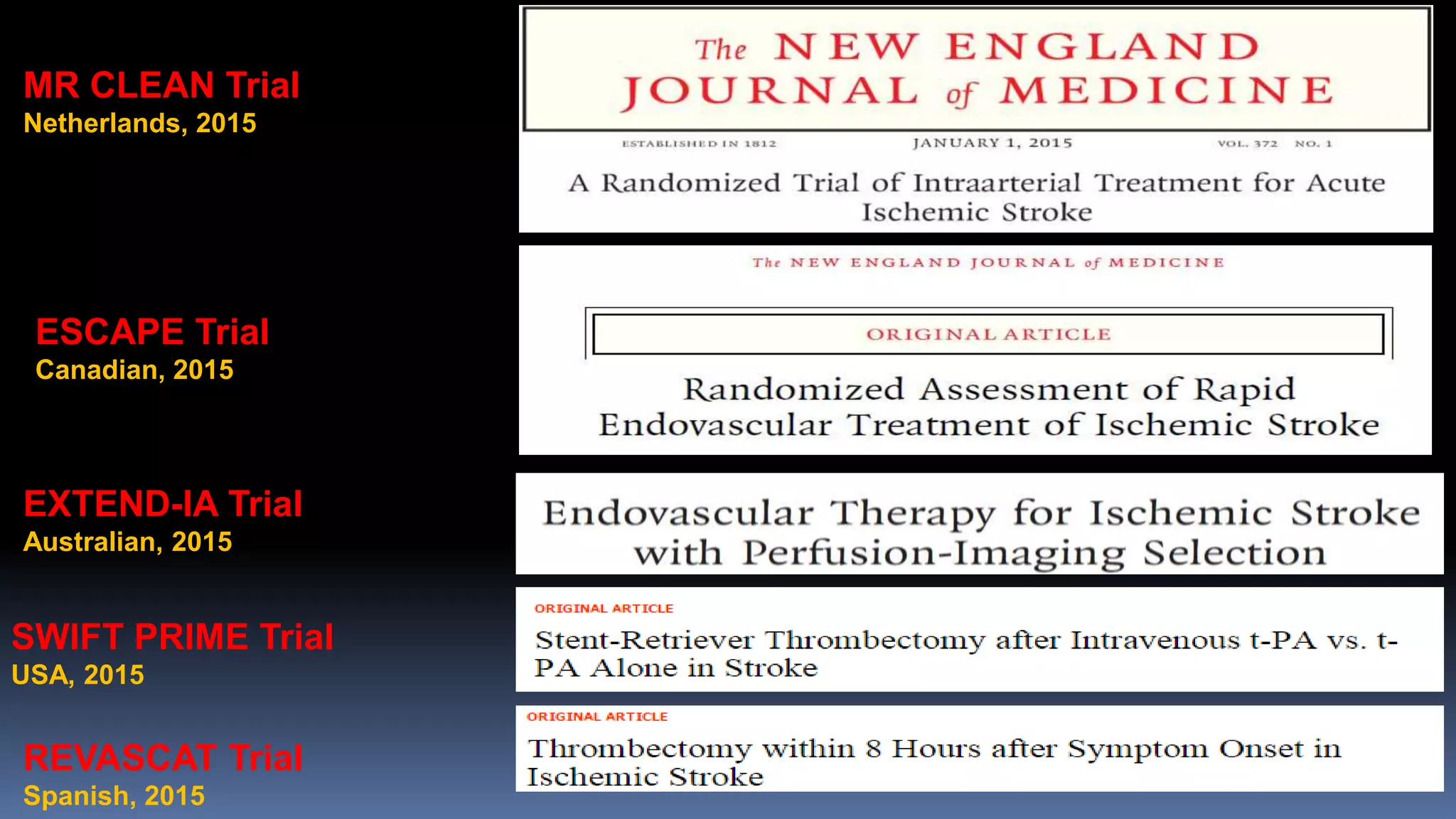
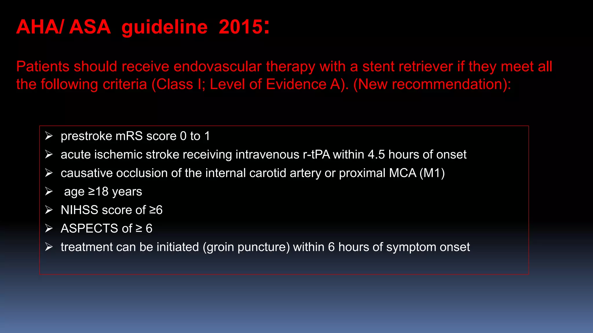
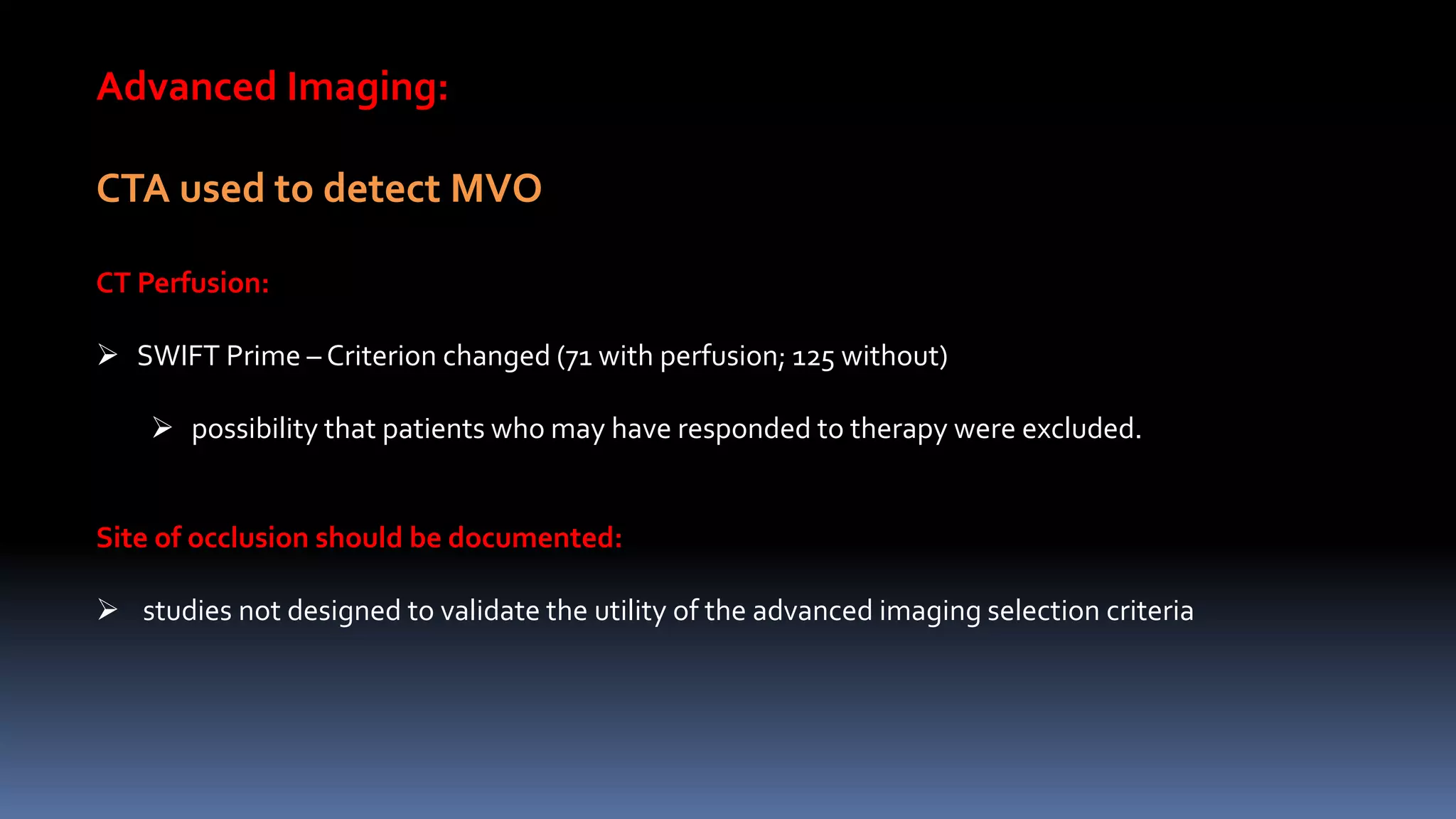
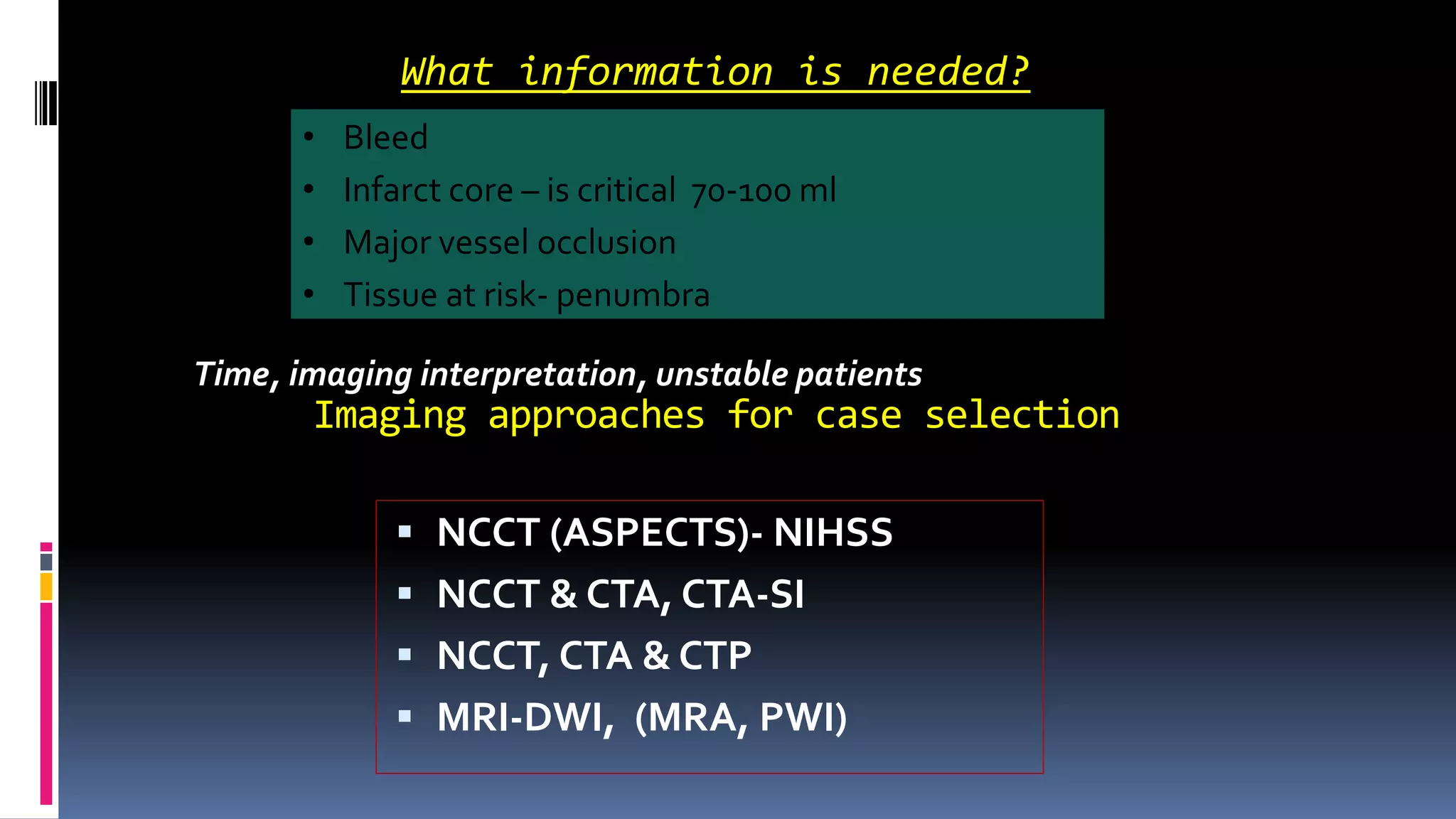
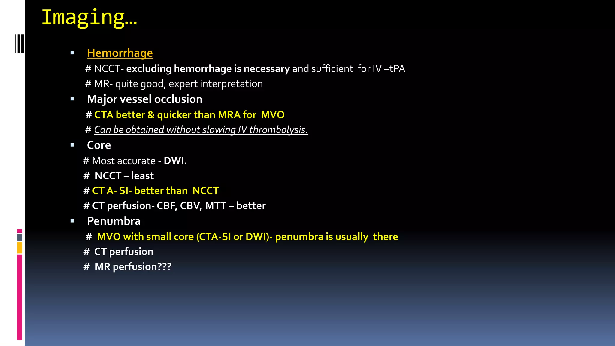
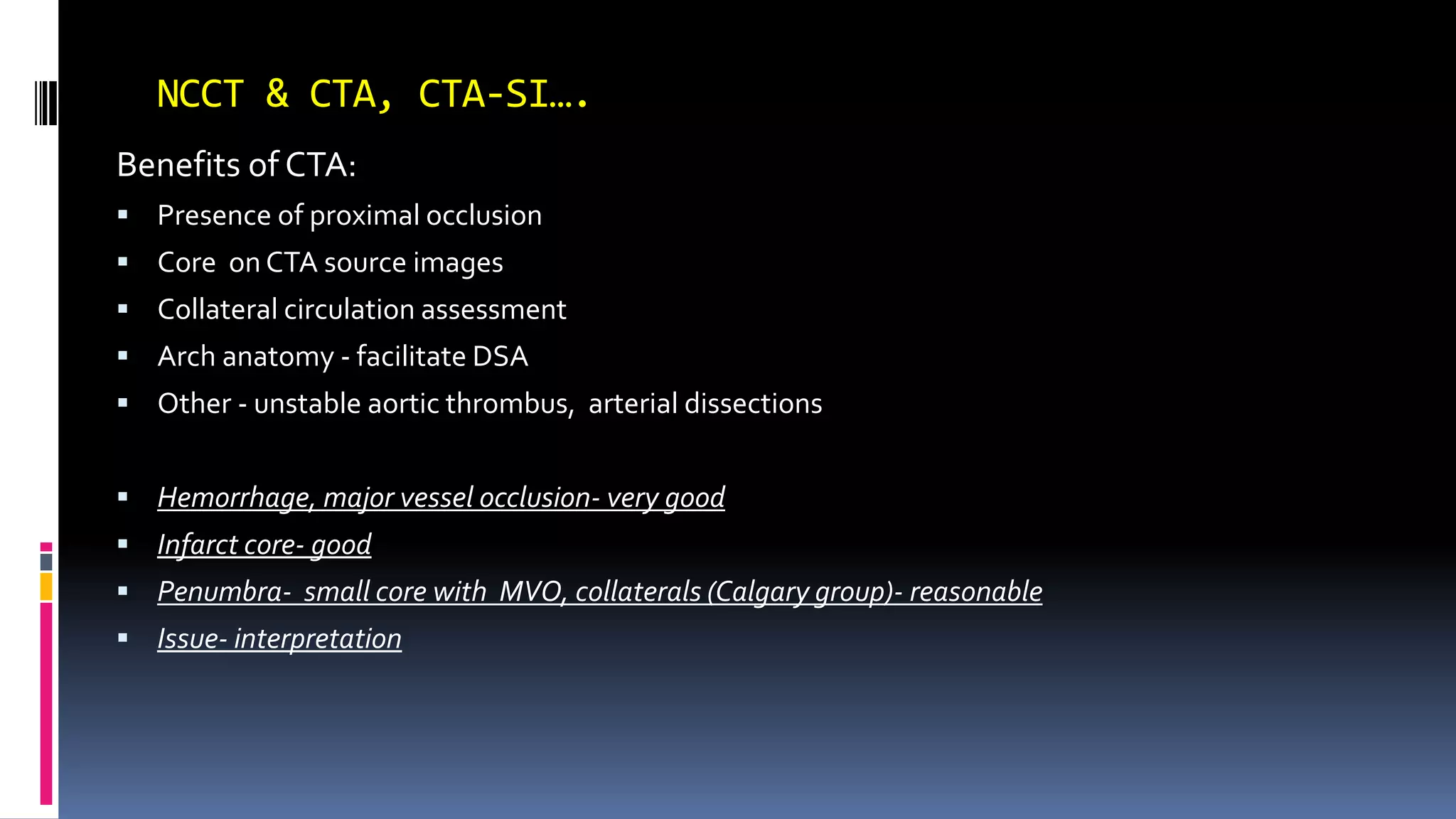
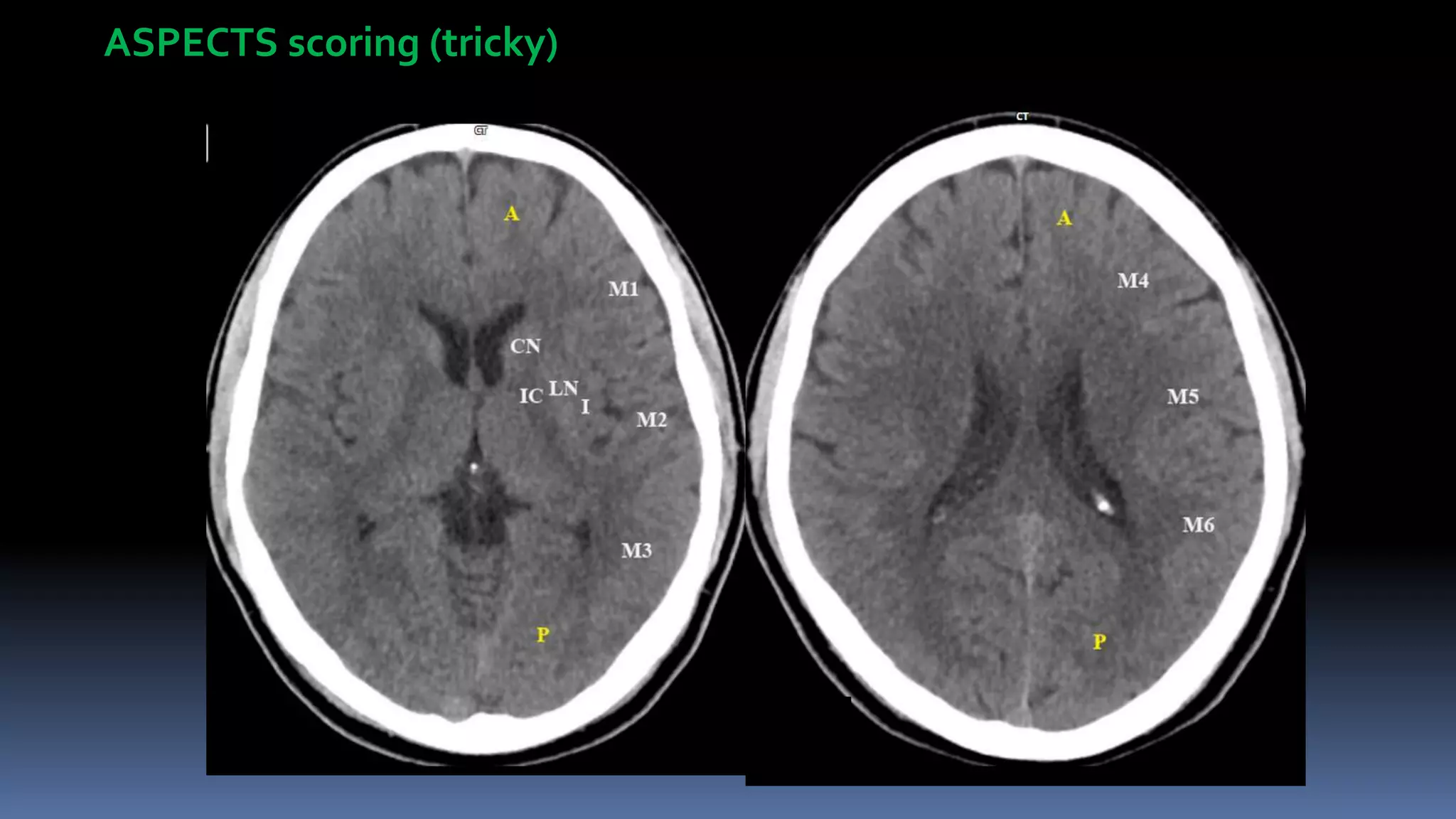
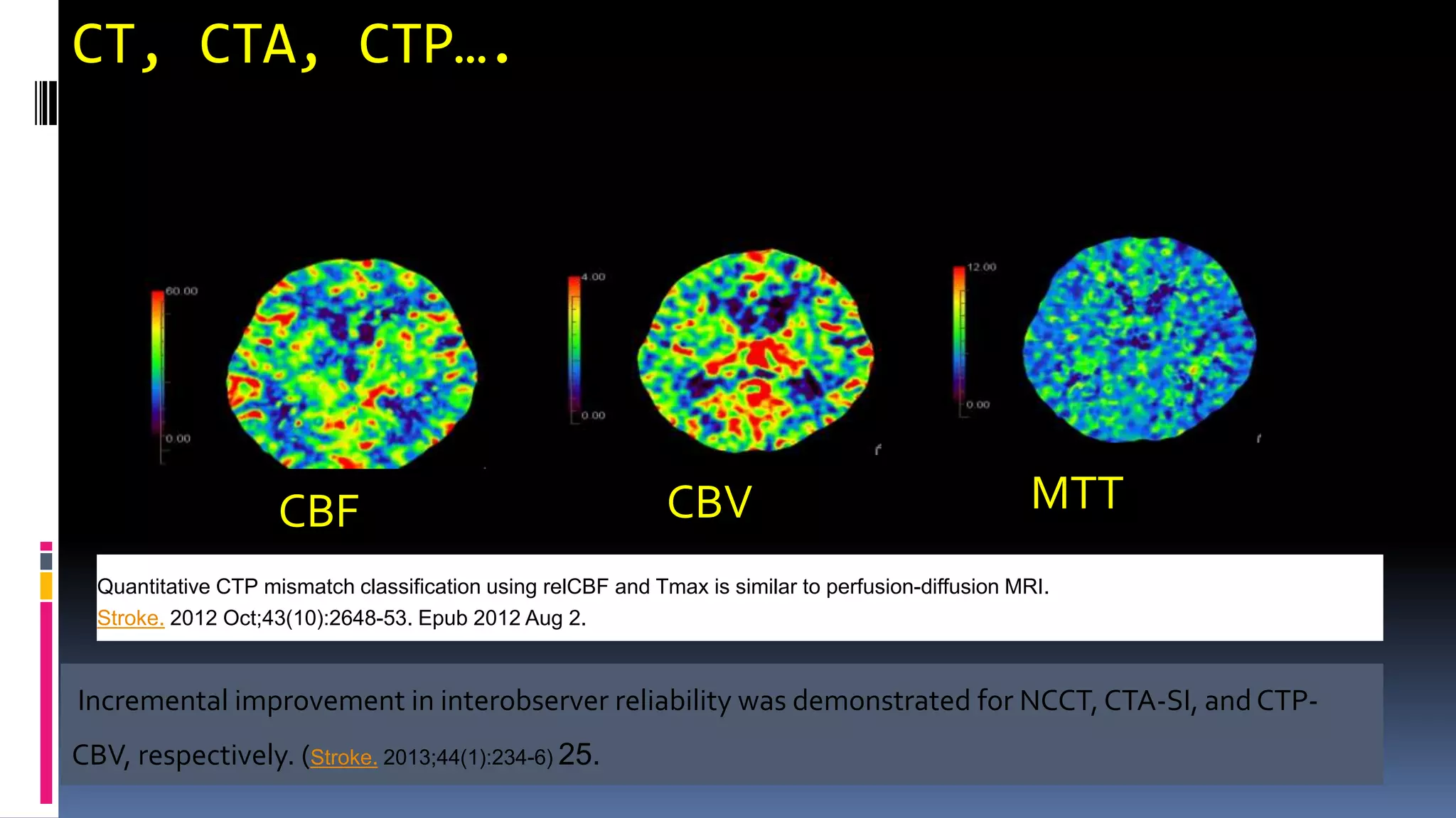
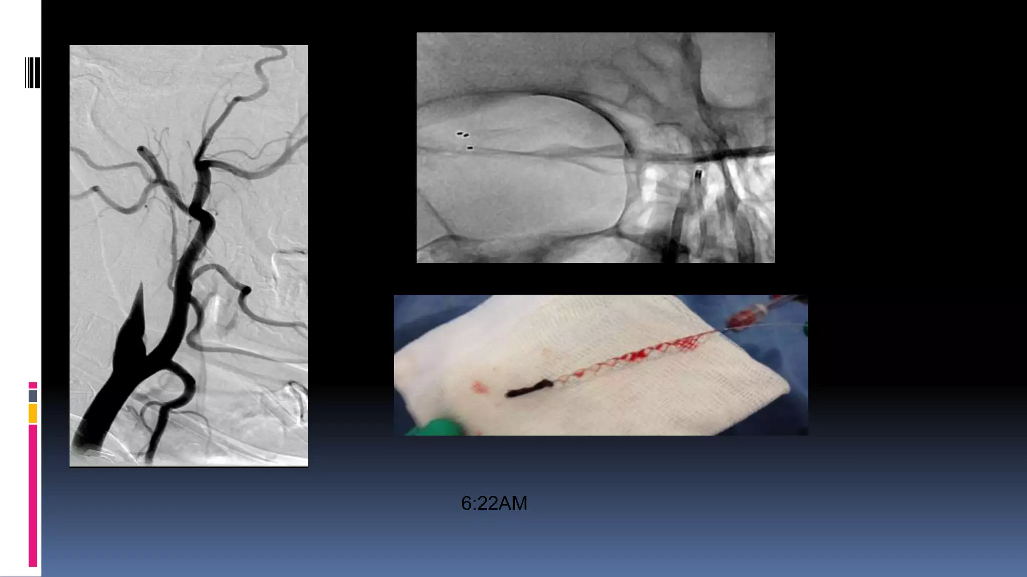
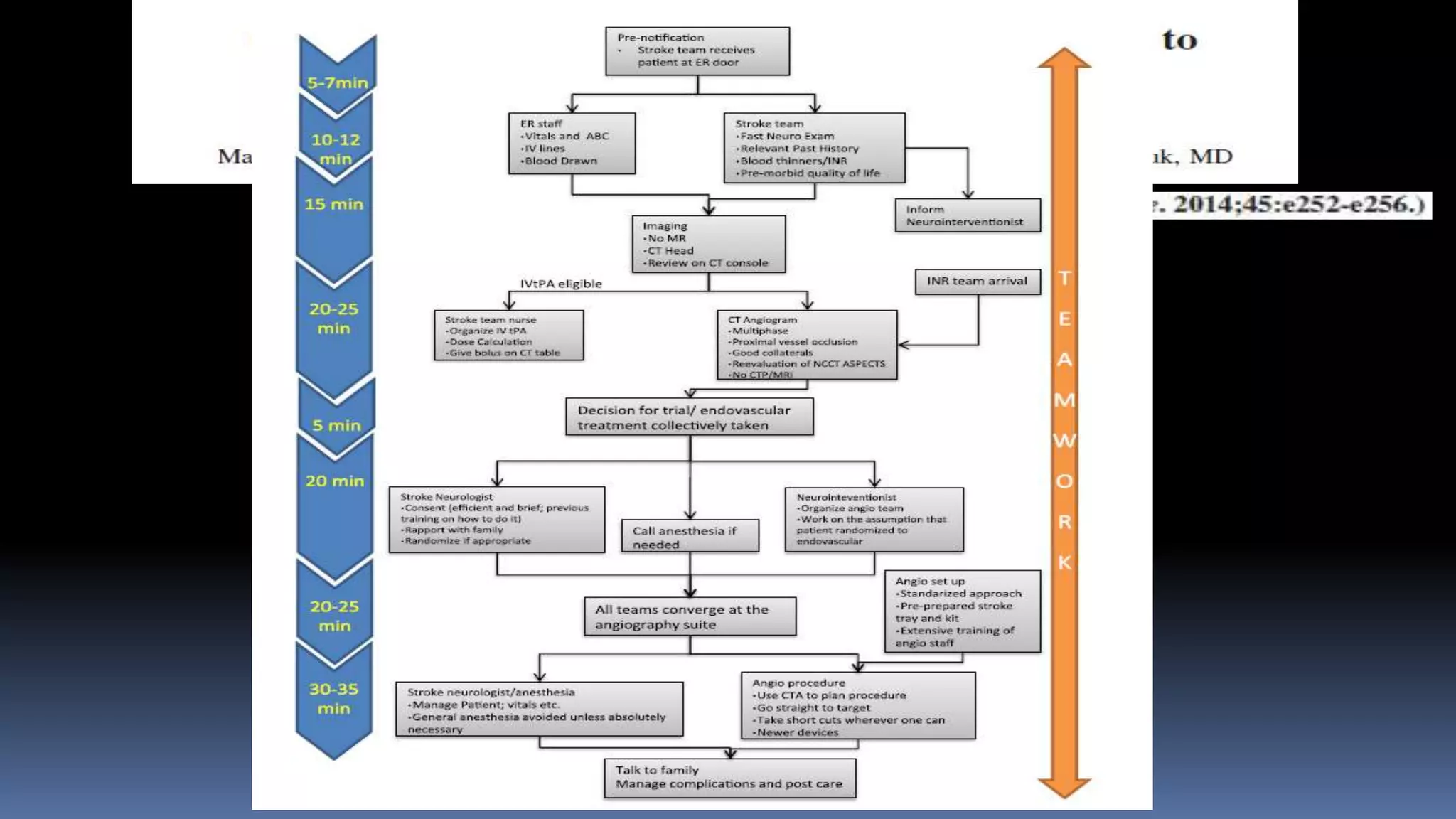
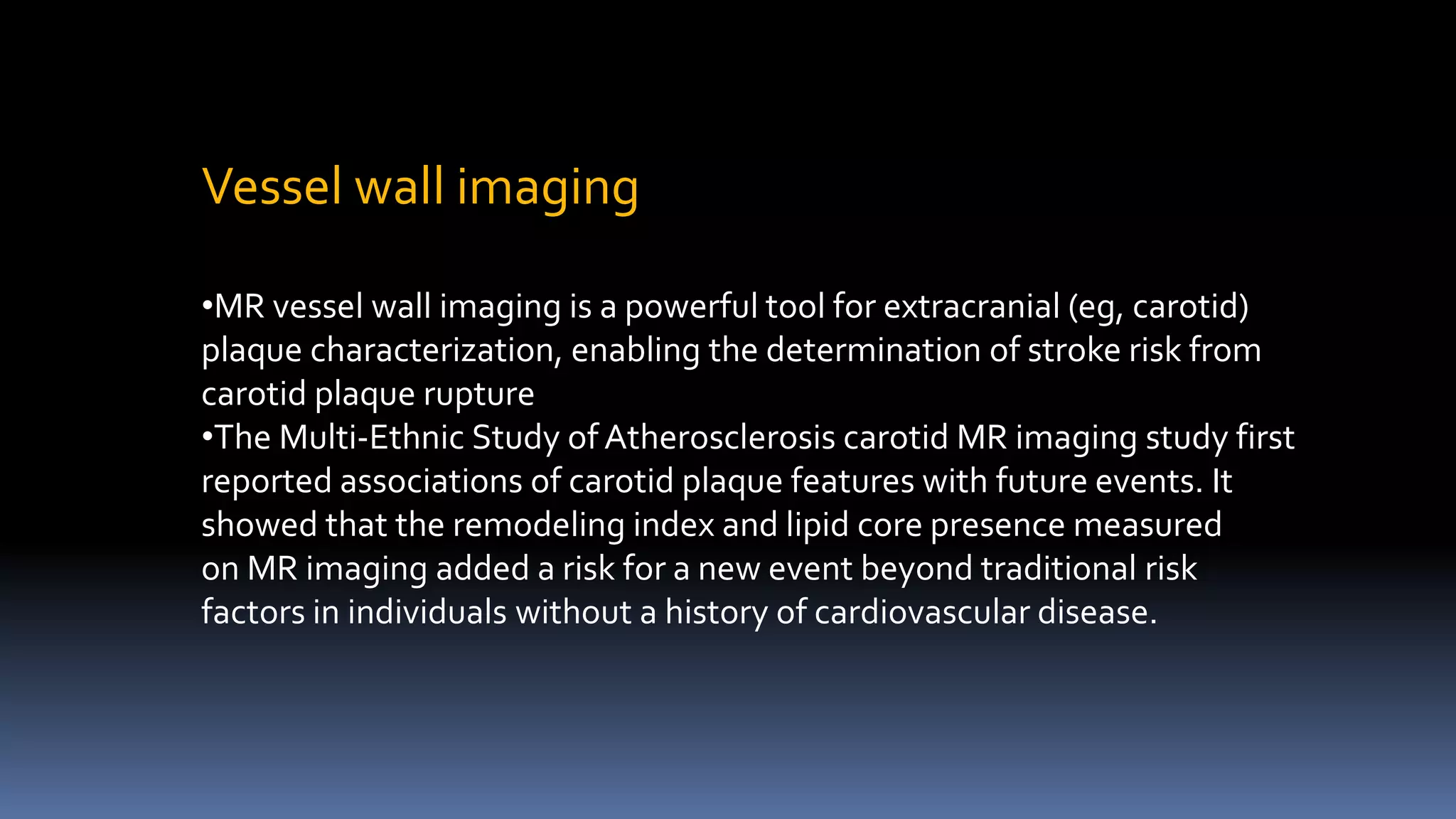
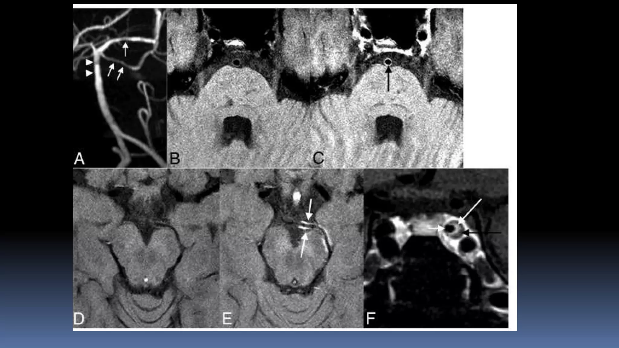
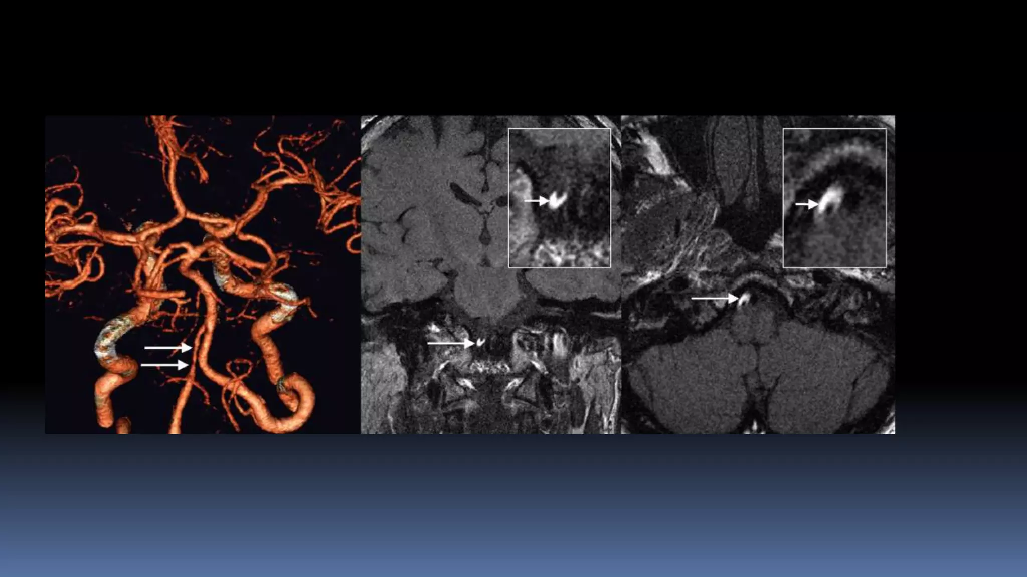
ADVANCES IN IMAGING OF
ISCHAEMIC STROKE IMAGING
Vipul Gupta
Neurointerventional Surgery
(Interventional Neuroradiology)
Artemis Hospital, Gurgaon
Advances in Stroke imaging …
 Acute stroke imaging – clinical approach
 Vessel wall imaging
 Neurointerventional suite imaging
 TCD – integration in clinical practice
MR CLEAN Trial
Netherlands, 2015
ESCAPE Trial
Canadian, 2015
EXTEND-IA Trial
Australian, 2015
SWIFT PRIME Trial
USA, 2015
REVASCAT Trial
Spanish, 2015
AHA/ ASA guideline 2015:
Patients should receive endovascular therapy with a stent retriever if they meet all
the following criteria (Class I; Level of Evidence A). (New recommendation):
 prestroke mRS score 0 to 1
 acute ischemic stroke receiving intravenous r-tPA within 4.5 hours of onset
 causative occlusion of the internal carotid artery or proximal MCA (M1)
 age ≥18 years
 NIHSS score of ≥6
 ASPECTS of ≥ 6
 treatment can be initiated (groin puncture) within 6 hours of symptom onset
Advanced Imaging:
CTA used to detect MVO
CT Perfusion:
 SWIFT Prime – Criterion changed (71 with perfusion; 125 without)
 possibility that patients who may have responded to therapy were excluded.
Site of occlusion should be documented:
 studies not designed to validate the utility of the advanced imaging selection criteria
Imaging approaches for case selection
 NCCT (ASPECTS)- NIHSS
 NCCT & CTA, CTA-SI
 NCCT, CTA & CTP
 MRI-DWI, (MRA, PWI)
What information is needed?
• Bleed
• Infarct core – is critical 70-100 ml
• Major vessel occlusion
• Tissue at risk- penumbra
Time, imaging interpretation, unstable patients
Imaging…
 Hemorrhage
# NCCT- excluding hemorrhage is necessary and sufficient for IV –tPA
# MR- quite good, expert interpretation
 Major vessel occlusion
# CTA better & quicker than MRA for MVO
# Can be obtained without slowing IV thrombolysis.
 Core
# Most accurate - DWI.
# NCCT – least
# CT A- SI- better than NCCT
# CT perfusion- CBF, CBV, MTT – better
 Penumbra
# MVO with small core (CTA-SI or DWI)- penumbra is usually there
# CT perfusion
# MR perfusion???
NCCT & CTA, CTA-SI….
Benefits of CTA:
 Presence of proximal occlusion
 Core on CTA source images
 Collateral circulation assessment
 Arch anatomy - facilitate DSA
 Other - unstable aortic thrombus, arterial dissections
 Hemorrhage, major vessel occlusion- very good
 Infarct core- good
 Penumbra- small core with MVO, collaterals (Calgary group)- reasonable
 Issue- interpretation
Left terminal ICA with Bovine arch Type 2 and type 3 arches
Extra stiff
wire for
exchange
Penumbra 3 Max and 4 Max
DAC 0.044 to cross loop
Contralateral approach
Penumbra 3Max through PCOM/ Solumbra
•68/M, DM, HTN, CAD, underwent PTCA to LAD
•Admitted for surgery of aortic stenosis.
•Double anti-platelets was stopped
•Patient developed acute onset right side weakness
with aphasia.
IV- tPA given, no improvement
Futile recanalization….
ASPECTS scoring (tricky)
Good collaterals by the Miteff method (OR, 3.341; 95% CI, 1.203–5.099; P .014) was the independent predictor of
good outcome amongst various collateral grading scales.
Arterial Collateral status – penumbra,
retention of penumbra
Miteff system.A, Contrast opacification all sylvian branches. B, Some vessels can be seen at the Sylvian fissure.C, distal
cortical filling alone
Multipha
se CTA
Better able to predict outcomes than single phase and perfusionCT
CT, CTA, CTP….
CT perfusion imaging
MTTCBF CBV
Quantitative CTP mismatch classification using relCBF and Tmax is similar to perfusion-diffusion MRI.
Stroke. 2012 Oct;43(10):2648-53. Epub 2012 Aug 2.
Incremental improvement in interobserver reliability was demonstrated for NCCT, CTA-SI, and CTP-
CBV, respectively. (Stroke. 2013;44(1):234-6) 25.
 Left hemiplegia, left UL and LL 0/5
 5:14AM
6:22AM
8:07 AM
Patient made gradual recovery
Left LL 4/5 and UL 3/5 - 30 day follow up mRS at 90
days- 0
Patient presented with in 2 hours
Futile IV tpa
• 60 years old female.
• h/o hypertension and hypothyroidism
• Acute onset left hemiparesis and left facial weakness
• CT Brain , CTP and CTA done 6 1/2 hours after ictus.
CT, CTA, CTP
 Hemorrhage, major vessel occlusion- very good
 Infarct core- CBV, good ? Better than SI
 Penumbra- CBF & MTT vs CBV
 Over all sensitivity and specificity – 80-90%
 Interpretation – convenient in emergency, technical issues are
there
MRI
Adv
Best for core – DWI
No radiation or contrast
Can do MRA and perfusionimaging
But
•Time…..time…..time…..
•Shifting, checking forCI, MRA and perfusion time
•Sick restless patients
•MRA- not good enough
•Key- should not be delaying IV tPA
•Having one protocol for all acute stroke patients
•Every 30 min delay – 10% decrease possibility of good
outcome (IMS-II)
Hand PJ, Wardlaw JM, Rowat AM, et al. Magnetic resonance brain imaging in patients with acute stroke: feasibility and patient related difficulties. J Neurol Neurosurg Psychiatry
2005;76:1525–27
Goyal M AJNR 33 August 2012
Every 30-min delay in angiographic reperfusion reduced the relative likelihood of a
good clinical outcome by 12% in adjusted analysis.
Conclusion
• Improving door to puncture time may be the key
SNIS – 2015 …
• Target Door to puncture < 60 min
Door to recanalization <90 min
• Small steps make a big difference!!!
Thrombus imaging
•Length
•Type
•Fragmentation
Vessel wall imaging
•MR vessel wall imaging is a powerful tool for extracranial (eg, carotid)
plaque characterization, enabling the determination of stroke risk from
carotid plaque rupture
•The Multi-Ethnic Study of Atherosclerosis carotid MR imaging study first
reported associations of carotid plaque features with future events. It
showed that the remodeling index and lipid core presence measured
on MR imaging added a risk for a new event beyond traditional risk
factors in individuals without a history of cardiovascular disease.
ICAD
DynaCT stents
syngo® Neuro PBV IR
Neuro Parenchymal Blood Volume1. DynaCT – Mask Acquisition
2. Steady State
Contrast Injection
3. DynaCT –
Fill Acquisition
Segmentation
of Bone and
Air
–
Subtracted Image
remove
Detection of
Arterial
Input normalize
Smoothing
0
10
5
mL/100g
PBV Map
Case study 1:
65 male with vascular risk factors
Diagnosed with asymptomatic carotid
stenosis
Underwent VMR testing
Why?
Meta-analysis - odds ratio of 3.86 (95%
CI, 1.99–7.48) for stroke risk
Technique:
Breath hold at end of inspiration for 30
seconds
Uncooperative patients – re breath
Always compare with the opposite side
Formula – MFV (end) – MFV (start)/ MFV (
start) x 100/ seconds of breath holding
< 0.6 is impaired
Vasomotor reserve testing
Criterion for MES
 3 db higher than background
 Unidirectional (spatial > 7.5 mm and
temporal > 30 ms)
 MCA positive slope
 ACA negative slope
Case study 2
60 male with left sided minor stroke
Started on dual antiplatelet
Had a further event
Planned for MES testing
Study 3
 61 year male patient
presented in ER with c/o
severe headache a/w nausea
since one day.
 NCCT Head shows SAH
Pulsatility index
•ICP
•Distal spasm
For more information on:
STROKE & NEUROVASCULAR INTERVENTIONS:
URL:
www.sanif.co.in
Facebook:
https://www.facebook.com/strokeawarenessindia
https://www.facebook.com/vipul.gupta.35175
Twitter
https://twitter.com/drvipulgupta25
LinkedIN
https://in.linkedin.com/pub/dr-vipul-gupta/51/8a1/25a
YouTube
Channel: Stroke & Neurovascular Interventions
www.youtube.com/c/StrokeNeurovascularInterventionsfoundation
DrVipulGupta
Thank you ….

Advances in Imaging of ischaAemic stroke

  • 1.
    ADVANCES IN IMAGINGOF ISCHAEMIC STROKE IMAGING Vipul Gupta Neurointerventional Surgery (Interventional Neuroradiology) Artemis Hospital, Gurgaon
  • 2.
    Advances in Strokeimaging …  Acute stroke imaging – clinical approach  Vessel wall imaging  Neurointerventional suite imaging  TCD – integration in clinical practice
  • 3.
    MR CLEAN Trial Netherlands,2015 ESCAPE Trial Canadian, 2015 EXTEND-IA Trial Australian, 2015 SWIFT PRIME Trial USA, 2015 REVASCAT Trial Spanish, 2015
  • 4.
    AHA/ ASA guideline2015: Patients should receive endovascular therapy with a stent retriever if they meet all the following criteria (Class I; Level of Evidence A). (New recommendation):  prestroke mRS score 0 to 1  acute ischemic stroke receiving intravenous r-tPA within 4.5 hours of onset  causative occlusion of the internal carotid artery or proximal MCA (M1)  age ≥18 years  NIHSS score of ≥6  ASPECTS of ≥ 6  treatment can be initiated (groin puncture) within 6 hours of symptom onset
  • 5.
    Advanced Imaging: CTA usedto detect MVO CT Perfusion:  SWIFT Prime – Criterion changed (71 with perfusion; 125 without)  possibility that patients who may have responded to therapy were excluded. Site of occlusion should be documented:  studies not designed to validate the utility of the advanced imaging selection criteria
  • 6.
    Imaging approaches forcase selection  NCCT (ASPECTS)- NIHSS  NCCT & CTA, CTA-SI  NCCT, CTA & CTP  MRI-DWI, (MRA, PWI) What information is needed? • Bleed • Infarct core – is critical 70-100 ml • Major vessel occlusion • Tissue at risk- penumbra Time, imaging interpretation, unstable patients
  • 7.
    Imaging…  Hemorrhage # NCCT-excluding hemorrhage is necessary and sufficient for IV –tPA # MR- quite good, expert interpretation  Major vessel occlusion # CTA better & quicker than MRA for MVO # Can be obtained without slowing IV thrombolysis.  Core # Most accurate - DWI. # NCCT – least # CT A- SI- better than NCCT # CT perfusion- CBF, CBV, MTT – better  Penumbra # MVO with small core (CTA-SI or DWI)- penumbra is usually there # CT perfusion # MR perfusion???
  • 8.
    NCCT & CTA,CTA-SI…. Benefits of CTA:  Presence of proximal occlusion  Core on CTA source images  Collateral circulation assessment  Arch anatomy - facilitate DSA  Other - unstable aortic thrombus, arterial dissections  Hemorrhage, major vessel occlusion- very good  Infarct core- good  Penumbra- small core with MVO, collaterals (Calgary group)- reasonable  Issue- interpretation
  • 9.
    Left terminal ICAwith Bovine arch Type 2 and type 3 arches Extra stiff wire for exchange Penumbra 3 Max and 4 Max DAC 0.044 to cross loop Contralateral approach Penumbra 3Max through PCOM/ Solumbra
  • 10.
    •68/M, DM, HTN,CAD, underwent PTCA to LAD •Admitted for surgery of aortic stenosis. •Double anti-platelets was stopped •Patient developed acute onset right side weakness with aphasia. IV- tPA given, no improvement
  • 12.
  • 13.
  • 14.
    Good collaterals bythe Miteff method (OR, 3.341; 95% CI, 1.203–5.099; P .014) was the independent predictor of good outcome amongst various collateral grading scales. Arterial Collateral status – penumbra, retention of penumbra
  • 15.
    Miteff system.A, Contrastopacification all sylvian branches. B, Some vessels can be seen at the Sylvian fissure.C, distal cortical filling alone
  • 16.
    Multipha se CTA Better ableto predict outcomes than single phase and perfusionCT
  • 17.
    CT, CTA, CTP…. CTperfusion imaging MTTCBF CBV Quantitative CTP mismatch classification using relCBF and Tmax is similar to perfusion-diffusion MRI. Stroke. 2012 Oct;43(10):2648-53. Epub 2012 Aug 2. Incremental improvement in interobserver reliability was demonstrated for NCCT, CTA-SI, and CTP- CBV, respectively. (Stroke. 2013;44(1):234-6) 25.
  • 18.
     Left hemiplegia,left UL and LL 0/5  5:14AM
  • 19.
  • 20.
    8:07 AM Patient madegradual recovery Left LL 4/5 and UL 3/5 - 30 day follow up mRS at 90 days- 0
  • 21.
    Patient presented within 2 hours Futile IV tpa
  • 22.
    • 60 yearsold female. • h/o hypertension and hypothyroidism • Acute onset left hemiparesis and left facial weakness • CT Brain , CTP and CTA done 6 1/2 hours after ictus.
  • 24.
    CT, CTA, CTP Hemorrhage, major vessel occlusion- very good  Infarct core- CBV, good ? Better than SI  Penumbra- CBF & MTT vs CBV  Over all sensitivity and specificity – 80-90%  Interpretation – convenient in emergency, technical issues are there
  • 25.
    MRI Adv Best for core– DWI No radiation or contrast Can do MRA and perfusionimaging But •Time…..time…..time….. •Shifting, checking forCI, MRA and perfusion time •Sick restless patients •MRA- not good enough •Key- should not be delaying IV tPA •Having one protocol for all acute stroke patients •Every 30 min delay – 10% decrease possibility of good outcome (IMS-II) Hand PJ, Wardlaw JM, Rowat AM, et al. Magnetic resonance brain imaging in patients with acute stroke: feasibility and patient related difficulties. J Neurol Neurosurg Psychiatry 2005;76:1525–27 Goyal M AJNR 33 August 2012
  • 26.
    Every 30-min delayin angiographic reperfusion reduced the relative likelihood of a good clinical outcome by 12% in adjusted analysis.
  • 28.
    Conclusion • Improving doorto puncture time may be the key SNIS – 2015 … • Target Door to puncture < 60 min Door to recanalization <90 min • Small steps make a big difference!!!
  • 29.
  • 30.
    Vessel wall imaging •MRvessel wall imaging is a powerful tool for extracranial (eg, carotid) plaque characterization, enabling the determination of stroke risk from carotid plaque rupture •The Multi-Ethnic Study of Atherosclerosis carotid MR imaging study first reported associations of carotid plaque features with future events. It showed that the remodeling index and lipid core presence measured on MR imaging added a risk for a new event beyond traditional risk factors in individuals without a history of cardiovascular disease.
  • 34.
  • 37.
  • 38.
    syngo® Neuro PBVIR Neuro Parenchymal Blood Volume1. DynaCT – Mask Acquisition 2. Steady State Contrast Injection 3. DynaCT – Fill Acquisition Segmentation of Bone and Air – Subtracted Image remove Detection of Arterial Input normalize Smoothing 0 10 5 mL/100g PBV Map
  • 39.
    Case study 1: 65male with vascular risk factors Diagnosed with asymptomatic carotid stenosis Underwent VMR testing Why? Meta-analysis - odds ratio of 3.86 (95% CI, 1.99–7.48) for stroke risk Technique: Breath hold at end of inspiration for 30 seconds Uncooperative patients – re breath Always compare with the opposite side Formula – MFV (end) – MFV (start)/ MFV ( start) x 100/ seconds of breath holding < 0.6 is impaired
  • 40.
  • 41.
    Criterion for MES 3 db higher than background  Unidirectional (spatial > 7.5 mm and temporal > 30 ms)  MCA positive slope  ACA negative slope Case study 2 60 male with left sided minor stroke Started on dual antiplatelet Had a further event Planned for MES testing
  • 43.
    Study 3  61year male patient presented in ER with c/o severe headache a/w nausea since one day.  NCCT Head shows SAH Pulsatility index •ICP •Distal spasm
  • 44.
    For more informationon: STROKE & NEUROVASCULAR INTERVENTIONS: URL: www.sanif.co.in Facebook: https://www.facebook.com/strokeawarenessindia https://www.facebook.com/vipul.gupta.35175 Twitter https://twitter.com/drvipulgupta25 LinkedIN https://in.linkedin.com/pub/dr-vipul-gupta/51/8a1/25a YouTube Channel: Stroke & Neurovascular Interventions www.youtube.com/c/StrokeNeurovascularInterventionsfoundation DrVipulGupta
  • 45.