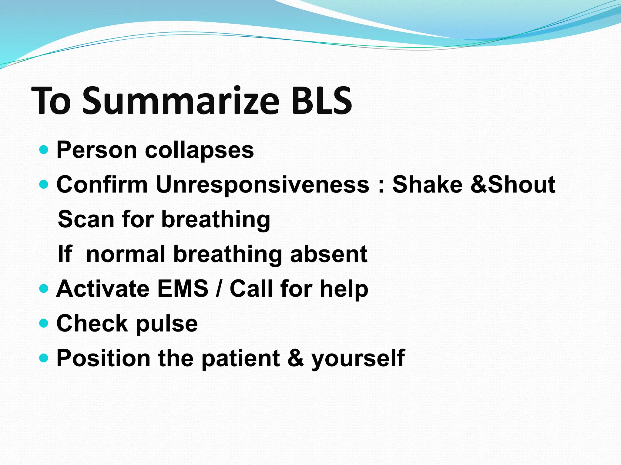
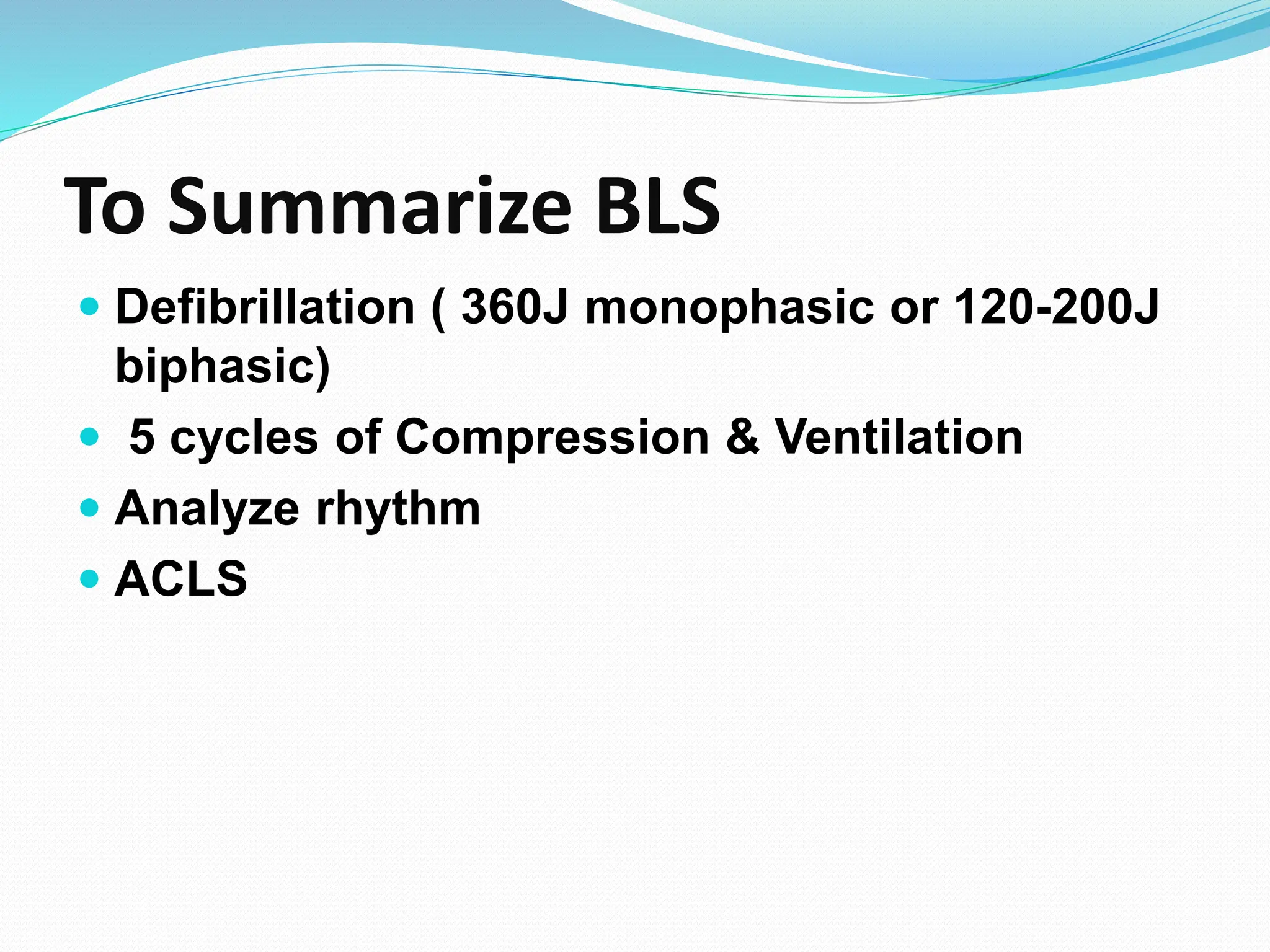
1. Dr Aggy S. Valentine MD, DA
Senior Consultant in Anaesthesia
Kerala State Health Services
2. CARDIO PULMONARY RESUSCITATION
Symptomatic therapy aimed at sustaining vital organ
function until natural cardiac function is restored
Basic Life Support (BLS)
Advanced Cardiac Life Support (ACLS)
Post Resuscitation Support
3. BLS is to maintain cardiac output and vital organ
perfusion.
ACLS is to diagnose cause of arrest and proceed
to the cause specific treatment while
continuining BLS.
4. Basic Life Support is the
foundation for saving lives
following cardiac arrest.
5. TIME IS CRITICAL
4 – 6 min, Irreversible neurological deficit.
RESUSCITATE HEART , RESTORE
BRAIN
Move quickly, calmly, do a systematic and
effective resuscitation
8. Code Blue
To provide immediate life
saving measures in case of life
threatening emergencies.
9. Scope of Code Blue
It is an event of utmost emergency, a
mode of alerting all medical, nursing,
paramedical and allied health-care
services and other personnel (house-
keeping, bio-medical, transport,
maintenance, security, etc)
10. Victims of cardiac arrest need immediate CPR.
Start CPR within 10 seconds of finding a patient
in cardiac arrest.
Inform and then start CPR
Trolley should reach within 2 minutes.
11. Adult BLS Sequence
Before approaching the victim, the rescuer must
ensure that the scene is safe.
Immediate recognition and activation of the
emergency response system.
Tap the victim on the shoulder and ask “Are you
alright?”
If victim responds but is injured, leave victim to
phone for help.
12. Then come back as soon as possible to check on
the patient.
If a lone rescuer finds an unresponsive patient ( no
movement in response to stimulation, gasping, no
breathing ) alert emergency services.
Return to patient, provide CPR, defibrillation if
needed.
If two or more rescuers present, one begins CPR
while the other phones for help.
13. Pulse Check
Should take not more than 10 seconds.
If no pulse felt
within that
period, start
chest
compression.
14. “A-B-C” to “C-A-B”
This development came in the 2010 CPCR
guidelines
Adults and Pediatric patients
15.
16. Chest Compressions
Consists of forceful, rhythmic application of pressure
over lower half of sternum.
All patients in cardiac arrest should receive
chest compressions
17. How does chest compression
work?
Chest compressions increase intrathoracic pressure
and directly compresses the heart. This generates
blood flow and oxygen delivery to the heart and
brain.
18.
19.
20.
21.
22. Effective Chest Compressions
Push hard and push fast.
100-120 compressions per minute.
Compression depth – 5cms.
Allow complete recoil of chest after each
compression to allow heart to fill completely.
Avoid interruptions in compression.
Rotate compressor every two minutes.
23. Compression/Ventilation ratio – 30:2
Two breaths should take no less than
10 seconds.
One breath should take just over 1
second.
26. Rescue Breaths
Mouth to mouth or Bag mask.
Deliver each rescue breath over 1 second.
Give a sufficient tidal volume to produce
visible chest rise.
Use a compression/ventilation ratio of 30
chest compressions to 2 ventilations
30. Early Defibrillation
Early Defibrillation with AED
Follow AED prompts.
Interval from collapse to defibrillation is one of the
most important determinants of survival from cardiac
arrest.
Resume chest compressions immediately after the
shock.
31. Defibrillation
Defibrillation stuns the heart, briefly stopping cardiac
electrical activity.
If the heart if still viable, its normal pace maker may
resume firing and produce an effective rhythm.
32.
33. Steps for using AED
• Switch power On.
• Attach paddles.
• Analyze rhythm ( done by AED ).
• Deliver shock ( if advised by AED ).
• Biphasic: 120 – 200J
Monophasic: 300J
• Resume CPR immediately after shock.
36. To Summarize BLS
Person collapses
Confirm Unresponsiveness : Shake &Shout
Scan for breathing
If normal breathing absent
Activate EMS / Call for help
Check pulse
Position the patient & yourself
37. To Summarize BLS
Start Chest Compression
After 30 chest compression Open Airway by
Triple maneuver
Two rescue breaths
Continue Chest Compression & Ventilation
(30:2) till AED / Manual Defibrillation
38. To Summarize BLS
Defibrillation ( 360J monophasic or 120-200J
biphasic)
5 cycles of Compression & Ventilation
Analyze rhythm
ACLS
39. ACLS-2015
Advanced cardiac life support or advanced
cardiovascular life support (ACLS) refers to a set of
clinical interventions for the urgent treatment of
cardiac arrest, stroke and other life-threatening
medical emergencies, as well as the knowledge and
skills to deploy those interventions.
40. IMPORTANCE OF BLS IN ACLS
ACLS is built heavily upon the foundation of BLS
48. Asystole
P, QRS, T- waves: Complete absent of
cardiac electrical activity
Complete absent of effective cardiac
pumping function
49. Pulseless Electrical Activity (PEA)
PEA rhythm occurs when any organized heart
rhythm that is observed on the electrocardiogram
(ECG) does not produce a pulse.
Performing a pulse check after a rhythm/monitor
check will ensure that you identify PEA in every
situation
52. Failure of a single adequate shock to restore a pulse
should be followed by continued CPR and second
shock delivered after five cycles of CPR
If cardiac arrest still persist- patient is intubated and
IV/IO access achieved
57. Routes of administration
Peripheral IV – easiest to insert during CPR, must
followed by 20 ml NS push
Central IV – fast onset of action, but do not wait or
waste time for CV line
Intraosseous – alternative IV route in peds, also in
adult
Intratracheally (down an ET tube)- not recommended
now a days
58.
59. Epinephrine (Adrenaline)
• It increases heart rate, stroke volume and blood
pressure
• May help in asystole, PEA and symptomatic
bradycardia
• IV Dose: 1 mg every 3-5 minutes
60. Amiodarone
• Indications: Vtach, Vfib
• IV Dose:
300 mg in 20-30 ml of N/S or D5W
Supplemental dose of 150 mg in 20-30 ml of N/S
Followed with continuous infusion of 1 mg/min
for 6 hours then 0.5mg/min to a maximum daily
dose of 2 grams
• Contraindications:
Cardiogenic shock, profound Sinus Bradycardia,
2nd and 3rd degree blocks that do not have a
pacemake
61. Lidocaine
• Indications:
PVCs, Vtach, Vfib
Can be toxic so no longer given
prophylactically
• IV dose :
1-1.5 mg/kg bolus then continuous infusion
of 2-4mg/min
Can be given down ET tube
• Signs of toxicity:
slurred speech, seizures, altered
consciousness
62. Oxygen
• FIO2 100%
• Assist Ventilation
• O2 Toxicity should not be a concern during ACLS
IV Fluids
Volume Expanders
• crystalloids , e.g. Ringer’s lactate, N/S
63. Vasopressin (ADH)- is out according to 2015
guidelines for ACLS
Sodium Bicarbonate
• Used for METABOLIC acidosis / hyperkalemia
• IV Dose: 1 mEq/kg
• Side effects:
Metabolic alkalosis
Increased CO2 production
64. Monitoring during CPR
Physiologic parameters
Monitoring of PETCO2 (35 to 40 mmHg)
Abrupt and sustained increase is a sensitive
indicator of ROSC that can be monitored without
interrupting chest compressions
65. ROSC
Abrupt and sustained increase in ETCO2 (typically
greater than 40mm Hg)
Palpable pulse.
Assess BP. Maintain BP with fluids and vasopressor
Obtain 12- lead ECG.
Maintain SpO2 greater than 94%
66.
67. Does the patient follow commands ?
No – Consider induced hypothermia
Yes – STEMI or high suspicion of AMI
Advanced critical care
Coronary reperfusion