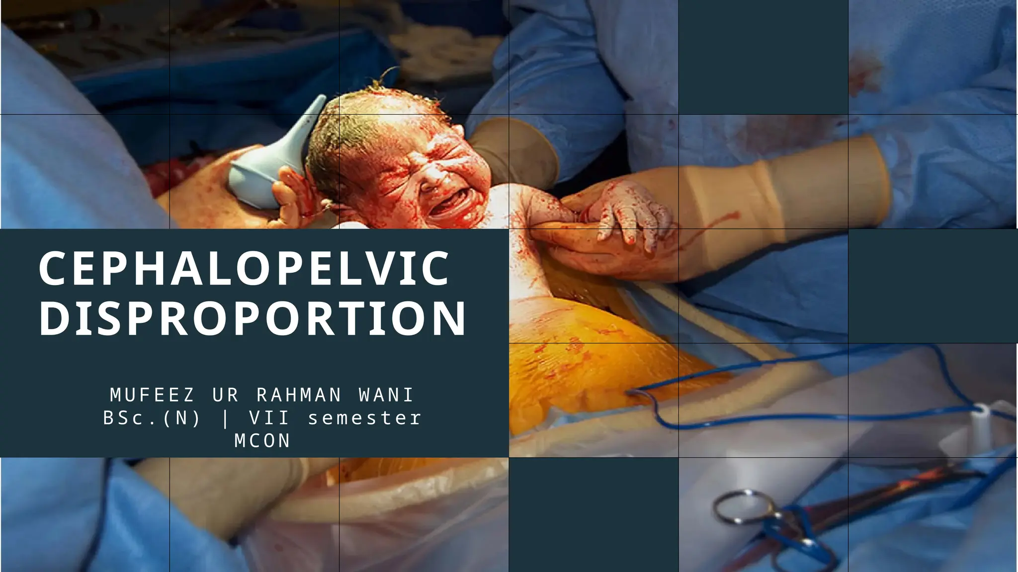
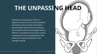
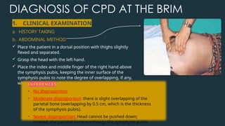
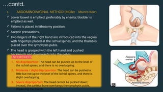
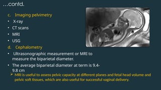
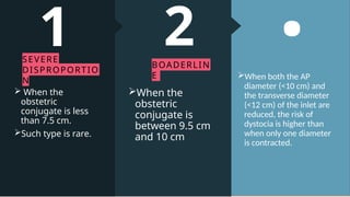
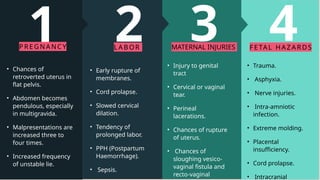
CEPHALOPELVIC
DISPROPORTION
M U F E E Z U R R A H M A N W A N I
B S c . ( N ) | V I I s e m e s t e r
M C O N
Cephalopelvic Disproportion (CPD) is a
significant obstetric concern, often identified
during labor when the baby's head fails to
descend despite strong uterine contractions.
This condition makes normal vaginal delivery
difficult or impossible and can lead to serious
complications such as prolonged labor, fetal
distress, and maternal exhaustion if not
managed properly.
THE UNPASSING HEAD
CPD
CONTENTS
1 2 3 4 5
DEFINITION
AND TYPES
ETIOLOGY DIAGNOSIS EFFECTS TREATMENT
A 2k25
DEFINITION
Cephalopelvic disproportion (CPD)
refers to the disparity in the relationship
between the fetal head and the maternal
pelvis.
Cephalopelvic disproportion (CPD) is
defined as the presence of a disparity
between the diameters of the fetal head
and the dimensions of the maternal
pelvis.
Can be due to abnormal fetal size,
reduced pelvic capacity or, more
commonly, a combination of both.
3
ed outlet
uberous
or less.
the outlet
vere
ead to
ed
annot be
eneath the
chial
elvis
m =
less.
SITES / TYPES
OF CEPHALOPELVIC
DISPROPORTION
3
PELVIC
OUTLET
• Due to a contracted outlet
 the interischial tuberous
diameter is 8 cm or less.
• Disproportion at the outlet may
not cause severe dystocia but
can lead to perineal tears.
• The head is pushed backwards
as it cannot be accommodated
beneath the symphysis pubis.
2
MIDPELV
IS
• ​Due to a contracted
midpelvis
 The sum of the interischial
and posterior sagittal
diameters of the midpelvis
(normal: 10.0 cm + 5 cm =
15.0 cm) is 13.5 cm or less.
1
PELVIC
INLET
 Occurs due to a contracted
pelvic inlet ;
• obstetric conjugate is <10 cm,
• the greatest transverse diameter
is <12 cm,
• or the diagonal conjugate is <11
cm.
 It can also be associated with
macrosomia (big baby, >4000g).
 Marked asynclitism of the
presenting head during labor
suggests inlet contraction.
INLET
CONTRACTI
ON
MIDPELVIC
CONTRACTIO
N
OUTLET
CONTRACTI
ON
AP diameter < 10
cm
Inter spinuous
diameter <8 cm
Interischial
tuberous diameter
8 cm
≤
Transverse
diameter < 12 cm
The sum of the
interischial and
posterior sagittal
diameters of the
midpelvis 13.5 cm
≤
or less.
Diagonal conjugate
< 11 cm
TABLE 1 : DIAMETERS IN A CONTRACTED
PELVIS
• From a clinical perspective, identifying CPD is more logical than concentrating entirely on
measurements of a given pelvis, as the fetal head is the best pelvimeter.
• Disproportion may be limited to one or more planes. The absence of CPD at the brim usually,
but not always, negates its presence at the mid-pelvic plane. Isolated outlet contraction
without midpelvic contraction is a rarity.
ETIOLOGY
• Genetic variables
• Maternal diabetes
• Excessive gestational weight
gain
• Multiparity
• Post-Term pregnancy
• Maternal obesity
• Overdue pregnancy
• Male child
• History of previous macrosomia
• Congenital heart diseases, esp.
Transposition of Greater Arteries
• Advanced maternal age
MACROSOMIA:
… c o n t d .
CONTRACTED PELVIS:
1. Nutritional and Environmental
factors: Rachitic or
osteomalacic pelvis.
2. Diseases and injuries: Which affect the bones
of the pelvis
• fracture, tumor, and tubercular arthritis.
• spinal deformities like kyphosis, scoliosis,
spondylolisthesis
• coccygeal deformity, or lower limb diseases
like poliomyelitis or hip joint diseases
…contd.
3.Asymmetrical pelvis:
a. Naegele's pelvis: Produced due to arrested
development of one ala of the sacrum.
b. Robert's pelvis (transversely
contracted pelvis): Both alae of the
sacrum are absent and the sacrum fused
with innominate bones.
4. Small stature women ( < 5 ft.)
Contracted pelvis;
INDICATIONS / POINTERS
TOWARDS CPD
• Prolonged labor despite augmentation.
• Head remaining high at full dilation.
• The cervix is loosely applied to the head.
• Excessive caput or irreducible moulding of the fetal
head.
DIAGNOSIS OF CPD AT THE BRIM
1. CLINICAL EXAMINATION
a. HISTORY TAKING
b. ABDOMINAL METHOD
 Place the patient in a dorsal position with thighs slightly
flexed and separated.
 Grasp the head with the left hand.
 Place the index and middle finger of the right hand above
the symphysis pubis, keeping the inner surface of the
symphysis pubis to note the degree of overlapping, if any,
when the head is pushed downwards and backwards.
I N F E R E N C E S
• No disproportion
• Moderate disproportion: there is slight overlapping of the
parietal bone (overlapping by 0.5 cm, which is the thickness
of the symphysis pubis).
• Severe disproportion: Head cannot be pushed down;
instead, the parietal bone overhangs the symphysis pubis.
…contd.
c. ABDOMINOVAGINAL METHOD (Müller - Munro Kerr)
 Lower bowel is emptied, preferably by enema; bladder is
emptied as well.
 Patient is placed in lithotomy position.
 Aseptic precautions.
 Two fingers of the right hand are introduced into the vagina
with fingertips placed at the ischial spines, and the thumb is
placed over the symphysis pubis.
 The head is grasped with the left hand and pushed
backwards and downwards into the pelvis.
I N F E R E N C E S
1. No disproportion: The head can be pushed up to the level of
the ischial spines, and there is no overlapping.
2. Moderate / slight disproportion: The head can be pushed a
little but not up to the level of the ischial spines, and there is
slight overlapping.
3. Severe disproportion: The head cannot be pushed down;
instead, the parietal bone overhangs the symphysis pubis,
displacing the thumb
…contd.
c. Imaging pelvimetry
• X-ray
• CT scans
• MRI
• USG
d. Cephalometry
• Ultrasonographic measurement or MRI to
measure the biparietal diameter.
• The average biparietal diameter at term is 9.4-
9.8 cm
 MRI is useful to assess pelvic capacity at different planes and fetal head volume and
pelvic soft tissues, which are also useful for successful vaginal delivery.
.he AP
0 cm) and
e diameter
he inlet are
risk of
gher than
ne diameter
.
2RLIN
s
5 cm
O
.
DEGREE OF
DISPROPORTION AND
CONTRACTED PELVIS
.
When both the AP
diameter (<10 cm) and
the transverse diameter
(<12 cm) of the inlet are
reduced, the risk of
dystocia is higher than
when only one diameter
is contracted.
2
BOADERLIN
E
When the
obstetric
conjugate is
between 9.5 cm
and 10 cm
1
SEVERE
DISPROPORTIO
N
 When the
obstetric
conjugate is less
than 7.5 cm.
Such type is rare.
4
S
s.
c infection.
ing.
ufficiency.
e.
ial
ract
l
ns.
e of
hing
ula
INJURIES
tion.
ged
Y
n
EFFECTS OF
CONTRACTED PELVIS
AND CPD
4
FE TAL HA Z A R DS
• Trauma.
• Asphyxia.
• Nerve injuries.
• Intra-amniotic
infection.
• Extreme molding.
• Placental
insufficiency.
• Cord prolapse.
• Intracranial
• Injury to genital
tract
• Cervical or vaginal
tear.
• Perineal
lacerations.
• Chances of rupture
of uterus.
• Chances of
sloughing vesico-
vaginal fistula and
recto-vaginal
3
MATERNAL INJURIES
2
LA B OR
• Early rupture of
membranes.
• Cord prolapse.
• Slowed cervical
dilation.
• Tendency of
prolonged labor.
• PPH (Postpartum
Haemorrhage).
• Sepsis.
1
PR E G N ANC Y
• Chances of
retroverted uterus in
flat pelvis.
• Abdomen becomes
pendulous, especially
in multigravida.
• Malpresentations are
increased three to
four times.
• Increased frequency
of unstable lie.
MANAGEMENT
• Minor to moderate degrees of pelvic
contraction.
• More theoretical
• One must be certain of the gestational
age.
1. PREMATURE INDUCTION OF LABOR;
2. ELECTIVE CESAREAN SECTION
• Most commonly done
• Indicated in;
 major degrees of inlet contraction (true
conjugate of <9 cm)
 moderate degree of inlet contraction with
outlet contraction
 associated with malpresentations
 special cases such as elderly primigravida
INDICATI
d midpelvic
t
on.
e of
ating factors
rly
vida,
entation,
turity,
disorders
-eclampsia,
, TB, etc.).
bility of
n section
f previous
ATIO
used
vic
ntly
t
h as
of
oman
C-
vis
n
althy
N
3.TRAIL LABOR
CONTRAINDICATI
ONS
• Associated midpelvic
and outlet
contraction.
 Presence of
complicating factors
like elderly
primigravida,
malpresentation,
post-maturity,
medical disorders
(e.g., pre-eclampsia,
diabetes, TB, etc.).
 Unavailability of
cesarean section
facility.
 Failure of previous
trial.
INDICATIO
NS
• Originally used
to test pelvic
adequacy.
• Subsequantly
used to test
numerous
factors such as
to test the
adequacy of
scar in a woman
with prior C-
Section
(TOLACS)
AIMS
• To check the
adequary of pelvis
• Avoiding
unnecessary
cesarean section
• Delivering a healthy
baby
DEFINITION
It is the conduction of
spontaneous labor in
a minor to moderate
degree of
cephalopelvic
disproportion in an
institution under
supervision with
watchful expectancy,
hoping for vaginal
delivery, and avoiding
an unnecessary
cesarean section and
delivering a healthy
baby.
… c o n t d .
CONDUCTION OF TRAIL LABOR
• Careful supervision and arrangement for operative delivery.
• The labor should ideally be spontaneous. But in case of failure of labor
to start even on due date, induction of labor may be done.
• NPO, hydration is maintained via IV drip.
• Epidural analgesia (ideally).
• Careful mapping using a partograph.
• If there is failure to progress due to inadequate uterine contraction,
augmentation of labor may be done by amniotomy along with
oxytocin. On no account should the procedure be employed before the
cervical dilatation is less than 3 cm.
• After the ROM, pelvic examination is to be done; (a) to exclude cord
prolapse; (b) to note the color of liquor; (c) to assess pelvis once more;
(d) to note the condition of cervix, including presence of the presenting
SUCCESSFUL
TRAIL
 If a healthy baby is
born spontaneously
or by forceps or by
ventouse with the
mother in good
condition, it is called
a successful trial.
 Delivery by cesarean
section or delivery of
a dead baby
spontaneously or by
craniotomy is
considered a failed
trial.
T ER MI NAT I
ON
1. Spontaneous
delivery with or
without episiotomy.
2. Forceps or
ventouse (30%) -
difficult forceps
delivery is to be
avoided.
3. Cesarean section
(40%) - judicial
decision must be
made promptly.
UNFAVORABLE
FEATURES
1. Abnormal uterine
contractions.
2. Cervical dilation <1
cm/hr (protracted
active phase).
3. Descent of head <1
cm/hr
4. Arrest of cervical
dilation.
5. Early rupture of
membranes.
6. Formation of caput
and evidence of
excessive moulding.
7. Fetal distress
SUCCESSFUL
OUTCOME
1. Degree of pelvic
contraction.
2. Shape of pelvis - flat
pelvis is better than
android or generally
contracted pelvis.
3. Favourable vertex
presentation.
4. Intact membranes
till full cervical
dilation.
5. Effective uterine
contractions.
6. Emotional support
to the woman
NURSING MANAGEMENT
II. HISTORY AND PHYSICAL ASSESSMENT
III. NURSING DIAGNOSIS
1. Ineffective labor progression related to cephalopelvic
disproportion as evidenced by non-progress of labor, abnormal uterine
contractions, protracted active phase, and arrest of cervical dilation.
 INTERVENTIONS :-
 Monitor progress of labor by plotting partograph.
 Assist with positioning to facilitate fetal descent.
 Provide pain management.
 Make judicial timely decision about cesarean section in
collaboration with healthcare team members.
 Encourage to do deep breathing exercises.
 Ensure suspension of oral feeding.
 Encourage the mother and provide emotional support.
 Provide comfort measures.
…contd.
2. Risk for fetal injury related to prolonged labor and
obstruction.
INTERVENTIONS :-
 Perform NST to assess fetal well-being.
 Provide left lateral position to avoid distress.
 Assess FHR manually or electronically, note variability,
periodic changes, and baseline rate.
 Note uterine pressure during resting and contractile
phases via intrauterine pressure catheter.
 Provide oxygen to the mother.
 Monitor fetal descent and overall progress of labor.
 Note the colour of amniotic fluid.
 Make all necessary arrangements for operational
delivery.
 Observe for cord prolapse, excessive moulding, or
occurrence of caput.
 Document all events.
…contd.
3. Risk for maternal injury related to mechanical
obstruction of labor secondary to cephalopelvic
disproportion.
 INTERVENTIONS :
 Review history of labor onset and duration.
 Evaluate level of fatigue as well as activity level.
 Assess uterine contractions, frequency, and strength.
 Examine condition of cervix.
 Assess amniotic fluid for color and amount.
 Assess degree of dehydration.
 Make necessary arrangements of surgical delivery.
 Assist in making decisions regarding cesarean section.
 Provide comfortable measures.
 Be available and provide emotional support.
 Administer analgesics and other medications as
…contd.
IV. EXPECTED
OUTCOME
• Client accomplishes normal
progression of labor.
• Fetal distress and injuries are
protected.
• Maternal injuries are prevented.
• Alleviation of pain during
prolonged labor.
• Reduction in anxiety
• Best possible intervention
/method of delivery is decided as
indicated by individual condition .
Cephalopelvic disproportion remains a
significant concern in obstetric practice,
particularly in ensuring safe labor and
delivery. It increases the risk of perinatal
mortality and morbidity, as well as maternal
mortality and morbidity. Early diagnosis,
close monitoring during labor, and prompt
surgical interventions can significantly
improve the intrapartum management and
ensure safer outcomes for both mother and
baby.
BEYOND THE OBSTRUCTION
A 25-year-old primigravida, low risk, has come for routine ANC visit at 38 weeks. She is 5'
tall. The fetus is average size, in cephalic presentation but the head has not gone into the
pelvis. On pelvic assessment the sacral promontory was tipped with ease and there was
mild cephalopelvic disproportion at the level of the inlet. The rest of the cavity is spacious,
IID is adequate, and outlet is adequate. What is the management plan for her;
a. Plan and Elective Cesarean at 39 weeks.
b. Plan for Cesarean once she goes into labor.
c. Plan Induction and trial of labor.
d. Allow to go into spontaneous labor and trial of labor
QUESTION
Q : A female with a H/O NVD of a normal sized baby 3 years back has no chances of
development of CPD. ( TRUE / FALSE )
Q : A "contracted pelvis" is anatomically defined as:
a. Reduced pelvic capacity
b. At least one pelvic diameter below normal by 1 cm
≥
c. All diameters normal
d. All diameters increased
Q: A key clinical sign of CPD during labor is:
e. Maternal hypertension
f. Fetal head not descending despite strong contractions
g. Premature cervical dilation
h. Excess amniotic fluid only
QUESTION
CEPHALOPELVIC DISPROPORTION (Mufeez).pptx

CEPHALOPELVIC DISPROPORTION (Mufeez).pptx

  • 1.
    CEPHALOPELVIC DISPROPORTION M U FE E Z U R R A H M A N W A N I B S c . ( N ) | V I I s e m e s t e r M C O N
  • 2.
    Cephalopelvic Disproportion (CPD)is a significant obstetric concern, often identified during labor when the baby's head fails to descend despite strong uterine contractions. This condition makes normal vaginal delivery difficult or impossible and can lead to serious complications such as prolonged labor, fetal distress, and maternal exhaustion if not managed properly. THE UNPASSING HEAD
  • 3.
    CPD CONTENTS 1 2 34 5 DEFINITION AND TYPES ETIOLOGY DIAGNOSIS EFFECTS TREATMENT A 2k25
  • 4.
    DEFINITION Cephalopelvic disproportion (CPD) refersto the disparity in the relationship between the fetal head and the maternal pelvis. Cephalopelvic disproportion (CPD) is defined as the presence of a disparity between the diameters of the fetal head and the dimensions of the maternal pelvis. Can be due to abnormal fetal size, reduced pelvic capacity or, more commonly, a combination of both.
  • 5.
    3 ed outlet uberous or less. theoutlet vere ead to ed annot be eneath the chial elvis m = less. SITES / TYPES OF CEPHALOPELVIC DISPROPORTION
  • 6.
    3 PELVIC OUTLET • Due toa contracted outlet  the interischial tuberous diameter is 8 cm or less. • Disproportion at the outlet may not cause severe dystocia but can lead to perineal tears. • The head is pushed backwards as it cannot be accommodated beneath the symphysis pubis. 2 MIDPELV IS • ​Due to a contracted midpelvis  The sum of the interischial and posterior sagittal diameters of the midpelvis (normal: 10.0 cm + 5 cm = 15.0 cm) is 13.5 cm or less. 1 PELVIC INLET  Occurs due to a contracted pelvic inlet ; • obstetric conjugate is <10 cm, • the greatest transverse diameter is <12 cm, • or the diagonal conjugate is <11 cm.  It can also be associated with macrosomia (big baby, >4000g).  Marked asynclitism of the presenting head during labor suggests inlet contraction.
  • 7.
    INLET CONTRACTI ON MIDPELVIC CONTRACTIO N OUTLET CONTRACTI ON AP diameter <10 cm Inter spinuous diameter <8 cm Interischial tuberous diameter 8 cm ≤ Transverse diameter < 12 cm The sum of the interischial and posterior sagittal diameters of the midpelvis 13.5 cm ≤ or less. Diagonal conjugate < 11 cm TABLE 1 : DIAMETERS IN A CONTRACTED PELVIS • From a clinical perspective, identifying CPD is more logical than concentrating entirely on measurements of a given pelvis, as the fetal head is the best pelvimeter. • Disproportion may be limited to one or more planes. The absence of CPD at the brim usually, but not always, negates its presence at the mid-pelvic plane. Isolated outlet contraction without midpelvic contraction is a rarity.
  • 8.
    ETIOLOGY • Genetic variables •Maternal diabetes • Excessive gestational weight gain • Multiparity • Post-Term pregnancy • Maternal obesity • Overdue pregnancy • Male child • History of previous macrosomia • Congenital heart diseases, esp. Transposition of Greater Arteries • Advanced maternal age MACROSOMIA:
  • 9.
    … c on t d . CONTRACTED PELVIS: 1. Nutritional and Environmental factors: Rachitic or osteomalacic pelvis. 2. Diseases and injuries: Which affect the bones of the pelvis • fracture, tumor, and tubercular arthritis. • spinal deformities like kyphosis, scoliosis, spondylolisthesis • coccygeal deformity, or lower limb diseases like poliomyelitis or hip joint diseases
  • 10.
    …contd. 3.Asymmetrical pelvis: a. Naegele'spelvis: Produced due to arrested development of one ala of the sacrum. b. Robert's pelvis (transversely contracted pelvis): Both alae of the sacrum are absent and the sacrum fused with innominate bones. 4. Small stature women ( < 5 ft.) Contracted pelvis;
  • 11.
    INDICATIONS / POINTERS TOWARDSCPD • Prolonged labor despite augmentation. • Head remaining high at full dilation. • The cervix is loosely applied to the head. • Excessive caput or irreducible moulding of the fetal head.
  • 12.
    DIAGNOSIS OF CPDAT THE BRIM 1. CLINICAL EXAMINATION a. HISTORY TAKING b. ABDOMINAL METHOD  Place the patient in a dorsal position with thighs slightly flexed and separated.  Grasp the head with the left hand.  Place the index and middle finger of the right hand above the symphysis pubis, keeping the inner surface of the symphysis pubis to note the degree of overlapping, if any, when the head is pushed downwards and backwards. I N F E R E N C E S • No disproportion • Moderate disproportion: there is slight overlapping of the parietal bone (overlapping by 0.5 cm, which is the thickness of the symphysis pubis). • Severe disproportion: Head cannot be pushed down; instead, the parietal bone overhangs the symphysis pubis.
  • 13.
    …contd. c. ABDOMINOVAGINAL METHOD(Müller - Munro Kerr)  Lower bowel is emptied, preferably by enema; bladder is emptied as well.  Patient is placed in lithotomy position.  Aseptic precautions.  Two fingers of the right hand are introduced into the vagina with fingertips placed at the ischial spines, and the thumb is placed over the symphysis pubis.  The head is grasped with the left hand and pushed backwards and downwards into the pelvis. I N F E R E N C E S 1. No disproportion: The head can be pushed up to the level of the ischial spines, and there is no overlapping. 2. Moderate / slight disproportion: The head can be pushed a little but not up to the level of the ischial spines, and there is slight overlapping. 3. Severe disproportion: The head cannot be pushed down; instead, the parietal bone overhangs the symphysis pubis, displacing the thumb
  • 14.
    …contd. c. Imaging pelvimetry •X-ray • CT scans • MRI • USG d. Cephalometry • Ultrasonographic measurement or MRI to measure the biparietal diameter. • The average biparietal diameter at term is 9.4- 9.8 cm  MRI is useful to assess pelvic capacity at different planes and fetal head volume and pelvic soft tissues, which are also useful for successful vaginal delivery.
  • 15.
    .he AP 0 cm)and e diameter he inlet are risk of gher than ne diameter . 2RLIN s 5 cm O . DEGREE OF DISPROPORTION AND CONTRACTED PELVIS
  • 16.
    . When both theAP diameter (<10 cm) and the transverse diameter (<12 cm) of the inlet are reduced, the risk of dystocia is higher than when only one diameter is contracted. 2 BOADERLIN E When the obstetric conjugate is between 9.5 cm and 10 cm 1 SEVERE DISPROPORTIO N  When the obstetric conjugate is less than 7.5 cm. Such type is rare.
  • 17.
  • 18.
    4 FE TAL HAZ A R DS • Trauma. • Asphyxia. • Nerve injuries. • Intra-amniotic infection. • Extreme molding. • Placental insufficiency. • Cord prolapse. • Intracranial • Injury to genital tract • Cervical or vaginal tear. • Perineal lacerations. • Chances of rupture of uterus. • Chances of sloughing vesico- vaginal fistula and recto-vaginal 3 MATERNAL INJURIES 2 LA B OR • Early rupture of membranes. • Cord prolapse. • Slowed cervical dilation. • Tendency of prolonged labor. • PPH (Postpartum Haemorrhage). • Sepsis. 1 PR E G N ANC Y • Chances of retroverted uterus in flat pelvis. • Abdomen becomes pendulous, especially in multigravida. • Malpresentations are increased three to four times. • Increased frequency of unstable lie.
  • 19.
    MANAGEMENT • Minor tomoderate degrees of pelvic contraction. • More theoretical • One must be certain of the gestational age. 1. PREMATURE INDUCTION OF LABOR; 2. ELECTIVE CESAREAN SECTION • Most commonly done • Indicated in;  major degrees of inlet contraction (true conjugate of <9 cm)  moderate degree of inlet contraction with outlet contraction  associated with malpresentations  special cases such as elderly primigravida
  • 20.
    INDICATI d midpelvic t on. e of atingfactors rly vida, entation, turity, disorders -eclampsia, , TB, etc.). bility of n section f previous ATIO used vic ntly t h as of oman C- vis n althy N 3.TRAIL LABOR
  • 21.
    CONTRAINDICATI ONS • Associated midpelvic andoutlet contraction.  Presence of complicating factors like elderly primigravida, malpresentation, post-maturity, medical disorders (e.g., pre-eclampsia, diabetes, TB, etc.).  Unavailability of cesarean section facility.  Failure of previous trial. INDICATIO NS • Originally used to test pelvic adequacy. • Subsequantly used to test numerous factors such as to test the adequacy of scar in a woman with prior C- Section (TOLACS) AIMS • To check the adequary of pelvis • Avoiding unnecessary cesarean section • Delivering a healthy baby DEFINITION It is the conduction of spontaneous labor in a minor to moderate degree of cephalopelvic disproportion in an institution under supervision with watchful expectancy, hoping for vaginal delivery, and avoiding an unnecessary cesarean section and delivering a healthy baby.
  • 22.
    … c on t d . CONDUCTION OF TRAIL LABOR • Careful supervision and arrangement for operative delivery. • The labor should ideally be spontaneous. But in case of failure of labor to start even on due date, induction of labor may be done. • NPO, hydration is maintained via IV drip. • Epidural analgesia (ideally). • Careful mapping using a partograph. • If there is failure to progress due to inadequate uterine contraction, augmentation of labor may be done by amniotomy along with oxytocin. On no account should the procedure be employed before the cervical dilatation is less than 3 cm. • After the ROM, pelvic examination is to be done; (a) to exclude cord prolapse; (b) to note the color of liquor; (c) to assess pelvis once more; (d) to note the condition of cervix, including presence of the presenting
  • 23.
    SUCCESSFUL TRAIL  If ahealthy baby is born spontaneously or by forceps or by ventouse with the mother in good condition, it is called a successful trial.  Delivery by cesarean section or delivery of a dead baby spontaneously or by craniotomy is considered a failed trial. T ER MI NAT I ON 1. Spontaneous delivery with or without episiotomy. 2. Forceps or ventouse (30%) - difficult forceps delivery is to be avoided. 3. Cesarean section (40%) - judicial decision must be made promptly. UNFAVORABLE FEATURES 1. Abnormal uterine contractions. 2. Cervical dilation <1 cm/hr (protracted active phase). 3. Descent of head <1 cm/hr 4. Arrest of cervical dilation. 5. Early rupture of membranes. 6. Formation of caput and evidence of excessive moulding. 7. Fetal distress SUCCESSFUL OUTCOME 1. Degree of pelvic contraction. 2. Shape of pelvis - flat pelvis is better than android or generally contracted pelvis. 3. Favourable vertex presentation. 4. Intact membranes till full cervical dilation. 5. Effective uterine contractions. 6. Emotional support to the woman
  • 24.
    NURSING MANAGEMENT II. HISTORYAND PHYSICAL ASSESSMENT III. NURSING DIAGNOSIS 1. Ineffective labor progression related to cephalopelvic disproportion as evidenced by non-progress of labor, abnormal uterine contractions, protracted active phase, and arrest of cervical dilation.  INTERVENTIONS :-  Monitor progress of labor by plotting partograph.  Assist with positioning to facilitate fetal descent.  Provide pain management.  Make judicial timely decision about cesarean section in collaboration with healthcare team members.  Encourage to do deep breathing exercises.  Ensure suspension of oral feeding.  Encourage the mother and provide emotional support.  Provide comfort measures.
  • 25.
    …contd. 2. Risk forfetal injury related to prolonged labor and obstruction. INTERVENTIONS :-  Perform NST to assess fetal well-being.  Provide left lateral position to avoid distress.  Assess FHR manually or electronically, note variability, periodic changes, and baseline rate.  Note uterine pressure during resting and contractile phases via intrauterine pressure catheter.  Provide oxygen to the mother.  Monitor fetal descent and overall progress of labor.  Note the colour of amniotic fluid.  Make all necessary arrangements for operational delivery.  Observe for cord prolapse, excessive moulding, or occurrence of caput.  Document all events.
  • 26.
    …contd. 3. Risk formaternal injury related to mechanical obstruction of labor secondary to cephalopelvic disproportion.  INTERVENTIONS :  Review history of labor onset and duration.  Evaluate level of fatigue as well as activity level.  Assess uterine contractions, frequency, and strength.  Examine condition of cervix.  Assess amniotic fluid for color and amount.  Assess degree of dehydration.  Make necessary arrangements of surgical delivery.  Assist in making decisions regarding cesarean section.  Provide comfortable measures.  Be available and provide emotional support.  Administer analgesics and other medications as
  • 27.
    …contd. IV. EXPECTED OUTCOME • Clientaccomplishes normal progression of labor. • Fetal distress and injuries are protected. • Maternal injuries are prevented. • Alleviation of pain during prolonged labor. • Reduction in anxiety • Best possible intervention /method of delivery is decided as indicated by individual condition .
  • 28.
    Cephalopelvic disproportion remainsa significant concern in obstetric practice, particularly in ensuring safe labor and delivery. It increases the risk of perinatal mortality and morbidity, as well as maternal mortality and morbidity. Early diagnosis, close monitoring during labor, and prompt surgical interventions can significantly improve the intrapartum management and ensure safer outcomes for both mother and baby. BEYOND THE OBSTRUCTION
  • 30.
    A 25-year-old primigravida,low risk, has come for routine ANC visit at 38 weeks. She is 5' tall. The fetus is average size, in cephalic presentation but the head has not gone into the pelvis. On pelvic assessment the sacral promontory was tipped with ease and there was mild cephalopelvic disproportion at the level of the inlet. The rest of the cavity is spacious, IID is adequate, and outlet is adequate. What is the management plan for her; a. Plan and Elective Cesarean at 39 weeks. b. Plan for Cesarean once she goes into labor. c. Plan Induction and trial of labor. d. Allow to go into spontaneous labor and trial of labor QUESTION
  • 31.
    Q : Afemale with a H/O NVD of a normal sized baby 3 years back has no chances of development of CPD. ( TRUE / FALSE ) Q : A "contracted pelvis" is anatomically defined as: a. Reduced pelvic capacity b. At least one pelvic diameter below normal by 1 cm ≥ c. All diameters normal d. All diameters increased Q: A key clinical sign of CPD during labor is: e. Maternal hypertension f. Fetal head not descending despite strong contractions g. Premature cervical dilation h. Excess amniotic fluid only QUESTION