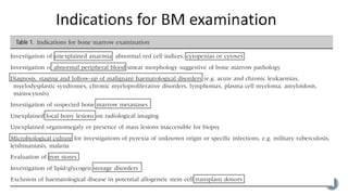
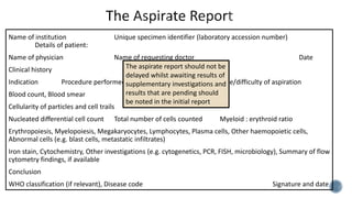
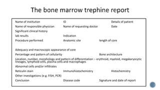
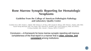
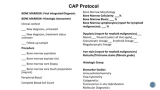
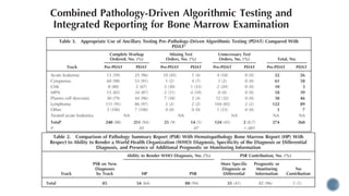
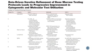
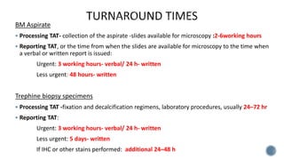
-Munira Saeed
1. Introduction
2. The Bone Marrow Aspirate
3. Bone Marrow Trephine Biopsy
4. Reporting system
5. Verbal Reports
6. Turnaround Times
7. Storage
8. External Quality Assurance
 Bone marrow (BM) aspirate and trephine biopsy examination is essential for the diagnosis &
management of blood and BM disorders
 The lack of uniformity can lead to inconsistencies in disease diagnosis and classification, and
thereby affect treatment and clinical outcomes
 In an attempt to standardize the indications for BM examination, the specimens required
and report format, a set of consensus guidelines and recommendations have been made
 Clinical indications - specimens required known
 Thrombocytopenia / coagulopathy (platelet transfusion/reversal of anticoagulation)
 A blood count and smear obtained/ 2days
 Informed consent
 Adequate sedation and analgesia
 Either (aspirate or biopsy) may be performed first - 0.5–1 cm from first site using the
respective needles- avoids hemorrhagic biopsy/ aspirate clotting
Jamshidi needle J- needle Moeller needle OnControl
 Posterior iliac crest- preferred
 Anterior iliac crest- immobile
 Medial tibial surface- infants
 Sternal aspirate- immobile,
radiotherapy pelvis, ‘dry tap’ ,biopsy
is not required
X Cardiac tamponade
X bone resorption
• 10- or 20-ml plastic
syringe, to
provide adequate
negative pressure,
attached to the
aspiration needle
• To preserve
morphology, the
syringe should not
contain anticoagulant
• Second syringe -
additional samples for
supplementary tests,
such as flow cytometry &
cytogenetics
• It is suggested that these
samples be collected
with all BM aspirates
• ≈0.5 ml of the first
draw of the aspirate-
smears by the
bedside;
• With increasing
volumes drawn=
progressive dilution of
the aspirate with PB
• Additional aspirate into
a EDTA tube to make
smears, in case the
sample clots rapidly
• amt of aspirate: amt of
EDTA - minimize EDTA
induced artefacts
A minimum of 6 smears Particle clot
section
2 particle squash (‘crush’) slide
The weight of the second
slide on the first is
sufficient to squash the
marrow particles;
no downward force should
be applied
Least possible amount of blood
should be included in the clot
sample
 2 smears and 1 squash slide - Romanowsky stain
 1 smear and 1squash slide - Prussian Blue (Perls’ reaction) + Safranin-O/ Kernecht Red
 Coverslipped
 Additional slides - cytochemistry (e.g. MPO, NSE), IHC, FISH, or archived as unfixed, unstained
smears, as required
 Low power(10X) - no & cellularity of particles, no. of megakaryocytes, abnormal cells
 Smear - higher power – morphology- cytological detail, parasites or cell inclusions- particularly
cellular detail & differential counts
 Squash - cellularity, megakaryocyte numbers, focal disease, fibrotic marrows, abnormal cells
No particles, megakaryocytes, precursors → ‘blood tap’ or peripheral blood
No particles, megakaryocytes/precursor cells present → dilute sample → qualitative evaluation
Particles with↓ cellularity → qualitative description
 Blood count and peripheral smear stained - reviewed in conjunction with aspirate
 Haemopoietic activity - compare proportions of different cell lineages & quantify abnormal cells
 Cell trails behind particles- minimally diluted with PB - well dispersed, least smudged cells
≥ 300 cells if not essential to diagnosis
≥ 500 cells ≥2 smears for precise %abnormal cell type for diagnosis and disease
No. increased -count another smear, or second observer -abnormal cell count -critical
threshold for disease stratification/ low threshold (e.g. 5%) /patchy involvement
 Total no. of cells counted - stated in the report
Comprise
blast cells, promyelocytes,
myelocytes,
metamyelocytes, band
forms, segmented
neutrophils, eosinophils,
basophils
Mast cells, promonocytes
and monocytes
lymphocytes, plasma cells
and erythroblasts
Not include
megakaryocytes,
macrophages,
osteoblasts,
osteoclasts,
stromal cells,
smudged cells or
non-haemopoietic
cells- Lymphoid
aggregates
The myeloid:
erythroid (M:E)
ratio calculated
Flow
cytometric DC-
not to be used as
surrogates for
smear NDC
Flow cytometry
and morphology-
complementary
methods- different
and valuable
information,
degree of
correlation varies
greatly.
 Prussian Blue - all initial BM aspirates
 ‘Dry tap’- core biopsy section -less reliable-decalcification removes
iron; sideroblast – imprints
 Iron store - macrophages- several particles; graded subjectively-
absent, reduced, normal, increased, or markedly increased
 Sideroblast - Total number(normal, reduced or increased)
frequency
location of granules (cytoplasmic/perinuclear)
 Ring sideroblasts - ≥5 granules – encircling ≥1/3nucleus - 100
erythroblasts
Name of institution Unique specimen identifier (laboratory accession number)
Details of patient:
Name of physician Name of requesting doctor Date
Clinical history
Indication Procedure performed site Ease/difficulty of aspiration
Blood count, Blood smear
Cellularity of particles and cell trails
Nucleated differential cell count Total number of cells counted Myeloid : erythroid ratio
Erythropoiesis, Myelopoiesis, Megakaryocytes, Lymphocytes, Plasma cells, Other haemopoietic cells,
Abnormal cells (e.g. blast cells, metastatic infiltrates)
Iron stain, Cytochemistry, Other investigations (e.g. cytogenetics, PCR, FISH, microbiology), Summary of flow
cytometry findings, if available
Conclusion
WHO classification (if relevant), Disease code Signature and date
The aspirate report should not be
delayed whilst awaiting results of
supplementary investigations and
results that are pending should
be noted in the initial report
Imprint
If no aspirate, imprint-
examine cell composition and
cytologic detail and a NDC can
be performed
Length- least 2 cm, shrinks
by ≈20% after processing
Longer in focal lesion -
bilateral - increases the
yield
labelled -name, id, date and
time of collection
.
Ischemic time
to be
monitored
Decalcification
• Shorter TAT methods not
recommended for special
studies
• Decalcification should be
followed by careful
rinsing of about 10 min
to remove decalcification
reagent
• Combined decalcifying &
fixative solutions are not
recommended
Fixation
• Heating and stirring both improve
fixation and should always be
considered
• Selection depends on the desirable
TAT
• Bouin’s solution contains picric acid –
explosive, a recent study showed that
Bouin’s did not provide IHC results
comparable with formalin
• Fixatives containing mercury (Zenker’s
and B5) suitable for IHC but are toxic
Staining
 Should be stained with H&E
 Giemsa staining may be carried out
in addition to H&E stains- identifying
plasma cells, mast cells, lymphoid
cells, eosinophils & for distinguishing
myeloblasts from proerythroblasts
 One section may be stained for
reticulin by the silver impregnation
method
 Embedded in paraffin wax / plastic
 Recommended thickness of 2 -3µ &
even
 ≥6 sections at three levels into the
cross-sectional diameter
 serial sections be mounted stepwise
on glass slides
 2-4 sections reviewed
 Overall marrow architecture and cellularity, focal lesions and patchy infiltrates
 The % cellularity- proportion of cells occupying the total marrow cavity, BM cellularity age
adjusted
 Low power - adequacy, pattern, cellularity, presence of focal lesions, megakaryocyte
number, abnormal cell clusters and location, bone structure , and osteoclastic and
osteoblastic activity
 High power - haemopoietic activity and cytological detail.
 Higher power - fine cytological details, e.g. intracellular granules, Auer rods
 Reticulin - quantified by grading from 0 to 3 (European consensus scoring system) or
alternative system, the system used must be stated
 Focal increase in reticulin (e.g. seen after therapy) should be commented on if
necessary for diagnosis
 Fiber density should be assessed only in hematopoietic areas.
 In grades MF-2 or MF-3 an additional trichrome stain is recommended by WHO
1)Pre analytical -time of procurement of the BM samples and ends by cutting the embedded tissue
onto glass slides
2)Analytical -protocols used for IHC staining (including antigen retrieval, primary antibody incubation,
incubation with detection system, color development for visualization of immunological reaction),
counterstaining, and it ends with cover slipping
3)Post analytical- interpretation of the IHC results by the (hemato)pathologist
4)Quality assurance - positive and negative controls, participation in PT provided by EQA programs,
use of flow cytometry (FCM) to validate IHC, and training and education
 Lack of standardization of the pre-analytical component- prevents building EQA/PT programs for
BM IHC
Class I (lesser risk):
IHC both interpreted and used by
pathologists:
 Qualitative- for diagnostic purposes-
determination of cell lineage
 Validation -medical director of
laboratory.
 Test performance characteristics-
descriptive- ‘positive’ or ‘negative’.
Class II (greater risk):
IHC prognostic/predictive nature-therapies /
decision-making used by treating physician
 Qualitative and quantitative- prognostic
and/or predictive markers.
 Validation- published guidelines for each
marker.
 Test performance characteristics-
descriptive/qualitative (positive vs. negative) or
(semi) quantitative (% positivity or H-score or
other results of a specific scoring system).
At present, only few Class II- relevant to BM IHC-
metastatic breast cancer, metastatic lung cancer,
or NPM-1 when molecular studies may not be
available
Recommendations
 1. CAP-ACP classification is recommended to properly design and follow QA
requirements, Class II IHC tests need higher level of QA
 2. All Class II IHC tests follow national and/or international guidelines for
recommended protocol for validation
 3. If not available, medical director prepares design for Class II markers, according to
published guidelines for validation
 4. Class II IHC tests need to be run with calibrated positive control and reagent
negative controls, used. It is recommended that (calibrated) controls are placed on
the same slide as the patient’s sample (so-called ‘on-slide’ controls)
 5. Reagent negative controls are generally not recommended for Class I IHC tests
Analytical Standards Recommendations
 1.IHC protocols need to be validated
 2.Monitoring of protocol performance
characteristics using appropriate calibrated
controls is recommended.
 3. SOPs for each IHC test that is performed in
the laboratory need to be developed
Post Analytical Standards Recommendations
 1. All BM aspirate results should be available to (hemato)pathologists directly or
indirectly smears
 2. Interpretation of IHC results should be performed in consideration of the sample
adequacy.
 3. It is recommended that the interpretation starts with the evaluation of the
external control(s).
 4. Evaluation of the patient sample starts with detection and evaluation of an internal
positive control if such is present
Quality Assurance Recommendations
 Positive and negative controls - Published guidelines should be followed whenever
possible
 Participation in proficiency testing (PT), provided by external quality assurance
programs - recommended that laboratories participate only if the EQA program
provides samples with identical or nearly identical BM tissue processing
 Use of flow cytometry to validate IHC- If FCM is available and the disease state is such
that it enables straight forward interpretation of IHC results, corresponding IHC
testing can be evaluated to compare the number of cells and the levels of expression
between the two methods
Name of institution ID Details of patient
Name of responsible physician Name of requesting doctor Date
Significant clinical history
lab results Indication
Procedure performed Anatomic site length of core
Adequacy and macroscopic appearance of core
Percentage and pattern of cellularity Bone architecture
Location, number, morphology and pattern of differentiation :- erythroid, myeloid, megakaryocytic
lineages, lymphoid cells, plasma cells and macrophages
Abnormal cells and/or infiltrates
Reticulin stain Immunohistochemistry Histochemistry
Other investigations (e.g. FISH, PCR)
Conclusion Disease code Signature and date of report
Conclusion.—A framework for bone marrow synoptic reporting will improve
completeness of the final report in a manner that is clear, concise, and
consistent among institutions
BONE MARROW: Final Integrated Diagnosis
BONE MARROW: Histologic Assessment
Clinical context
___ New diagnosis, untreated
___ New diagnosis, treatment status
unknown
___ Follow up sample
Procedure
___ Bone marrow aspiration
___ Bone marrow aspirate clot
___ Bone marrow core biopsy
___ Bone marrow core touch preparation
(imprint)
Peripheral Blood
Complete Blood Cell Count
Bone Marrow Morphology
Bone Marrow Cellularity: ___%
Bone Marrow Blasts: ___ %
Bone Marrow Lymphocytes (report for lymphoid
malignancies): ___ %
Dysplasia (report for myeloid malignancies)___
Absent___ Present (select all that apply)___
Granulocytic lineage___ Erythroid lineage___
Megakaryocytic lineage
Iron stain (report for myeloid malignancies)
Reticulin/Trichrome stains (fibrosis grade)
Histologic Group
Biomarker Studies
Immunohistochemistry
Flow Cytometry
Cytogenetics
Fluorescence in situ Hybridization
Molecular Diagnostics
CAP Protocol
When a verbal report is given to a clinician, a comment should be added to indicate the
name of the pathologist providing the information, to whom the report was given, and the
time and date
BM Aspirate
 Processing TAT- collection of the aspirate -slides available for microscopy :2-6working hours
 Reporting TAT, or the time from when the slides are available for microscopy to the time when
a verbal or written report is issued:
Urgent: 3 working hours- verbal/ 24 h- written
Less urgent: 48 hours- written
Trephine biopsy specimens
 Processing TAT -fixation and decalcification regimens, laboratory procedures, usually 24–72 hr
 Reporting TAT:
Urgent: 3 working hours- verbal/ 24 h- written
Less urgent: 5 days- written
If IHC or other stains performed: additional 24–48 h
 Spare BM aspirate slides may be wrapped tightly in aluminum foil for storage at -20 Celsius to
preserve cellular antigens.
Unfixed and unstained aspirate smears stored at room temp for long periods may give variable
results on Giemsa staining
Aspirate slides fixed in absolute methanol preserve DNA for future FISH, or DNA extraction and
subsequent PCR amplification.
 The duration of storage of BM specimens and reports should comply with national regulatory
guidelines
 Where these are not available, BM slides should be stored for at least 20 years, or indefinitely,
if possible
 Digital images and electronic reports may be stored indefinitely.
Participation in external quality assurance (EQA) schemes for both technical and interpretative
elements of BM examination are encouraged and recommended to ensure accuracy,
reproducibility and standardization.
 ICSH guidelines for the standardization of bone marrow specimens and reports, LEE ET AL,
Int. Jnl. Lab. Hem. 2008, 30, 349–364
 ICSH guidelines for the standardization of bone marrow immunohistochemistry,
TORLAKOVIC ET AL, Int. Jnl. Lab. Hem. 2015, 37, 431–449
 Protocol for the Examination of Hematologic Malignancies in Bone Marrow, Version: Bone
Marrow 4.0.0.0 Protocol Posting Date: February 2019
 Combined Pathology-Driven Algorithmic Testing and Reporting for Bone Marrow
Examination, Pearson et al, Arch Pathol Lab Med, ahead of print
 Bone Marrow Synoptic Reporting for Hematologic Neoplasms, Cordelia Sever et al, Arch
Pathol Lab Med—Vol 140, September 2016
 Data-Driven Iterative Refinement of Bone Marrow Testing Protocols Leads to Progressive
Improvement in Cytogenetic and Molecular Test Utilization, Seegmiller et al, Am J Clin
Pathol 2016;146:585-593
 Diagnostic testing managed by hematopathology specialty and other laboratories: costs
and patient diagnostic outcomes, Engel-Nitz et al, BMC Clinical Pathology 2014
 Making the most of bone marrow trephine biopsy, Wilkins et al, (2009) Histopathology 55,
631–640
 Optimal processing of bone marrow trephine biopsy: the Hammersmith Protocol, Naresh et
al, J Clin Pathol 2006;59:903–911
 Bone Marrow Biopsy Needle Type Affects Core BiopsySpecimen Length and Quality and
Aspirate Hemodilution,Brestoff et al, Am J Clin Pathol February 2019;151:185-193
 Comparison of the bone marrow trephine sample quality between OnControl drill system and
the Jamshidi needle, Forwood et al, Int J Lab Hematol. 2019;1–7
 Bone marrow solid core biopsy needle: a critical assessment of the utility, benefits and
limitations of the instruments employed in current day haematology and oncology,Islam A. J
Clin Pathol 2017
 Immunohistochemistry of Bone-Marrow Biopsy, STEFAN0 ET
AL, (1997) Immunohistochemistry of Bone-Marrow Biopsy, Leukemia &
Lymphoma, 26:sup1, 69-75

Standardization of bone marrow specimen processing; immunohistochemistry and reporting

  • 1.
  • 2.
    1. Introduction 2. TheBone Marrow Aspirate 3. Bone Marrow Trephine Biopsy 4. Reporting system 5. Verbal Reports 6. Turnaround Times 7. Storage 8. External Quality Assurance
  • 3.
     Bone marrow(BM) aspirate and trephine biopsy examination is essential for the diagnosis & management of blood and BM disorders  The lack of uniformity can lead to inconsistencies in disease diagnosis and classification, and thereby affect treatment and clinical outcomes  In an attempt to standardize the indications for BM examination, the specimens required and report format, a set of consensus guidelines and recommendations have been made
  • 5.
     Clinical indications- specimens required known  Thrombocytopenia / coagulopathy (platelet transfusion/reversal of anticoagulation)  A blood count and smear obtained/ 2days  Informed consent  Adequate sedation and analgesia  Either (aspirate or biopsy) may be performed first - 0.5–1 cm from first site using the respective needles- avoids hemorrhagic biopsy/ aspirate clotting
  • 7.
    Jamshidi needle J-needle Moeller needle OnControl
  • 8.
     Posterior iliaccrest- preferred  Anterior iliac crest- immobile  Medial tibial surface- infants  Sternal aspirate- immobile, radiotherapy pelvis, ‘dry tap’ ,biopsy is not required X Cardiac tamponade X bone resorption
  • 9.
    • 10- or20-ml plastic syringe, to provide adequate negative pressure, attached to the aspiration needle • To preserve morphology, the syringe should not contain anticoagulant
  • 10.
    • Second syringe- additional samples for supplementary tests, such as flow cytometry & cytogenetics • It is suggested that these samples be collected with all BM aspirates • ≈0.5 ml of the first draw of the aspirate- smears by the bedside; • With increasing volumes drawn= progressive dilution of the aspirate with PB • Additional aspirate into a EDTA tube to make smears, in case the sample clots rapidly • amt of aspirate: amt of EDTA - minimize EDTA induced artefacts
  • 11.
    A minimum of6 smears Particle clot section 2 particle squash (‘crush’) slide The weight of the second slide on the first is sufficient to squash the marrow particles; no downward force should be applied Least possible amount of blood should be included in the clot sample
  • 12.
     2 smearsand 1 squash slide - Romanowsky stain  1 smear and 1squash slide - Prussian Blue (Perls’ reaction) + Safranin-O/ Kernecht Red  Coverslipped  Additional slides - cytochemistry (e.g. MPO, NSE), IHC, FISH, or archived as unfixed, unstained smears, as required
  • 13.
     Low power(10X)- no & cellularity of particles, no. of megakaryocytes, abnormal cells  Smear - higher power – morphology- cytological detail, parasites or cell inclusions- particularly cellular detail & differential counts  Squash - cellularity, megakaryocyte numbers, focal disease, fibrotic marrows, abnormal cells No particles, megakaryocytes, precursors → ‘blood tap’ or peripheral blood No particles, megakaryocytes/precursor cells present → dilute sample → qualitative evaluation Particles with↓ cellularity → qualitative description  Blood count and peripheral smear stained - reviewed in conjunction with aspirate
  • 14.
     Haemopoietic activity- compare proportions of different cell lineages & quantify abnormal cells  Cell trails behind particles- minimally diluted with PB - well dispersed, least smudged cells ≥ 300 cells if not essential to diagnosis ≥ 500 cells ≥2 smears for precise %abnormal cell type for diagnosis and disease No. increased -count another smear, or second observer -abnormal cell count -critical threshold for disease stratification/ low threshold (e.g. 5%) /patchy involvement  Total no. of cells counted - stated in the report
  • 15.
    Comprise blast cells, promyelocytes, myelocytes, metamyelocytes,band forms, segmented neutrophils, eosinophils, basophils Mast cells, promonocytes and monocytes lymphocytes, plasma cells and erythroblasts Not include megakaryocytes, macrophages, osteoblasts, osteoclasts, stromal cells, smudged cells or non-haemopoietic cells- Lymphoid aggregates The myeloid: erythroid (M:E) ratio calculated Flow cytometric DC- not to be used as surrogates for smear NDC Flow cytometry and morphology- complementary methods- different and valuable information, degree of correlation varies greatly.
  • 16.
     Prussian Blue- all initial BM aspirates  ‘Dry tap’- core biopsy section -less reliable-decalcification removes iron; sideroblast – imprints  Iron store - macrophages- several particles; graded subjectively- absent, reduced, normal, increased, or markedly increased  Sideroblast - Total number(normal, reduced or increased) frequency location of granules (cytoplasmic/perinuclear)  Ring sideroblasts - ≥5 granules – encircling ≥1/3nucleus - 100 erythroblasts
  • 17.
    Name of institutionUnique specimen identifier (laboratory accession number) Details of patient: Name of physician Name of requesting doctor Date Clinical history Indication Procedure performed site Ease/difficulty of aspiration Blood count, Blood smear Cellularity of particles and cell trails Nucleated differential cell count Total number of cells counted Myeloid : erythroid ratio Erythropoiesis, Myelopoiesis, Megakaryocytes, Lymphocytes, Plasma cells, Other haemopoietic cells, Abnormal cells (e.g. blast cells, metastatic infiltrates) Iron stain, Cytochemistry, Other investigations (e.g. cytogenetics, PCR, FISH, microbiology), Summary of flow cytometry findings, if available Conclusion WHO classification (if relevant), Disease code Signature and date The aspirate report should not be delayed whilst awaiting results of supplementary investigations and results that are pending should be noted in the initial report
  • 19.
    Imprint If no aspirate,imprint- examine cell composition and cytologic detail and a NDC can be performed Length- least 2 cm, shrinks by ≈20% after processing Longer in focal lesion - bilateral - increases the yield labelled -name, id, date and time of collection . Ischemic time to be monitored
  • 20.
    Decalcification • Shorter TATmethods not recommended for special studies • Decalcification should be followed by careful rinsing of about 10 min to remove decalcification reagent • Combined decalcifying & fixative solutions are not recommended Fixation • Heating and stirring both improve fixation and should always be considered • Selection depends on the desirable TAT • Bouin’s solution contains picric acid – explosive, a recent study showed that Bouin’s did not provide IHC results comparable with formalin • Fixatives containing mercury (Zenker’s and B5) suitable for IHC but are toxic
  • 21.
    Staining  Should bestained with H&E  Giemsa staining may be carried out in addition to H&E stains- identifying plasma cells, mast cells, lymphoid cells, eosinophils & for distinguishing myeloblasts from proerythroblasts  One section may be stained for reticulin by the silver impregnation method  Embedded in paraffin wax / plastic  Recommended thickness of 2 -3µ & even  ≥6 sections at three levels into the cross-sectional diameter  serial sections be mounted stepwise on glass slides
  • 22.
     2-4 sectionsreviewed  Overall marrow architecture and cellularity, focal lesions and patchy infiltrates  The % cellularity- proportion of cells occupying the total marrow cavity, BM cellularity age adjusted  Low power - adequacy, pattern, cellularity, presence of focal lesions, megakaryocyte number, abnormal cell clusters and location, bone structure , and osteoclastic and osteoblastic activity  High power - haemopoietic activity and cytological detail.  Higher power - fine cytological details, e.g. intracellular granules, Auer rods
  • 23.
     Reticulin -quantified by grading from 0 to 3 (European consensus scoring system) or alternative system, the system used must be stated  Focal increase in reticulin (e.g. seen after therapy) should be commented on if necessary for diagnosis  Fiber density should be assessed only in hematopoietic areas.  In grades MF-2 or MF-3 an additional trichrome stain is recommended by WHO
  • 24.
    1)Pre analytical -timeof procurement of the BM samples and ends by cutting the embedded tissue onto glass slides 2)Analytical -protocols used for IHC staining (including antigen retrieval, primary antibody incubation, incubation with detection system, color development for visualization of immunological reaction), counterstaining, and it ends with cover slipping 3)Post analytical- interpretation of the IHC results by the (hemato)pathologist 4)Quality assurance - positive and negative controls, participation in PT provided by EQA programs, use of flow cytometry (FCM) to validate IHC, and training and education  Lack of standardization of the pre-analytical component- prevents building EQA/PT programs for BM IHC
  • 25.
    Class I (lesserrisk): IHC both interpreted and used by pathologists:  Qualitative- for diagnostic purposes- determination of cell lineage  Validation -medical director of laboratory.  Test performance characteristics- descriptive- ‘positive’ or ‘negative’. Class II (greater risk): IHC prognostic/predictive nature-therapies / decision-making used by treating physician  Qualitative and quantitative- prognostic and/or predictive markers.  Validation- published guidelines for each marker.  Test performance characteristics- descriptive/qualitative (positive vs. negative) or (semi) quantitative (% positivity or H-score or other results of a specific scoring system). At present, only few Class II- relevant to BM IHC- metastatic breast cancer, metastatic lung cancer, or NPM-1 when molecular studies may not be available
  • 26.
    Recommendations  1. CAP-ACPclassification is recommended to properly design and follow QA requirements, Class II IHC tests need higher level of QA  2. All Class II IHC tests follow national and/or international guidelines for recommended protocol for validation  3. If not available, medical director prepares design for Class II markers, according to published guidelines for validation  4. Class II IHC tests need to be run with calibrated positive control and reagent negative controls, used. It is recommended that (calibrated) controls are placed on the same slide as the patient’s sample (so-called ‘on-slide’ controls)  5. Reagent negative controls are generally not recommended for Class I IHC tests
  • 27.
    Analytical Standards Recommendations 1.IHC protocols need to be validated  2.Monitoring of protocol performance characteristics using appropriate calibrated controls is recommended.  3. SOPs for each IHC test that is performed in the laboratory need to be developed
  • 28.
    Post Analytical StandardsRecommendations  1. All BM aspirate results should be available to (hemato)pathologists directly or indirectly smears  2. Interpretation of IHC results should be performed in consideration of the sample adequacy.  3. It is recommended that the interpretation starts with the evaluation of the external control(s).  4. Evaluation of the patient sample starts with detection and evaluation of an internal positive control if such is present
  • 29.
    Quality Assurance Recommendations Positive and negative controls - Published guidelines should be followed whenever possible  Participation in proficiency testing (PT), provided by external quality assurance programs - recommended that laboratories participate only if the EQA program provides samples with identical or nearly identical BM tissue processing  Use of flow cytometry to validate IHC- If FCM is available and the disease state is such that it enables straight forward interpretation of IHC results, corresponding IHC testing can be evaluated to compare the number of cells and the levels of expression between the two methods
  • 30.
    Name of institutionID Details of patient Name of responsible physician Name of requesting doctor Date Significant clinical history lab results Indication Procedure performed Anatomic site length of core Adequacy and macroscopic appearance of core Percentage and pattern of cellularity Bone architecture Location, number, morphology and pattern of differentiation :- erythroid, myeloid, megakaryocytic lineages, lymphoid cells, plasma cells and macrophages Abnormal cells and/or infiltrates Reticulin stain Immunohistochemistry Histochemistry Other investigations (e.g. FISH, PCR) Conclusion Disease code Signature and date of report
  • 32.
    Conclusion.—A framework forbone marrow synoptic reporting will improve completeness of the final report in a manner that is clear, concise, and consistent among institutions
  • 33.
    BONE MARROW: FinalIntegrated Diagnosis BONE MARROW: Histologic Assessment Clinical context ___ New diagnosis, untreated ___ New diagnosis, treatment status unknown ___ Follow up sample Procedure ___ Bone marrow aspiration ___ Bone marrow aspirate clot ___ Bone marrow core biopsy ___ Bone marrow core touch preparation (imprint) Peripheral Blood Complete Blood Cell Count Bone Marrow Morphology Bone Marrow Cellularity: ___% Bone Marrow Blasts: ___ % Bone Marrow Lymphocytes (report for lymphoid malignancies): ___ % Dysplasia (report for myeloid malignancies)___ Absent___ Present (select all that apply)___ Granulocytic lineage___ Erythroid lineage___ Megakaryocytic lineage Iron stain (report for myeloid malignancies) Reticulin/Trichrome stains (fibrosis grade) Histologic Group Biomarker Studies Immunohistochemistry Flow Cytometry Cytogenetics Fluorescence in situ Hybridization Molecular Diagnostics CAP Protocol
  • 36.
    When a verbalreport is given to a clinician, a comment should be added to indicate the name of the pathologist providing the information, to whom the report was given, and the time and date
  • 37.
    BM Aspirate  ProcessingTAT- collection of the aspirate -slides available for microscopy :2-6working hours  Reporting TAT, or the time from when the slides are available for microscopy to the time when a verbal or written report is issued: Urgent: 3 working hours- verbal/ 24 h- written Less urgent: 48 hours- written Trephine biopsy specimens  Processing TAT -fixation and decalcification regimens, laboratory procedures, usually 24–72 hr  Reporting TAT: Urgent: 3 working hours- verbal/ 24 h- written Less urgent: 5 days- written If IHC or other stains performed: additional 24–48 h
  • 38.
     Spare BMaspirate slides may be wrapped tightly in aluminum foil for storage at -20 Celsius to preserve cellular antigens. Unfixed and unstained aspirate smears stored at room temp for long periods may give variable results on Giemsa staining Aspirate slides fixed in absolute methanol preserve DNA for future FISH, or DNA extraction and subsequent PCR amplification.  The duration of storage of BM specimens and reports should comply with national regulatory guidelines  Where these are not available, BM slides should be stored for at least 20 years, or indefinitely, if possible  Digital images and electronic reports may be stored indefinitely.
  • 39.
    Participation in externalquality assurance (EQA) schemes for both technical and interpretative elements of BM examination are encouraged and recommended to ensure accuracy, reproducibility and standardization.
  • 40.
     ICSH guidelinesfor the standardization of bone marrow specimens and reports, LEE ET AL, Int. Jnl. Lab. Hem. 2008, 30, 349–364  ICSH guidelines for the standardization of bone marrow immunohistochemistry, TORLAKOVIC ET AL, Int. Jnl. Lab. Hem. 2015, 37, 431–449  Protocol for the Examination of Hematologic Malignancies in Bone Marrow, Version: Bone Marrow 4.0.0.0 Protocol Posting Date: February 2019  Combined Pathology-Driven Algorithmic Testing and Reporting for Bone Marrow Examination, Pearson et al, Arch Pathol Lab Med, ahead of print  Bone Marrow Synoptic Reporting for Hematologic Neoplasms, Cordelia Sever et al, Arch Pathol Lab Med—Vol 140, September 2016  Data-Driven Iterative Refinement of Bone Marrow Testing Protocols Leads to Progressive Improvement in Cytogenetic and Molecular Test Utilization, Seegmiller et al, Am J Clin Pathol 2016;146:585-593  Diagnostic testing managed by hematopathology specialty and other laboratories: costs and patient diagnostic outcomes, Engel-Nitz et al, BMC Clinical Pathology 2014
  • 41.
     Making themost of bone marrow trephine biopsy, Wilkins et al, (2009) Histopathology 55, 631–640  Optimal processing of bone marrow trephine biopsy: the Hammersmith Protocol, Naresh et al, J Clin Pathol 2006;59:903–911  Bone Marrow Biopsy Needle Type Affects Core BiopsySpecimen Length and Quality and Aspirate Hemodilution,Brestoff et al, Am J Clin Pathol February 2019;151:185-193  Comparison of the bone marrow trephine sample quality between OnControl drill system and the Jamshidi needle, Forwood et al, Int J Lab Hematol. 2019;1–7  Bone marrow solid core biopsy needle: a critical assessment of the utility, benefits and limitations of the instruments employed in current day haematology and oncology,Islam A. J Clin Pathol 2017  Immunohistochemistry of Bone-Marrow Biopsy, STEFAN0 ET AL, (1997) Immunohistochemistry of Bone-Marrow Biopsy, Leukemia & Lymphoma, 26:sup1, 69-75

Editor's Notes

  • #3 This topic will be covered under the following head
  • #5 Work up of stem cell transplant donors
  • #6 It is important tht the operator…. It is recommended that both BM aspirate and biopsy be routinely performed so that respective findings can be correlated. However, in some clinical situations, a BM aspirate alone may suffice If a trephine biopsy is indicated and not performed, or is not obtainable, a particle clot preparation is recommended. If an aspirate is unobtainable (e.g. in a ‘dry tap’), trephine imprints should be prepared
  • #7 The difff types
  • #8 .Triple- shorter and more crush artifact than hemodilute specimen., core-capturing device by the sleeve, OnControl system was associated with more crush artefact
  • #9 Sternal aspiration should only be performed by an experienced operator because of the risk of cardiac tamponade should not be attempted in patients with suspected plasma cell myeloma or other disorders associated with bone resorption
  • #11 . The samples may be discarded if the investigations are considered to be unnecessary after the BM slides have been reviewed.
  • #12 In oyr lab 12 smears are made...Topography maintained, May harbor diagnosis, Information before trephine is ready, An important advantage of particle clot preparations is the lack of decalcification-associated nucleic acid or protein, better preservation, IHC can be done
  • #13 Two air-dried smears and one squash slide should be fixed with fresh acetone-free absolute methanol and stained with a Romanowsky stain, such as May – Grunwald Giemsa (MGG) (Lewis, Bain & Bates,2006) or Wright Giemsa stain…. All BM smears should be coverslipped using a mounting medium that hardens and dries rapidly. Toxic – fume hood
  • #14 (e.g. lymphoma, plasma cell myeloma, mast cells, metastatic carcinoma, granulomas),
  • #15 Different cell lineages with known reference ranges
  • #17  A BM smear with increased iron stores should be included as a positive control
  • #20 Ischemic timebiopsy imprints (touch preps) be prepared at the bedside by trained professional assisting the procedure before the sample sample placed in the container with fixative immediately at the bedside…If transportation of unfixed sample is required, the transportation time of the sample to the laboratory needs to be monitored, and it should be as short as possible dehydration of the BM trephine biopsy needs to be prevented by transporting the sample in a closed container with physiological saline, PBS
  • #21 Fixation time should be maintained otherwise ZONING PHENOMENON occurs
  • #25 there is more than one tissue processing protocol (various combinations of various fixatives and decalcifying reagents with significant time range for both fixation and decalcification) that may produce excellent results for IHC testing, as well as good quality of DNA and RNA. However, the large number of protocols prevents standardization, may create difficulties for external consultation/review, and hinders development of EQA
  • #26 the Canadian Association of Pathologists (CAP-ACP) developed classification of IHC tests based on the ‘risk assessment’
  • #27 Canadian Association of Pathologists Due to higher risk for patient safety,…. that are used for stratification for definitive targeted therapy need to …. Calibrated positive controls need to include negative tissues, weakly positive tissues, and strongly positive tissues
  • #29 External controls ), by which it is determined that the proper antibody was applied and the test is properly calibrated
  • #32 Integerated reporting system be followed
  • #34 Plasma cell myeloma, lymphoma or metastatic cancer
  • #35 PEARSON ET AL,,, Conclusions.—Pathology-driven algorithmic testing with integrated reporting improves the pathologist’s ability to render a specific WHO diagnosis and improves test utilization. Clinicians prefer PDAT to clinician-ordered testing.
  • #36 Seegmiller et al…group of pathologists and clinical hematologists worked together to develop standard ordering protocols (SOPs) defining which cytogenetic and molecular tests should be performed on bone marrow biopsy specimens