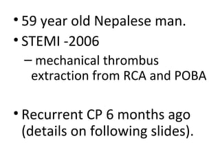
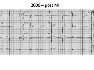
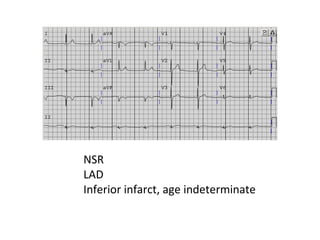
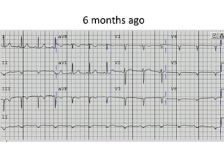
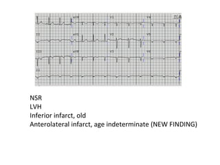
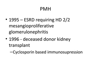
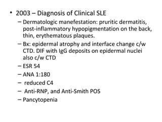
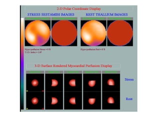
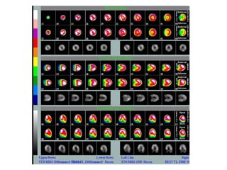
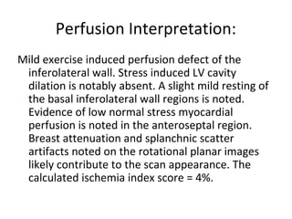
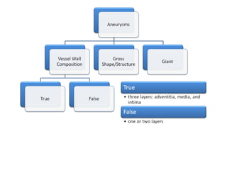
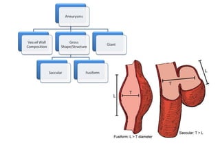
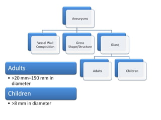
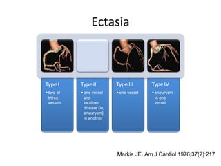
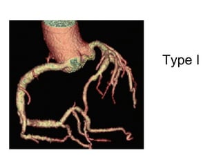
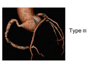
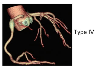
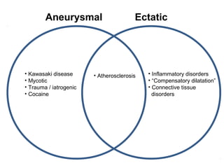
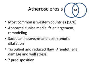
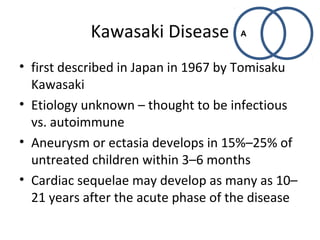
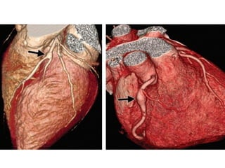
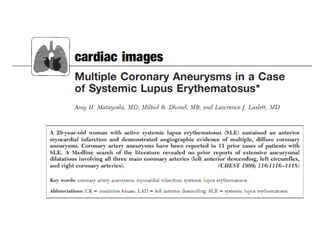
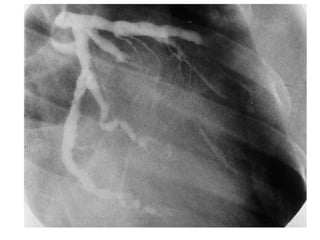
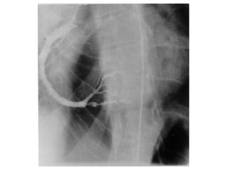
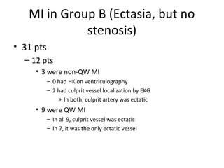
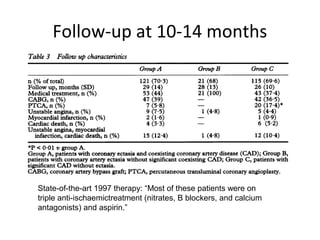
Cardiology Division 
Morning Report 
Michael G. Katz, M.D. 
Fellow in Cardiovascular Disease 
University of Rochester 
December 13, 2010
• 59 year old Nepalese man. 
• STEMI -2006 
– mechanical thrombus 
extraction from RCA and POBA 
• Recurrent CP 6 months ago 
(details on following slides).
2006 – post MI
NSR 
LAD 
Inferior infarct, age indeterminate
6 months ago
NSR 
LVH 
Inferior infarct, old 
Anterolateral infarct, age indeterminate (NEW FINDING)
PMH 
• 1995 – ESRD requiring HD 2/2 
mesangioproliferative 
glomerulonephritis 
• 1996 - deceased donor kidney 
transplant 
–Cyclosporin based immunosupression
• 2003 – Diagnosis of Clinical SLE 
– Dermatologic manefestation: pruritic dermatitis, 
post-inflammatory hypopigmentation on the back, 
thin, erythematous plaques. 
– Bx: epidermal atrophy and interface change c/w 
CTD. DIF with IgG deposits on epidermal nuclei 
also c/w CTD 
– ESR 54 
– ANA 1:180 
– reduced C4 
– Anti-RNP, and Anti-Smith POS 
– Pancytopenia
– Rash had excellent response to topical steroids 
and prednisone taper. 
– Started on hydroxychloroquine in an effort to 
reduce prednisone over time. 
– Plan to reduce prednisone hampered by skin 
changes / pain in fingers and hands. 
– CellCept added 2007 
– Essentially stable with waxing and waning skin 
symptoms.
Review of LHC from 2006 STEMI
• February 2010: several episodes of vague 
chest discomfort and palpitations. Several ED 
visits, where MI was ruled out. 
– Holter: occasional PVCs and PACs but no evidence 
of VT or sustained arrhythmia 
– SPECT nuclear stress:
Perfusion Interpretation: 
Mild exercise induced perfusion defect of the 
inferolateral wall. Stress induced LV cavity 
dilation is notably absent. A slight mild resting of 
the basal inferolateral wall regions is noted. 
Evidence of low normal stress myocardial 
perfusion is noted in the anteroseptal region. 
Breast attenuation and splanchnic scatter 
artifacts noted on the rotational planar images 
likely contribute to the scan appearance. The 
calculated ischemia index score = 4%.
LHC 2010
• At his post cath f/u visit: 
– Re-started on clopidogrel (which he had not been taking) 
– Continued on low-dose ASA 
– Started on TUMS and ranitidine (to avoid potential PPI 
interaction with clopidogrel) for empiric treatment of 
GERD. 
• October 2010 
– Dramatic improvement in “chest pressure” with antacid. 
– Self-discontinued clopidogrel after easy bruising on arms 
and hands. 
– TC 112 TG 78 HDL 46 LDL 50 TC/HDL 2.4 
– CRP 2
Meds 
• Amlodipine- 
Benazepril 
• Aspirin 81 
• Cellcept 
• Cyclosporine 
• Hydroxychloroquine 
• Lotrel 5/10 mg 
• Metoprolol 
• mycophenolate 
mofetil 
• Nitroglycerin 
• Pravastatin 
• Prednisone 
• Ranitidine 
• Sildenafil
• Brief summary 
• DDx
Coronary Aneurysms and Ectasia 
• Terminology and Classification 
• Etiology and pathogeneisis 
• Examples
Giovanni Morgagni 
1761 
Syphilitic aortitis
Aneuyrsm
Ectasia
Ectasia 
Markis JE. Am J Cardiol 1976;37(2):217
Type I
Type III
Type IV
Epidemiology 
• 0.5 – 5% prevalence 
• More frequent in men than women 
– 2.2% vs. 0.02% 
• Age predilection varies by etiology
Aneurysmal Ectatic 
• Kawasaki disease 
• Mycotic 
• Trauma / iatrogenic 
• Cocaine 
• Atherosclerosis • Inflammatory disorders 
• “Compensatory dilatation” 
• Connective tissue 
disorders
A/E Atherosclerosis 
• Most common is western countries (50%) 
• Abnormal tunica media  enlargement, 
remodeling 
• Saccular aneurysms and post-stenotic 
dilatation 
• Turbulent and reduced flow  endothelial 
damage and wall stress 
• ? predisposition
• Usually multiple and involve more than one 
coronary artery 
– As opposed to congenital, traumatic, or dissection 
• RCA (40%–61%), LAD (15%–32%), and LCx 
(15%–23%). LM involvement rare (0.1%–3.5%)
Kawasaki Disease 
A 
• first described in Japan in 1967 by Tomisaku 
Kawasaki 
• Etiology unknown – thought to be infectious 
vs. autoimmune 
• Aneurysm or ectasia develops in 15%–25% of 
untreated children within 3–6 months 
• Cardiac sequelae may develop as many as 10– 
21 years after the acute phase of the disease
• By 2 years after the onset of Kawasaki disease, 
49% of the patients have spontaneous 
regression of aneurysms 
• LM (12%), RCA (3%), both arteries in 8%
Misc. 
• Iatrogenic 
– Trauma from oversized balloon or high inflation pressures, coronary 
dissection; compounded by inadequate healing because of antiproliferative 
treatment with cortisone, colchicine, and antiinflammatory drugs 
• Mycotic 
– Infection with S.A., P.A., syphilis, Lyme disease 
– Microembolization to vasa vasorum, direct pathogen invasion of arterial wall, 
immune com- 
– plex deposition 
• Cocaine 
– Direct endothelial damage from severe episodic hypertension, 
vasoconstriction, and underlying atherosclerosis 
A
Inflammatory 
Disorders 
• Takayasu arteritis, SLE, RA, Wegener 
granulomatosis, GCA, Churg-Strauss 
syndrome, MPA, APS, Behçet syndrome, 
sarcoid, PAN, CREST syndrome, AS, Reiter 
syndrome, psoriatic arthritis 
• Inflammatory mediators: VCAM-1, ICAM-1, E 
selectin 
E
Takayasu arteritis 
• large-vessel vasculitis 
• young women 
• marked thinning of the tunica media 
• of the vessel, disruption of the elastic fibers, and 
thickening of the tunica adventitia and intima. 
• Coronary involvement: 12% 
• Signs and symptoms are due to ischemia secondary 
to arterial stenosis or occlusion 
• Aneurysms and ectatic collateral vessels as a 
compensatory mechanism
Compensatory 
dilatation 
• Fistula 
– Compensatory dilatation secondary to high-flow 
E 
state 
– originate from the RCA in 52% of cases; LAD 30%, 
and LCx 18%. 
– drainage is to right chambers, direct volume 
overload to the pulmonary vascular bed, the left 
atrium, and left ventricle; whereas if drainage to 
left cardiac chambers overload spares the 
pulmonary vasculature
• Coronary Artery Anomalies – eg. anomalous 
origin of the left coronary artery from the 
pulmonary artery (ALCAPA) syndrome (or 
Bland-White-Garland syndrome) 
– 1/300,000 live births 
– LM from PA  Ectatic RCA (due to elevate 
pressures)  R to L collaterals and steal 
phenomena in RCA terrirory 
E
• Connective Tissue Disorders 
– Ehlers-Danlos syndrome, Marfan syndrome, cystic 
medial necrosis 
– IL-6, C-reactive protein MMP-2, MMP-9 
E
• What is the most likely Dx? 
• W/u? 
• Antiplatelet therapy? 
• Anticoagulation?
• Group A –Ectasia and obstructive CAD 
• Group B – Ectasia, but no stenosis 
• Group C – Obstructive CAD, but no ectasia
Group A –Ectasia and obstructive 
CAD
In patients with ectasia, was it 
specifically associated with 
stenosis? – No.
MI in Group B (Ectasia, but no 
stenosis) 
• 31 pts 
– 12 pts 
• 3 were non-QW MI 
– 0 had HK on ventriculography 
– 2 had culprit vessel localization by EKG 
» In both, culprit artery was ectatic 
• 9 were QW MI 
– In all 9, culprit vessel was ectatic 
– In 7, it was the only ectatic vessel
Follow-up at 10-14 months 
State-of-the-art 1997 therapy: “Most of these patients were on 
triple anti-ischaemictreatment (nitrates, B blockers, and calcium 
antagonists) and aspirin.”

Coronary Artery Aneurysms and Ectasia

  • 1.
    Cardiology Division MorningReport Michael G. Katz, M.D. Fellow in Cardiovascular Disease University of Rochester December 13, 2010
  • 2.
    • 59 yearold Nepalese man. • STEMI -2006 – mechanical thrombus extraction from RCA and POBA • Recurrent CP 6 months ago (details on following slides).
  • 3.
  • 4.
    NSR LAD Inferiorinfarct, age indeterminate
  • 5.
  • 6.
    NSR LVH Inferiorinfarct, old Anterolateral infarct, age indeterminate (NEW FINDING)
  • 7.
    PMH • 1995– ESRD requiring HD 2/2 mesangioproliferative glomerulonephritis • 1996 - deceased donor kidney transplant –Cyclosporin based immunosupression
  • 8.
    • 2003 –Diagnosis of Clinical SLE – Dermatologic manefestation: pruritic dermatitis, post-inflammatory hypopigmentation on the back, thin, erythematous plaques. – Bx: epidermal atrophy and interface change c/w CTD. DIF with IgG deposits on epidermal nuclei also c/w CTD – ESR 54 – ANA 1:180 – reduced C4 – Anti-RNP, and Anti-Smith POS – Pancytopenia
  • 9.
    – Rash hadexcellent response to topical steroids and prednisone taper. – Started on hydroxychloroquine in an effort to reduce prednisone over time. – Plan to reduce prednisone hampered by skin changes / pain in fingers and hands. – CellCept added 2007 – Essentially stable with waxing and waning skin symptoms.
  • 10.
    Review of LHCfrom 2006 STEMI
  • 11.
    • February 2010:several episodes of vague chest discomfort and palpitations. Several ED visits, where MI was ruled out. – Holter: occasional PVCs and PACs but no evidence of VT or sustained arrhythmia – SPECT nuclear stress:
  • 14.
    Perfusion Interpretation: Mildexercise induced perfusion defect of the inferolateral wall. Stress induced LV cavity dilation is notably absent. A slight mild resting of the basal inferolateral wall regions is noted. Evidence of low normal stress myocardial perfusion is noted in the anteroseptal region. Breast attenuation and splanchnic scatter artifacts noted on the rotational planar images likely contribute to the scan appearance. The calculated ischemia index score = 4%.
  • 15.
  • 16.
    • At hispost cath f/u visit: – Re-started on clopidogrel (which he had not been taking) – Continued on low-dose ASA – Started on TUMS and ranitidine (to avoid potential PPI interaction with clopidogrel) for empiric treatment of GERD. • October 2010 – Dramatic improvement in “chest pressure” with antacid. – Self-discontinued clopidogrel after easy bruising on arms and hands. – TC 112 TG 78 HDL 46 LDL 50 TC/HDL 2.4 – CRP 2
  • 17.
    Meds • Amlodipine- Benazepril • Aspirin 81 • Cellcept • Cyclosporine • Hydroxychloroquine • Lotrel 5/10 mg • Metoprolol • mycophenolate mofetil • Nitroglycerin • Pravastatin • Prednisone • Ranitidine • Sildenafil
  • 18.
  • 19.
    Coronary Aneurysms andEctasia • Terminology and Classification • Etiology and pathogeneisis • Examples
  • 20.
    Giovanni Morgagni 1761 Syphilitic aortitis
  • 21.
  • 22.
  • 27.
    Ectasia Markis JE.Am J Cardiol 1976;37(2):217
  • 28.
  • 29.
  • 30.
  • 31.
    Epidemiology • 0.5– 5% prevalence • More frequent in men than women – 2.2% vs. 0.02% • Age predilection varies by etiology
  • 32.
    Aneurysmal Ectatic •Kawasaki disease • Mycotic • Trauma / iatrogenic • Cocaine • Atherosclerosis • Inflammatory disorders • “Compensatory dilatation” • Connective tissue disorders
  • 33.
    A/E Atherosclerosis •Most common is western countries (50%) • Abnormal tunica media  enlargement, remodeling • Saccular aneurysms and post-stenotic dilatation • Turbulent and reduced flow  endothelial damage and wall stress • ? predisposition
  • 34.
    • Usually multipleand involve more than one coronary artery – As opposed to congenital, traumatic, or dissection • RCA (40%–61%), LAD (15%–32%), and LCx (15%–23%). LM involvement rare (0.1%–3.5%)
  • 35.
    Kawasaki Disease A • first described in Japan in 1967 by Tomisaku Kawasaki • Etiology unknown – thought to be infectious vs. autoimmune • Aneurysm or ectasia develops in 15%–25% of untreated children within 3–6 months • Cardiac sequelae may develop as many as 10– 21 years after the acute phase of the disease
  • 36.
    • By 2years after the onset of Kawasaki disease, 49% of the patients have spontaneous regression of aneurysms • LM (12%), RCA (3%), both arteries in 8%
  • 38.
    Misc. • Iatrogenic – Trauma from oversized balloon or high inflation pressures, coronary dissection; compounded by inadequate healing because of antiproliferative treatment with cortisone, colchicine, and antiinflammatory drugs • Mycotic – Infection with S.A., P.A., syphilis, Lyme disease – Microembolization to vasa vasorum, direct pathogen invasion of arterial wall, immune com- – plex deposition • Cocaine – Direct endothelial damage from severe episodic hypertension, vasoconstriction, and underlying atherosclerosis A
  • 39.
    Inflammatory Disorders •Takayasu arteritis, SLE, RA, Wegener granulomatosis, GCA, Churg-Strauss syndrome, MPA, APS, Behçet syndrome, sarcoid, PAN, CREST syndrome, AS, Reiter syndrome, psoriatic arthritis • Inflammatory mediators: VCAM-1, ICAM-1, E selectin E
  • 40.
    Takayasu arteritis •large-vessel vasculitis • young women • marked thinning of the tunica media • of the vessel, disruption of the elastic fibers, and thickening of the tunica adventitia and intima. • Coronary involvement: 12% • Signs and symptoms are due to ischemia secondary to arterial stenosis or occlusion • Aneurysms and ectatic collateral vessels as a compensatory mechanism
  • 43.
    Compensatory dilatation •Fistula – Compensatory dilatation secondary to high-flow E state – originate from the RCA in 52% of cases; LAD 30%, and LCx 18%. – drainage is to right chambers, direct volume overload to the pulmonary vascular bed, the left atrium, and left ventricle; whereas if drainage to left cardiac chambers overload spares the pulmonary vasculature
  • 44.
    • Coronary ArteryAnomalies – eg. anomalous origin of the left coronary artery from the pulmonary artery (ALCAPA) syndrome (or Bland-White-Garland syndrome) – 1/300,000 live births – LM from PA  Ectatic RCA (due to elevate pressures)  R to L collaterals and steal phenomena in RCA terrirory E
  • 47.
    • Connective TissueDisorders – Ehlers-Danlos syndrome, Marfan syndrome, cystic medial necrosis – IL-6, C-reactive protein MMP-2, MMP-9 E
  • 48.
    • What isthe most likely Dx? • W/u? • Antiplatelet therapy? • Anticoagulation?
  • 54.
    • Group A–Ectasia and obstructive CAD • Group B – Ectasia, but no stenosis • Group C – Obstructive CAD, but no ectasia
  • 55.
    Group A –Ectasiaand obstructive CAD
  • 56.
    In patients withectasia, was it specifically associated with stenosis? – No.
  • 57.
    MI in GroupB (Ectasia, but no stenosis) • 31 pts – 12 pts • 3 were non-QW MI – 0 had HK on ventriculography – 2 had culprit vessel localization by EKG » In both, culprit artery was ectatic • 9 were QW MI – In all 9, culprit vessel was ectatic – In 7, it was the only ectatic vessel
  • 58.
    Follow-up at 10-14months State-of-the-art 1997 therapy: “Most of these patients were on triple anti-ischaemictreatment (nitrates, B blockers, and calcium antagonists) and aspirin.”

Editor's Notes

  • #11 1, 2, 3, 19, 61(second from the end)
  • #50 (Raymond de Vieussens, 1641 - 1715, French anatomist), collateral circulatory connection between the conus artery of the right coronary and a proximal right ventricular branch of the left anterior descending artery. This collateral circle provides flow to reconstitute a proximally occluded left anterior descending artery or less frequently a proximally occluded right coronary artery. This collateral channel is demonstrated on selective coronary arteriography.