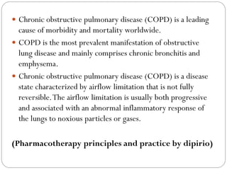
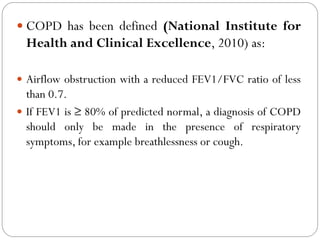
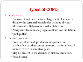
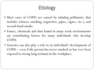
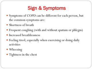
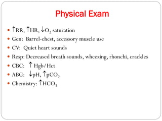
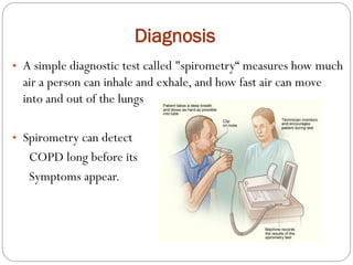
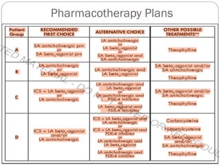
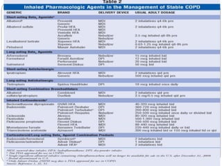
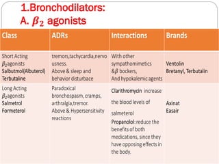
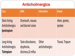
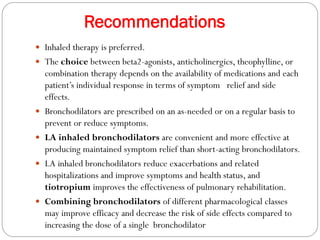
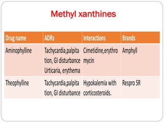
Chronic Obstructive Pulmonary Disease (COPD) is a progressive lung disease characterized by limited airflow. It is caused most often by smoking or long-term exposure to lung irritants. Symptoms include shortness of breath, cough, wheezing, and fatigue. Diagnosis involves spirometry testing showing reduced airflow. Treatment focuses on bronchodilators, inhaled corticosteroids, pulmonary rehabilitation, oxygen therapy, and managing exacerbations. The goal is improving symptoms, lung function and quality of life.
Chronic obstructive pulmonary disorders COPD is a [preventable and treatable disease with some significant extra pulmonary effects that may contribute to the severity in individual clients.
It is characterized by airflow limitation that is not completely reversible.
Chronic obstructive pulmonary disease (COPD)- Preeti sharmaEducate with smile
COPD is a type of obstructive lung disease and related conditions. it is very helpful presentation to you about information of COPD.
It includes all things that is definition, causes, symptoms, pathophysiology, diagnostic evaluation, types, treatment and role of nurses for COPD patient.
Emphysema-medical information |management |diagnosis | tests martinshaji
HAPPY PHARMACIST DAY
Emphysema is a lung condition that causes shortness of breath. In people with emphysema, the air sacs in the lungs (alveoli) are damaged. Over time, the inner walls of the air sacs weaken and rupture — creating larger air spaces instead of many small ones
please comment
thank you
What is COPD, what causes COPD? What is the pathophysiology?How can we diagnose COPD. What is it's classification depending on severity. How can we diagnose COPD clinically as well as under microscope.How can we treat and manage COPD with the help of medicine as well as socially. Let's discuss.
What is emphysema?
Emphysema is a condition that forms part of chronic obstructive pulmonary disease (COPD) and involves the enlargement of the air sacs in the lung.
The alveoli at the end of the bronchioles of the lung become enlarged because of the breakdown of their walls. The fewer and larger damaged sacs that result mean there is a reduced surface area for the exchange of oxygen into the blood and carbon dioxide out of it.
Definition
Emphysema is a condition in which the alveoli become stiff expands and continuously filled the air even after expiration. Emphysema is a chronic obstructive disease due to lack of elasticity in the lungs and alveoli surface area.
Classification
Panlobular (panacinar)
It is damage to the respiratory bronchi, alveolar ducts and alveoli. All air space in the little lobes much enlarged, with little inflammatory disease. The characteristics that have chest hyperinflation, and is characterized by dyspnea on exertion, and weight loss.
CENTRILOBULAR (CENTROACINAR)
The pathological changes mainly occur in the centre of the secondary lobes, and peripheral of acini remain good. Often there is chaos-ventilation perfusion ratio, which lead to hypoxia, hypercapnia (increased CO2 in the arterial blood), polycythaemia and heart failure episodes right. The condition leads to cyanosis, peripheral oedema, and respiratory failure.
CAUSES OF EMPHYSEMA
The biggest known cause or risk factor for emphysema - and for COPD - is smoking. Cigarette smoking is responsible for around 90% of cases of COPD. However, COPD will develop only in smokers who are genetically susceptible - smoking does not always lead to the disease.
- Video recording of this lecture in English language: https://youtu.be/lK81BzxMqdo
- Video recording of this lecture in Arabic language: https://youtu.be/Ve4P0COk9OI
- Link to download the book free: https://nephrotube.blogspot.com/p/nephrotube-nephrology-books.html
- Link to NephroTube website: www.NephroTube.com
- Link to NephroTube social media accounts: https://nephrotube.blogspot.com/p/join-nephrotube-on-social-media.html
Title: Sense of Smell
Presenter: Dr. Faiza, Assistant Professor of Physiology
Qualifications:
MBBS (Best Graduate, AIMC Lahore)
FCPS Physiology
ICMT, CHPE, DHPE (STMU)
MPH (GC University, Faisalabad)
MBA (Virtual University of Pakistan)
Learning Objectives:
Describe the primary categories of smells and the concept of odor blindness.
Explain the structure and location of the olfactory membrane and mucosa, including the types and roles of cells involved in olfaction.
Describe the pathway and mechanisms of olfactory signal transmission from the olfactory receptors to the brain.
Illustrate the biochemical cascade triggered by odorant binding to olfactory receptors, including the role of G-proteins and second messengers in generating an action potential.
Identify different types of olfactory disorders such as anosmia, hyposmia, hyperosmia, and dysosmia, including their potential causes.
Key Topics:
Olfactory Genes:
3% of the human genome accounts for olfactory genes.
400 genes for odorant receptors.
Olfactory Membrane:
Located in the superior part of the nasal cavity.
Medially: Folds downward along the superior septum.
Laterally: Folds over the superior turbinate and upper surface of the middle turbinate.
Total surface area: 5-10 square centimeters.
Olfactory Mucosa:
Olfactory Cells: Bipolar nerve cells derived from the CNS (100 million), with 4-25 olfactory cilia per cell.
Sustentacular Cells: Produce mucus and maintain ionic and molecular environment.
Basal Cells: Replace worn-out olfactory cells with an average lifespan of 1-2 months.
Bowman’s Gland: Secretes mucus.
Stimulation of Olfactory Cells:
Odorant dissolves in mucus and attaches to receptors on olfactory cilia.
Involves a cascade effect through G-proteins and second messengers, leading to depolarization and action potential generation in the olfactory nerve.
Quality of a Good Odorant:
Small (3-20 Carbon atoms), volatile, water-soluble, and lipid-soluble.
Facilitated by odorant-binding proteins in mucus.
Membrane Potential and Action Potential:
Resting membrane potential: -55mV.
Action potential frequency in the olfactory nerve increases with odorant strength.
Adaptation Towards the Sense of Smell:
Rapid adaptation within the first second, with further slow adaptation.
Psychological adaptation greater than receptor adaptation, involving feedback inhibition from the central nervous system.
Primary Sensations of Smell:
Camphoraceous, Musky, Floral, Pepperminty, Ethereal, Pungent, Putrid.
Odor Detection Threshold:
Examples: Hydrogen sulfide (0.0005 ppm), Methyl-mercaptan (0.002 ppm).
Some toxic substances are odorless at lethal concentrations.
Characteristics of Smell:
Odor blindness for single substances due to lack of appropriate receptor protein.
Behavioral and emotional influences of smell.
Transmission of Olfactory Signals:
From olfactory cells to glomeruli in the olfactory bulb, involving lateral inhibition.
Primitive, less old, and new olfactory systems with different path
Ozempic: Preoperative Management of Patients on GLP-1 Receptor Agonists Saeid Safari
Preoperative Management of Patients on GLP-1 Receptor Agonists like Ozempic and Semiglutide
ASA GUIDELINE
NYSORA Guideline
2 Case Reports of Gastric Ultrasound
MANAGEMENT OF ATRIOVENTRICULAR CONDUCTION BLOCK.pdfJim Jacob Roy
Cardiac conduction defects can occur due to various causes.
Atrioventricular conduction blocks ( AV blocks ) are classified into 3 types.
This document describes the acute management of AV block.
Couples presenting to the infertility clinic- Do they really have infertility...Sujoy Dasgupta
Dr Sujoy Dasgupta presented the study on "Couples presenting to the infertility clinic- Do they really have infertility? – The unexplored stories of non-consummation" in the 13th Congress of the Asia Pacific Initiative on Reproduction (ASPIRE 2024) at Manila on 24 May, 2024.
Anti ulcer drugs and their Advance pharmacology ||
Anti-ulcer drugs are medications used to prevent and treat ulcers in the stomach and upper part of the small intestine (duodenal ulcers). These ulcers are often caused by an imbalance between stomach acid and the mucosal lining, which protects the stomach lining.
||Scope: Overview of various classes of anti-ulcer drugs, their mechanisms of action, indications, side effects, and clinical considerations.
TEST BANK for Operations Management, 14th Edition by William J. Stevenson, Ve...kevinkariuki227
TEST BANK for Operations Management, 14th Edition by William J. Stevenson, Verified Chapters 1 - 19, Complete Newest Version.pdf
Factory Supply Best Quality Pmk Oil CAS 28578–16–7 PMK Powder in Stockrebeccabio
Factory Supply Best Quality Pmk Oil CAS 28578–16–7 PMK Powder in Stock
Telegram: bmksupplier
signal: +85264872720
threema: TUD4A6YC
You can contact me on Telegram or Threema
Communicate promptly and reply
Free of customs clearance, Double Clearance 100% pass delivery to USA, Canada, Spain, Germany, Netherland, Poland, Italy, Sweden, UK, Czech Republic, Australia, Mexico, Russia, Ukraine, Kazakhstan.Door to door service
Hot Selling Organic intermediates
2. Chronic obstructive pulmonary disease (COPD) is a leading
cause of morbidity and mortality worldwide.
COPD is the most prevalent manifestation of obstructive
lung disease and mainly comprises chronic bronchitis and
emphysema.
Chronic obstructive pulmonary disease (COPD) is a disease
state characterized by airflow limitation that is not fully
reversible.The airflow limitation is usually both progressive
and associated with an abnormal inflammatory response of
the lungs to noxious particles or gases.
(Pharmacotherapy principles and practice by dipirio)
3. COPD has been defined (National Institute for
Health and Clinical Excellence, 2010) as:
Airflow obstruction with a reduced FEV1/FVC ratio of less
than 0.7.
If FEV1 is ≥ 80% of predicted normal, a diagnosis of COPD
should only be made in the presence of respiratory
symptoms, for example breathlessness or cough.
4. COPD: An umbrella term
• Umbrella term used to describe progressive lung diseases
which include:
• Emphysema
• Chronic bronchitis
COPD is also known as:
Chronic Obstructive Lung Disease (COLD)
Chronic Lower Respiratory Disease (CLRD)
5. Types of COPD
Emphysema
Permanent and destructive enlargement of airspaces
distal to the terminal bronchioles without obvious
fibrosis and with loss of normal architecture
Always involves clinically significant airflow limitation.
“pink puffer”
Chronic Bronchitis
Presence of a cough productive of sputum not
attributable to other causes on most days for at least 3
months over 2 consecutive years
May be present in the absence of airflow limitation.
“blue bloater”
6. Etiology
Most cases of COPD are caused by inhaling pollutants; that
includes tobacco smoking (cigarettes, pipes, cigars, etc.), and
second-hand smoke.
Fumes, chemicals and dust found in many work environments
are contributing factors for many individuals who develop
COPD.
Genetics can also play a role in an individual’s development of
COPD—even if the person has never smoked or has ever been
exposed to strong lung irritants in the workplace.
7. Sign & Symptoms
Symptoms of COPD can be different for each person, but
the common symptoms are:
Shortness of breath
Frequent coughing (with and without sputum or phlegm)
Increased breathlessness
Feeling tired, especially when exercising or doing daily
activities
Wheezing
Tightness in the chest
8. Pathogenesis of COPD
Increased number of activated polymorphonuclear cells
and macrophages produce elastases (such as human
leukocyte elastase), resulting in lung destruction.
Increased oxidative stress caused by free radicals in
cigarette smoke, the oxidants released by phagocytes,
and polymorphonuclear leukocytes all may lead to
apoptosis or necrosis of exposed cells
10. Risk Factors
Smoking
COPD most often occurs in people 40 years of
age and older who have a history of smoking.
These may be individuals who are current or
former smokers. While not everybody who
smokes gets COPD, most of the individuals who
have COPD (about 90% of them) have smoked.
However, only one in five smokers will get
significant COPD.
11. Environmental Factors
COPD can also occur in those who have had long
term exposure and contact with harmful pollutants in
the workplace. Some of these harmful lung irritants
include certain chemicals, dust, or fumes. Heavy or
long-term contact with secondhand smoke or other
lung irritants in the home, such as organic cooking
fuel, may also cause COPD. Individuals who have
worked for many years around these irritants are at
risk for developing mild COPD
12. Genetic Factors
Even if an individual has never smoked or been exposed to
pollutants for an extended period of time, they can still
develop COPD. Alpha-1 Antitrypsin Deficiency (AATD) is
the most commonly known genetic risk factor for
emphysema. Alpha-1 Antitrypsin related COPD is caused by a
deficiency of the Alpha-1 Antitrypsin protein in the
bloodstream. Without the Alpha-1 Antitrypsin protein, white
blood cells begin to harm the lungs and lung deterioration
occurs. The World Health Organization and the American
Thoracic Society recommends that every individual diagnosed
with COPD be tested for Alpha-1.
13. Alpha 1-antitrypsin deficiency is a genetic condition
that is responsible for about 2% cases of COPD.
In this condition, the body does not make enough of
a protein, alpha 1-antitrypsin.
This protein protects the lungs from damage caused
by protease enzymes, such as elastase and trypsin,
that can be released as a result of an inflammatory
response to tobacco smoke.
Almost 90% of COPD deaths occur in low- and
middle-income countries, where effective strategies
for prevention and control are not always
implemented or accessible.
15. Diagnosis
• A simple diagnostic test called "spirometry“ measures how much
air a person can inhale and exhale, and how fast air can move
into and out of the lungs
• Spirometry can detect
COPD long before its
Symptoms appear.
20. Recommendations
Inhaled therapy is preferred.
The choice between beta2-agonists, anticholinergics, theophylline, or
combination therapy depends on the availability of medications and each
patient’s individual response in terms of symptom relief and side
effects.
Bronchodilators are prescribed on an as-needed or on a regular basis to
prevent or reduce symptoms.
LA inhaled bronchodilators are convenient and more effective at
producing maintained symptom relief than short-acting bronchodilators.
LA inhaled bronchodilators reduce exacerbations and related
hospitalizations and improve symptoms and health status, and
tiotropium improves the effectiveness of pulmonary rehabilitation.
Combining bronchodilators of different pharmacological classes
may improve efficacy and decrease the risk of side effects compared to
increasing the dose of a single bronchodilator
22. Recommendations
Methylxanthines are less effective and less well tolerated
than inhaled long-acting bronchodilators and are not
recommended if those drugs are available and
affordable.
Addition of theophylline to salmeterol produces a
greater increase in FEV1 and relief of breathlessness than
salmeterol alone. Low-dose theophylline reduces
exacerbations but does not improve post- bronchodilator
lung function.
24. Recommendations
• In COPD patients with FEV1 < 60% predicted, regular treatment
with inhaled corticosteroids improves symptoms, lung function,
and quality of life, and reduces the frequency of exacerbations.
• Inhaled corticosteroid therapy is associated with an
increased risk of pneumonia.
• Withdrawal from treatment with inhaled corticosteroids
may lead to exacerbations in some patients.
• Based on efficacy and side effects, inhaled bronchodilators
are preferred over oral bronchodilators.
• Long-term monotherapy with inhaled corticosteroids is
not recommended.
• Long-term treatment with oral corticosteroids is also not
recommended
26. Mucolytic agents
Patients with viscous sputum may benefit from
mucolytics (e.g. carbocysteine), but overall
benefits are very small.
27. Vaccines
Influenza vaccines can reduce serious illness and death in
COPD patients.
Vaccines containing inactivated viruses are recommended,
and should be given once each year.
Pneumococcal polysaccharide vaccine is recommended for
COPD patients 65 years and older, and has been shown to
reduce community-acquired pneumonia in those under age
65 with FEV1< 40% predicted.
Alpha-1 Antitrypsin AugmentationTherapy
Not recommended for patients with COPD that is unrelated to
alpha-1 antitrypsin deficiency.
28. Antibiotics
Not recommended except for treatment of infectious
exacerbations and other bacterial infections.
Drugs include doxycycline, trimethoprim-
sulfamethoxazole and amoxicillin-clavulanate
potassium.
Treatment with augmented penicillins,
fluoroquinolones, third-generation
cephalosporins or aminoglycosides may be
considered in patients with more severe exacerbations
29. Non pharmacological treatments
Rehabilitation
Oxygen therapy
Ventilatory support
Surgical treatments
Palliative Care, End-of-life Care, and Hospice Care
30. Rehabilitation
Patients at all stages of disease benefit from exercise training
programs with improvements in exercise tolerance and
symptoms of dyspnea and fatigue.
Benefits can be sustained even after a single pulmonary
rehabilitation program.
The minimum length of an effective rehabilitation program is
6 weeks; the longer the program continues, the more
effective the results.
31. Oxygen therapy
Long-term administration of oxygen (> 15 hours per day) to
patients with chronic respiratory failure has been shown to
increase survival in patients with severe, resting hypoxemia.
Long-term oxygen therapy is indicated for patients who have:
o PaO2 between 7.3 kPa (55 mmHg) and 8.0 kPa (60 mmHg).
32. Ventilatory support
Combination of non-invasive ventilation with long-term
oxygen therapy may be of some use in a selected subset
of patients, particularly in those with pronounced
daytime hypercapnia.
It may improve survival but does not improve quality of
life.
33. Surgical treatment
Advantage of lung volume reduction surgery (LVRS)
over medical therapy is more significant among patients
with upper-lobe predominant emphysema and low
exercise capacity prior to treatment.
LVRS is costly relative to health-care programs not
including surgery.
In appropriately selected patients with very severe
COPD, lung transplantation has been shown to
improve quality of life and functional capacity.