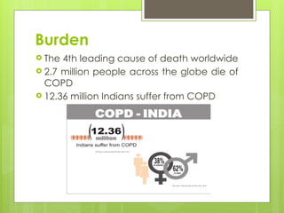
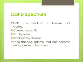
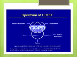
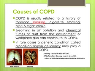
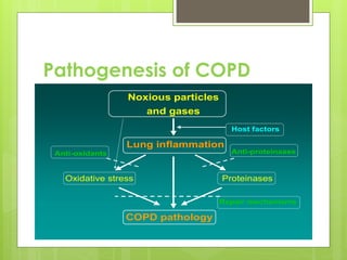
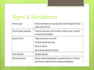
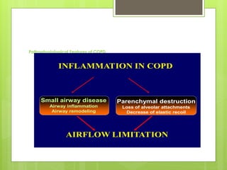
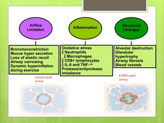
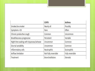
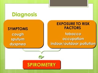
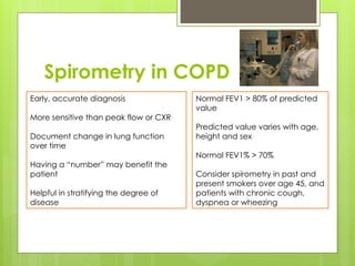
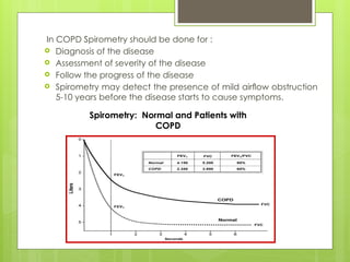
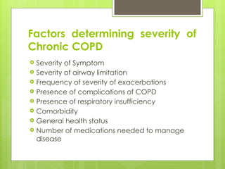
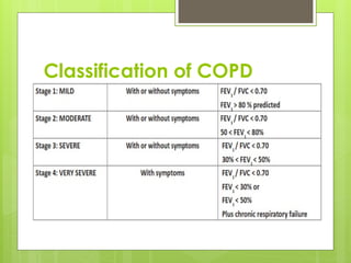
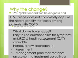
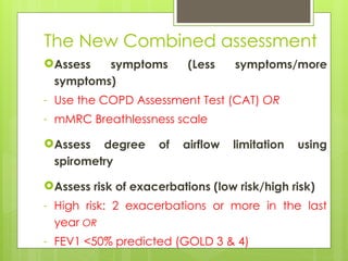
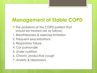
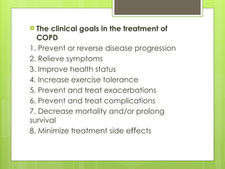
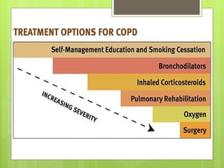
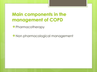
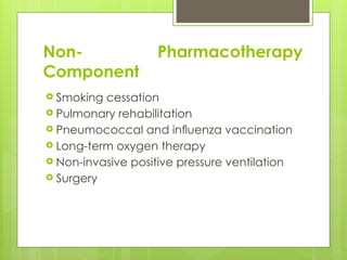
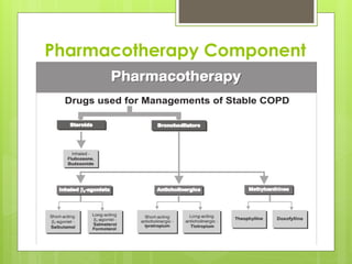
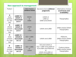
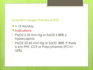
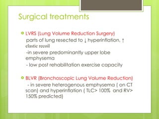
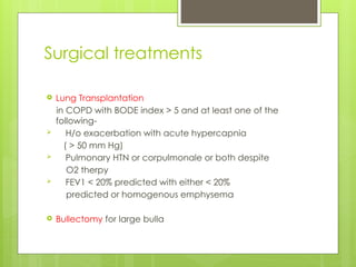
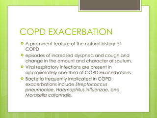
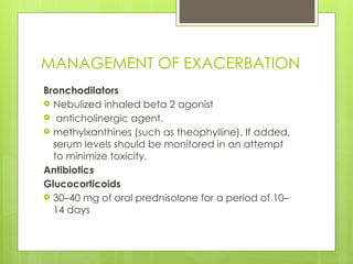
COPD
Dr. Vipin Goyal
Burden
 The 4th leading cause of death worldwide
 2.7 million people across the globe die of
COPD
 12.36 million Indians suffer from COPD
Definition
 A preventable and treatable disease
state characterized by persistent airflow
limitation that is not fully reversible.
 The airflow limitation is usually progressive
and is associated with an abnormal
chronic inflammatory response of the
lungs and airways to noxious particles or
gases, primarily caused by cigarette
smoking.
 Although COPD affects the lungs, it also
produces significant systemic
consequences.
 preventable and treatable: COPD can be
prevented if the risk factors, especially
cigarette smoke are avoided and it is
treatable with the new treatments available
 not fully reversible: there is some reversibility,
which can be treated with bronchodilators
 progressive: with each exacerbation
causing a further deterioration in the lung
functions, which never come back to what it
was prior to the exacerbation. Also the
elastin that is present in the alveolar walls,
once destroyed by the enzymes will never
be synthesized again
COPD Spectrum
COPD is a spectrum of diseases that
includes:
 Chronic bronchitis
 Emphysema
 Small airway disease
 Long-standing asthma that has become
unresponsive to treatment
Clinical observations led to suggestions that
there were two distinct type of patients
 TYPE-A fighter is pink and puffing. Although the
person is breathless, arterial tensions of oxygen
and carbon dioxide are normal and there is no
cor pulmonale. These individuals were thought to
be suffering predominantly from emphysema.
 TYPE-B non-fighter, on the other hand, is blue and
bloated. The person does not appear to be
breathless but has marked arterial hypoxemia,
carbon dioxide retention, secondary
polycythemia and cor pulmonale. These patients
were thought to be suffering predominantly from
chronic bronchitis
Causes of COPD
 COPD is usually related to a history of
tobacco smoking, cigarette smoking,
pipe & cigar smoke.
 Breathing in air pollution and chemical
fumes or dust from the environment or
workplace also can contribute to COPD.
 In rare cases a genetic condition called
alpha1-antitrypsin deficiency may play a
role in causing COPD
Non-Smoker Smoker
 Smoking causes 80-90% of COPD.
 50% of smokers develop chronic bronchitis
 15-20% of smokers develop clinical airflow obstruction
Pathogenesis of COPD
Noxious particles
and gases
Lung inflammation
Host factors
COPD pathology
Proteinases
Oxidative stress
Anti-proteinases
Anti-oxidants
Repair mechanisms
Signs & Symptoms
Pathophysiological Features of COPD
INFLAMMATION IN COPD
Small airway disease
Airway inflammation
Airway remodeling
Parenchymal destruction
Loss of alveolar attachments
Decrease of elastic recoil
AIRFLOW LIMITATION
Bronchoconstriction
Mucus hyper secretion
Loss of elastic recoil
Airway narrowing
Dynamic hyperinflation
during exercise
Oxidative stress
 Neutrophils
 Macrophages
 CD8+ lymphocytes
 IL-8 and TNF-
Protease/antiprotease
imbalance
Alveolar destruction
Glandular
hypertrophy
Airway fibrosis
Blood vessels
Airflow
Limitation
Inflammation Structural
Changes
normal small
airway
COPD small
airway
Differential Diagnosis: COPD and Asthma
Diagnosis
SYMPTOMS
cough
sputum
dyspnea
EXPOSURE TO RISK
FACTORS
tobacco
occupation
indoor/outdoor pollution
SPIROMETRY
è
Spirometry in COPD
Early, accurate diagnosis
More sensitive than peak flow or CXR
Document change in lung function
over time
Having a “number” may benefit the
patient
Helpful in stratifying the degree of
disease
Normal FEV1 > 80% of predicted
value
Predicted value varies with age,
height and sex
Normal FEV1% > 70%
Consider spirometry in past and
present smokers over age 45, and
patients with chronic cough,
dyspnea or wheezing
In COPD Spirometry should be done for :
 Diagnosis of the disease
 Assessment of severity of the disease
 Follow the progress of the disease
 Spirometry may detect the presence of mild airflow obstruction
5-10 years before the disease starts to cause symptoms.
Spirometry: Normal and Patients with
COPD
Factors determining severity of
Chronic COPD
 Severity of Symptom
 Severity of airway limitation
 Frequency of severity of exacerbations
 Presence of complications of COPD
 Presence of respiratory insufficiency
 Comorbidity
 General health status
 Number of medications needed to manage
disease
Classification of COPD
The GOLD Initiative 2011
A Change of
Paradigm
The change
2010
Assessing
impact of the
disease on lung
function (FEV1)
2011
Assessing
impact of the
disease on
FEV1;
Symptoms &
health status;
the risk of future
events
(exacerbations
& mortality)
A move beyond the “One size fits all”
approach
Why the change?
 FEV1- ‘gold standard’ for the diagnosis and
assessment of COPD severity
 But alone, it is an unreliable marker of
 severity of breathlessness
 exercise limitation
 health status impairment
 exacerbation frequency
What do we have today?
Easy to use questionnaires for symptoms
(mMRC) & health assessment (CAT)
available
Hence, a new approach to
• Assessment
• Management (one that matches
assessment to treatment objectives)
FEV1 alone does not completely capture
the heterogeneity that exists among
patients with COPD
The New Combined assessment
Assess symptoms (Less symptoms/more
symptoms)
- Use the COPD Assessment Test (CAT) OR
- mMRC Breathlessness scale
Assess degree of airflow limitation using
spirometry
Assess risk of exacerbations (low risk/high risk)
- High risk: 2 exacerbations or more in the last
year OR
- FEV1 <50% predicted (GOLD 3 & 4)
Grade Description of Breathlessness Group
0 I only get breathless with strenuous exercise. A or C
1 I get short of breath when hurrying on level ground or walking up a
slight hill.
A or C
2 On level ground, I walk slower than people of the same age because
of breathlessness, or have to stop for breath when walking at my own
pace.
B or D
3 I stop for breath after walking about 100 yards or after a few minutes
on level ground.
B or D
4 I am too breathless to leave the house or I am breathless when
dressing.
B or D
The Modified Medical Research Council Dyspnea Scale, or MMRC, uses a simple
grading system to assess a patient's level of dyspnea -- shortness of breath.
The MMRC is used to help calculate BODE Index, a tool for predicting life expectancy of
someone with COPD.
MMRC Dyspnea Scale
COPD Assessment Test (CAT)
 8 item unidimensional measure of health
status impairment in COPD.
 Score 0-40
 0 to 9 – less symptoms- Group A or C
 > 10 - More symptoms- Group B or D
Less symptoms More symptoms
High risk
Low risk
Risk
(GOLD
Classification
of
Airflow
Limitation)
Risk
(Exacerbation
history)
> 2
1
0
(C)
(Less
symptoms,
high risk)
mMRC 0-1
CAT < 10
4
3
2
1
mMRC > 2
CAT > 10
Symptoms
(mMRC or CAT score)
The A, B, C and D of COPD
(A)
(Less
symptoms,
low risk)
(B)
(More
symptoms,
low risk)
(D)
(More
symptoms,
high risk)
Management of Stable COPD
 The problems of the COPD patient that
should be treated are as follows:
1. Breathlessness & exercise limitation
2. Frequent exacerbations
3. Respiratory failure
4. Cor pulmonale
5. Under nutrition
6. Chronic productive cough
7. Anxiety & depression
 The clinical goals in the treatment of
COPD
1. Prevent or reverse disease progression
2. Relieve symptoms
3. Improve health status
4. Increase exercise tolerance
5. Prevent and treat exacerbations
6. Prevent and treat complications
7. Decrease mortality and/or prolong
survival
8. Minimize treatment side e ects
ff
Main components in the
management of COPD
 Pharmacotherapy
 Non pharmacological management
Non- Pharmacotherapy
Component
 Smoking cessation
 Pulmonary rehabilitation
 Pneumococcal and influenza vaccination
 Long-term oxygen therapy
 Non-invasive positive pressure ventilation
 Surgery
Smoking cessation
 Strategy-
ASK
ADVISE
ASSESS
ASSIST
ARRANGE
 Pharmacotherapy-
NRT – Nicotine gum, patch, lozenges, inhaler, nasal
spray
Varenicline
Bupriopion
Nortriptyline
Pharmacotherapy Component
Patient First choice
(Evidence based)
Second choice (Clinical
judgment)
Alternative choices
(cost issues and
availability)
A
(Less
sympto
ms, low
risk)
mMRC 0-1
GOLD1,2
Exacerbati
on 0-1
SAMA
or
SABA
LAMA or
LABA or
SABA and SAMA
Theophylline
B
(More
sympto
ms, low
risk)
mMRC >2
GOLD1,2
Exacerbati
on 0-1
LAMA
or
LABA
LAMA and LABA
SABA and/or SAMA
Theophylline
C
(Less
sympto
ms, high
risk)
mMRC 0-1
GOLD3,4
Exacerbati
on ≥ 2
ICS + LABA
or
LAMA
LAMA and LABA
PDE4-inh.
SABA and/or SAMA
Theophylline
D
(More
sympto
ms, high
risk)
mMRC >2
GOLD3,4
Exacerbati
on ≥ 2
ICS + LABA
or
LAMA
ICS and LAMA or
ICS + LABA and LAMA or
ICS+LABA and PDE4-inh.
or
LAMA and LABA or
LAMA and PDE4-inh.
Carbocysteine
SABA and/or SAMA
Theophylline
New approach to management
Long-term oxygen therapy (LTOT)
 > 15 Hrs/day
 Indications
 PaO2 ≤ 55 mm Hg or SaO2 ≤ 88% ±
hypercapnia
 PaO2 55-60 mm Hg or SaO2 88% if there
is e/o PHT, CCF or Polycythemia (PCV>
55%)
Surgical treatments
 LVRS (Lung Volume Reduction Surgery)
parts of lung resected to ↓ hyperinflation, ↑
elastic recoil
-in severe predominantly upper lobe
emphysema
- low post rehabilitation exercise capacity
 BLVR (Bronchoscopic Lung Volume Reduction)
- in severe heterogenous emphysema ( on CT
scan) and hyperinflation ( TLC> 100% and RV>
150% predicted)
Surgical treatments
 Lung Transplantation
in COPD with BODE index > 5 and at least one of the
following-
 H/o exacerbation with acute hypercapnia
( > 50 mm Hg)
 Pulmonary HTN or corpulmonale or both despite
O2 therpy
 FEV1 < 20% predicted with either < 20%
predicted or homogenous emphysema
 Bullectomy for large bulla
COPD EXACERBATION
 A prominent feature of the natural history of
COPD
 episodes of increased dyspnea and cough and
change in the amount and character of sputum.
 Viral respiratory infections are present in
approximately one-third of COPD exacerbations.
 Bacteria frequently implicated in COPD
exacerbations include Streptococcus
pneumoniae, Haemophilus influenzae, and
Moraxella catarrhalis.
MANAGEMENT OF EXACERBATION
Bronchodilators
 Nebulized inhaled beta 2 agonist
 anticholinergic agent.
 methylxanthines (such as theophylline). If added,
serum levels should be monitored in an attempt
to minimize toxicity.
Antibiotics
Glucocorticoids
 30–40 mg of oral prednisolone for a period of 10–
14 days
Oxygen
 keep arterial saturations around 90%
Mechanical Ventilatory Support
 The initiation of noninvasive positive-pressure
ventilation (NIPPV) in patients with respiratory
failure, defined as PaCO2 >45 mmHg, results in
a significant reduction in mortality rate, need
for intubation, complications of therapy, and
hospital length of stay.
 Invasive (conventional) mechanical ventilation
via an endotracheal tube is indicated for
patients with
 severe respiratory distress despite initial therapy,
 life-threatening hypoxemia,
 severe hypercarbia and/or
 acidosis,
 markedly impaired mental status,
 respiratory arrest,
 hemodynamic instability, or
 other complications.
Chronic obstructive pulmonary disease COPD

Chronic obstructive pulmonary disease COPD

  • 1.
  • 2.
    Burden  The 4thleading cause of death worldwide  2.7 million people across the globe die of COPD  12.36 million Indians suffer from COPD
  • 3.
    Definition  A preventableand treatable disease state characterized by persistent airflow limitation that is not fully reversible.  The airflow limitation is usually progressive and is associated with an abnormal chronic inflammatory response of the lungs and airways to noxious particles or gases, primarily caused by cigarette smoking.  Although COPD affects the lungs, it also produces significant systemic consequences.
  • 4.
     preventable andtreatable: COPD can be prevented if the risk factors, especially cigarette smoke are avoided and it is treatable with the new treatments available  not fully reversible: there is some reversibility, which can be treated with bronchodilators  progressive: with each exacerbation causing a further deterioration in the lung functions, which never come back to what it was prior to the exacerbation. Also the elastin that is present in the alveolar walls, once destroyed by the enzymes will never be synthesized again
  • 5.
    COPD Spectrum COPD isa spectrum of diseases that includes:  Chronic bronchitis  Emphysema  Small airway disease  Long-standing asthma that has become unresponsive to treatment
  • 7.
    Clinical observations ledto suggestions that there were two distinct type of patients  TYPE-A fighter is pink and puffing. Although the person is breathless, arterial tensions of oxygen and carbon dioxide are normal and there is no cor pulmonale. These individuals were thought to be suffering predominantly from emphysema.  TYPE-B non-fighter, on the other hand, is blue and bloated. The person does not appear to be breathless but has marked arterial hypoxemia, carbon dioxide retention, secondary polycythemia and cor pulmonale. These patients were thought to be suffering predominantly from chronic bronchitis
  • 8.
    Causes of COPD COPD is usually related to a history of tobacco smoking, cigarette smoking, pipe & cigar smoke.  Breathing in air pollution and chemical fumes or dust from the environment or workplace also can contribute to COPD.  In rare cases a genetic condition called alpha1-antitrypsin deficiency may play a role in causing COPD Non-Smoker Smoker  Smoking causes 80-90% of COPD.  50% of smokers develop chronic bronchitis  15-20% of smokers develop clinical airflow obstruction
  • 9.
    Pathogenesis of COPD Noxiousparticles and gases Lung inflammation Host factors COPD pathology Proteinases Oxidative stress Anti-proteinases Anti-oxidants Repair mechanisms
  • 10.
  • 11.
    Pathophysiological Features ofCOPD INFLAMMATION IN COPD Small airway disease Airway inflammation Airway remodeling Parenchymal destruction Loss of alveolar attachments Decrease of elastic recoil AIRFLOW LIMITATION
  • 12.
    Bronchoconstriction Mucus hyper secretion Lossof elastic recoil Airway narrowing Dynamic hyperinflation during exercise Oxidative stress  Neutrophils  Macrophages  CD8+ lymphocytes  IL-8 and TNF- Protease/antiprotease imbalance Alveolar destruction Glandular hypertrophy Airway fibrosis Blood vessels Airflow Limitation Inflammation Structural Changes normal small airway COPD small airway
  • 13.
  • 14.
  • 15.
    Spirometry in COPD Early,accurate diagnosis More sensitive than peak flow or CXR Document change in lung function over time Having a “number” may benefit the patient Helpful in stratifying the degree of disease Normal FEV1 > 80% of predicted value Predicted value varies with age, height and sex Normal FEV1% > 70% Consider spirometry in past and present smokers over age 45, and patients with chronic cough, dyspnea or wheezing
  • 16.
    In COPD Spirometryshould be done for :  Diagnosis of the disease  Assessment of severity of the disease  Follow the progress of the disease  Spirometry may detect the presence of mild airflow obstruction 5-10 years before the disease starts to cause symptoms. Spirometry: Normal and Patients with COPD
  • 17.
    Factors determining severityof Chronic COPD  Severity of Symptom  Severity of airway limitation  Frequency of severity of exacerbations  Presence of complications of COPD  Presence of respiratory insufficiency  Comorbidity  General health status  Number of medications needed to manage disease
  • 18.
  • 19.
    The GOLD Initiative2011 A Change of Paradigm
  • 20.
    The change 2010 Assessing impact ofthe disease on lung function (FEV1) 2011 Assessing impact of the disease on FEV1; Symptoms & health status; the risk of future events (exacerbations & mortality) A move beyond the “One size fits all” approach
  • 21.
    Why the change? FEV1- ‘gold standard’ for the diagnosis and assessment of COPD severity  But alone, it is an unreliable marker of  severity of breathlessness  exercise limitation  health status impairment  exacerbation frequency What do we have today? Easy to use questionnaires for symptoms (mMRC) & health assessment (CAT) available Hence, a new approach to • Assessment • Management (one that matches assessment to treatment objectives) FEV1 alone does not completely capture the heterogeneity that exists among patients with COPD
  • 22.
    The New Combinedassessment Assess symptoms (Less symptoms/more symptoms) - Use the COPD Assessment Test (CAT) OR - mMRC Breathlessness scale Assess degree of airflow limitation using spirometry Assess risk of exacerbations (low risk/high risk) - High risk: 2 exacerbations or more in the last year OR - FEV1 <50% predicted (GOLD 3 & 4)
  • 23.
    Grade Description ofBreathlessness Group 0 I only get breathless with strenuous exercise. A or C 1 I get short of breath when hurrying on level ground or walking up a slight hill. A or C 2 On level ground, I walk slower than people of the same age because of breathlessness, or have to stop for breath when walking at my own pace. B or D 3 I stop for breath after walking about 100 yards or after a few minutes on level ground. B or D 4 I am too breathless to leave the house or I am breathless when dressing. B or D The Modified Medical Research Council Dyspnea Scale, or MMRC, uses a simple grading system to assess a patient's level of dyspnea -- shortness of breath. The MMRC is used to help calculate BODE Index, a tool for predicting life expectancy of someone with COPD. MMRC Dyspnea Scale
  • 24.
    COPD Assessment Test(CAT)  8 item unidimensional measure of health status impairment in COPD.  Score 0-40  0 to 9 – less symptoms- Group A or C  > 10 - More symptoms- Group B or D
  • 25.
    Less symptoms Moresymptoms High risk Low risk Risk (GOLD Classification of Airflow Limitation) Risk (Exacerbation history) > 2 1 0 (C) (Less symptoms, high risk) mMRC 0-1 CAT < 10 4 3 2 1 mMRC > 2 CAT > 10 Symptoms (mMRC or CAT score) The A, B, C and D of COPD (A) (Less symptoms, low risk) (B) (More symptoms, low risk) (D) (More symptoms, high risk)
  • 27.
    Management of StableCOPD  The problems of the COPD patient that should be treated are as follows: 1. Breathlessness & exercise limitation 2. Frequent exacerbations 3. Respiratory failure 4. Cor pulmonale 5. Under nutrition 6. Chronic productive cough 7. Anxiety & depression
  • 28.
     The clinicalgoals in the treatment of COPD 1. Prevent or reverse disease progression 2. Relieve symptoms 3. Improve health status 4. Increase exercise tolerance 5. Prevent and treat exacerbations 6. Prevent and treat complications 7. Decrease mortality and/or prolong survival 8. Minimize treatment side e ects ff
  • 30.
    Main components inthe management of COPD  Pharmacotherapy  Non pharmacological management
  • 31.
    Non- Pharmacotherapy Component  Smokingcessation  Pulmonary rehabilitation  Pneumococcal and influenza vaccination  Long-term oxygen therapy  Non-invasive positive pressure ventilation  Surgery
  • 32.
    Smoking cessation  Strategy- ASK ADVISE ASSESS ASSIST ARRANGE Pharmacotherapy- NRT – Nicotine gum, patch, lozenges, inhaler, nasal spray Varenicline Bupriopion Nortriptyline
  • 33.
  • 34.
    Patient First choice (Evidencebased) Second choice (Clinical judgment) Alternative choices (cost issues and availability) A (Less sympto ms, low risk) mMRC 0-1 GOLD1,2 Exacerbati on 0-1 SAMA or SABA LAMA or LABA or SABA and SAMA Theophylline B (More sympto ms, low risk) mMRC >2 GOLD1,2 Exacerbati on 0-1 LAMA or LABA LAMA and LABA SABA and/or SAMA Theophylline C (Less sympto ms, high risk) mMRC 0-1 GOLD3,4 Exacerbati on ≥ 2 ICS + LABA or LAMA LAMA and LABA PDE4-inh. SABA and/or SAMA Theophylline D (More sympto ms, high risk) mMRC >2 GOLD3,4 Exacerbati on ≥ 2 ICS + LABA or LAMA ICS and LAMA or ICS + LABA and LAMA or ICS+LABA and PDE4-inh. or LAMA and LABA or LAMA and PDE4-inh. Carbocysteine SABA and/or SAMA Theophylline New approach to management
  • 35.
    Long-term oxygen therapy(LTOT)  > 15 Hrs/day  Indications  PaO2 ≤ 55 mm Hg or SaO2 ≤ 88% ± hypercapnia  PaO2 55-60 mm Hg or SaO2 88% if there is e/o PHT, CCF or Polycythemia (PCV> 55%)
  • 36.
    Surgical treatments  LVRS(Lung Volume Reduction Surgery) parts of lung resected to ↓ hyperinflation, ↑ elastic recoil -in severe predominantly upper lobe emphysema - low post rehabilitation exercise capacity  BLVR (Bronchoscopic Lung Volume Reduction) - in severe heterogenous emphysema ( on CT scan) and hyperinflation ( TLC> 100% and RV> 150% predicted)
  • 37.
    Surgical treatments  LungTransplantation in COPD with BODE index > 5 and at least one of the following-  H/o exacerbation with acute hypercapnia ( > 50 mm Hg)  Pulmonary HTN or corpulmonale or both despite O2 therpy  FEV1 < 20% predicted with either < 20% predicted or homogenous emphysema  Bullectomy for large bulla
  • 38.
    COPD EXACERBATION  Aprominent feature of the natural history of COPD  episodes of increased dyspnea and cough and change in the amount and character of sputum.  Viral respiratory infections are present in approximately one-third of COPD exacerbations.  Bacteria frequently implicated in COPD exacerbations include Streptococcus pneumoniae, Haemophilus influenzae, and Moraxella catarrhalis.
  • 39.
    MANAGEMENT OF EXACERBATION Bronchodilators Nebulized inhaled beta 2 agonist  anticholinergic agent.  methylxanthines (such as theophylline). If added, serum levels should be monitored in an attempt to minimize toxicity. Antibiotics Glucocorticoids  30–40 mg of oral prednisolone for a period of 10– 14 days
  • 40.
    Oxygen  keep arterialsaturations around 90% Mechanical Ventilatory Support  The initiation of noninvasive positive-pressure ventilation (NIPPV) in patients with respiratory failure, defined as PaCO2 >45 mmHg, results in a significant reduction in mortality rate, need for intubation, complications of therapy, and hospital length of stay.
  • 41.
     Invasive (conventional)mechanical ventilation via an endotracheal tube is indicated for patients with  severe respiratory distress despite initial therapy,  life-threatening hypoxemia,  severe hypercarbia and/or  acidosis,  markedly impaired mental status,  respiratory arrest,  hemodynamic instability, or  other complications.

Editor's Notes

  • #19 The Global Initiative for Chronic Obstructive Lung Disease (GOLD) committee has recently released a new, extensively revised version of its existing guidelines on Chronic Obstructive Pulmonary Disease. It builds on the strengths of its original recommendations and incorporates newer scientific knowledge and evidence. The revised report brings in a more individualized approach to COPD classification and, based on this, a new paradigm for management of stable disease.
  • #20 In previous GOLD documents, recommendations for management of COPD were based solely on spirometric category. However, there is considerable evidence that the level of FEV1 is a poor descriptor of disease status and for this reason the management of stable COPD based on a strategy considering both disease impact (determined mainly by symptom burden and activity limitation) and future risk of disease progression (especially of exacerbations) is recommended.
  • #21 In previous GOLD documents, recommendations for management of COPD were based solely on spirometric category. However, there is considerable evidence that the level of FEV1 is a poor descriptor of disease status and for this reason the management of stable COPD based on a strategy considering both disease impact (determined mainly by symptom burden and activity limitation) and future risk of disease progression (especially of exacerbations) is recommended.
  • #22 In previous GOLD documents, recommendations for management of COPD were based solely on spirometric category. However, there is considerable evidence that the level of FEV1 is a poor descriptor of disease status and for this reason the management of stable COPD based on a strategy considering both disease impact (determined mainly by symptom burden and activity limitation) and future risk of disease progression (especially of exacerbations) is recommended. Assessment of COPD is now based on the patient’s symptoms, risk of exacerbations, the severity of the spirometric abnormality, and the identification of comorbidities.
  • #25 You need to assess the symptoms first (as per the COPD Assessment Test (CAT) or mMRC Breathlessness scale), followed by the risk of exacerbations (looking at the FEV1 and history of exacerbations (two or more exacerbations within the last year or an FEV1 < 50 % of predicted value are indicators of high risk). Based on this combined assessment, the GOLD report divides patients into four groups: A (less symptoms, low risk of exacerbations and other negative events); B (more symptoms, low risk); C (less symptoms, high risk); and D (more symptoms, high risk).
  • #34 The classes of medications commonly used in treating COPD are shown in the table below. The choice within each class depends on the availability and cost of medication and the patient’s response. Each treatment regimen needs to be patient-specific as the relationship between severity of symptoms, airflow limitation, and severity of exacerbations will differ between patients.