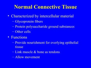
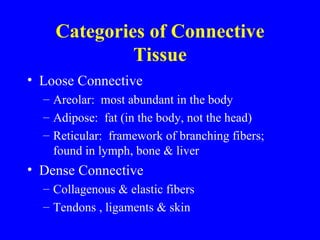
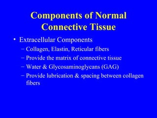
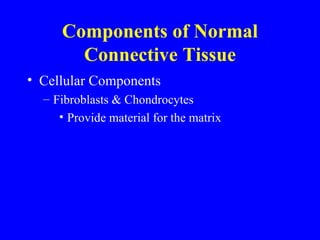
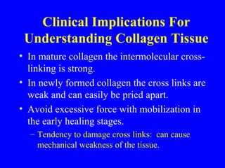
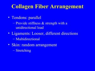
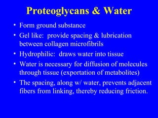
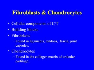
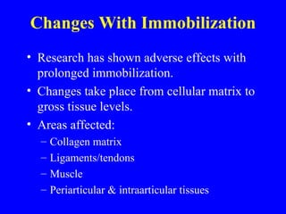
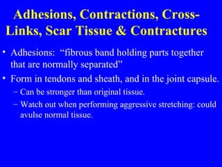
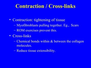
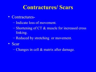
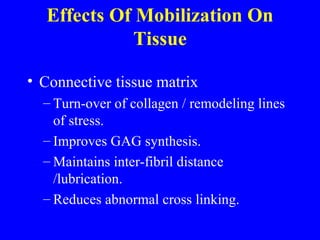
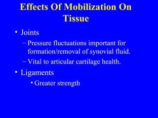
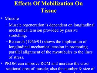
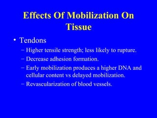
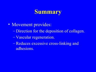
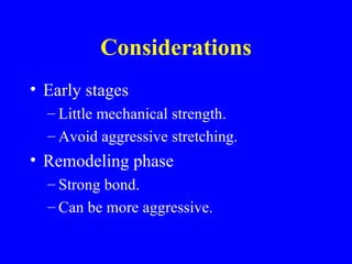
Changes In Tissue During Injury, Immobilization,and Remobilization
Part I:  Review Identify Normal Connective Tissue Components of Normal Connective Tissue Collagen and Elastin Proteoglycans & Water Fibroblasts and Chondrocytes Soft Tissue Changes Following Injury
Normal Connective Tissue Characterized by intercellular material Glycoprotein fibers Protein polysaccharide ground substances Other cells Functions Provide nourishment for overlying epithelial tissue Link muscle & bone as tendons Allow movement
Categories of Connective Tissue Loose Connective Areolar:  most abundant in the body Adipose:  fat (in the body, not the head) Reticular:  framework of branching fibers; found in lymph, bone & liver Dense Connective Collagenous & elastic fibers Tendons , ligaments & skin
Components of Normal Connective Tissue Extracellular Components Collagen, Elastin, Reticular fibers  Provide the matrix of connective tissue Water & Glycosaminoglycans (GAG) Provide lubrication & spacing between collagen fibers
Components of Normal Connective Tissue Cellular Components Fibroblasts & Chondrocytes Provide material for the matrix
 
Clinical Implications For Understanding Collagen Tissue In mature collagen the intermolecular cross-linking is strong. In newly formed collagen the cross links are weak and can easily be pried apart. Avoid excessive force with mobilization in the early healing stages. Tendency to damage cross links:  can cause mechanical weakness of the tissue .
Collagen Fiber Arrangement Tendons: parallel  Provide stiffness & strength with a unidirectional load Ligaments: Looser, different directions Multidirectional Skin: random arrangement Stretching
Proteoglycans & Water Form ground substance Gel like:  provide spacing & lubrication between collagen microfibrils Hydrophilic:  draws water into tissue Water is necessary for diffusion of molecules through tissue (exportation of metabolites) The spacing, along w/ water, prevents adjacent fibers from linking, thereby reducing friction.
Fibroblasts & Chondrocytes Cellular components of C/T Building blocks Fibroblasts  Found in ligaments, tendons,  fascia, joint capsules. Chondrocytes Found in the collagen matrix of articular cartilage.
Soft Tissues Changes Following Injury Two types of cellular events take place in order to repair tissue: Immunological Reparative
Immunological Immediate Prevents bacteria from entering  injured area. Macrophages & leukocytes
Soft Tissues Changes Following Injury Reparative Initiated about 48 hours after injury. Collagen initially held by blood clots begins to form a weak mesh. During this period adhesions of collagen have little mechanical strength. Collagen deposition Increases 5th day Peaks 14th day 120 days:  reduction of collagen turn over
Changes With Immobilization Research has shown adverse effects with prolonged immobilization. Changes take place from cellular matrix to gross tissue levels. Areas affected: Collagen matrix Ligaments/tendons Muscle Periarticular & intraarticular tissues
Effects of Immobilization  on Collagen Matrix Increase in production of collagen Deposits in random fashion, not along the lines of stress. Decrease in GAG & H2O Causes a loss of  lubrication and a closer  contact of fibers Abnormal cross-linking Restricted normal interfibril gliding
Immobilization On Ligaments & Tendons Ligaments Loss of strength/ stiffness at the insertion point. Harwood et al, 1990: atrophy of  the ligaments Tendon Atrophy  Gelberman 1986: extensive obliteration of space between tendon and sheath  Adhesions / impediment of glide between sheath & tendon
Effects of Immobilization  On Muscle 2º shortening of the muscle  Results in a 40% reduction of sarcomeres (Lederman)
Effects of Immobilization  On Periarticular & Intraarticular Structures Atrophy of the capsule, ligaments & synovial  membrane  Adhesions & abnormal cross-links Synovial tissue:  most sensitive to effects of immobilization,  undergoing fibrofatty changes Mature fibrofatty tissue will cover non-articulating areas and form adhesions.
Adhesions, Contractions, Cross-Links, Scar Tissue & Contractures Adhesions:  “fibrous band holding parts together that are normally separated” Form in tendons and sheath, and in the joint capsule. Can be stronger than original tissue. Watch out when performing aggressive stretching: could avulse normal tissue.
Contraction / Cross-links Contraction: tightening of tissue  Myofibroblasts pulling together. Eg.,  Scars ROM exercises prevent this. Cross-links Chemical bonds within & between the collagen molecules. Reduce tissue extensibility.
Contractures/ Scars Contractures-  Indicate loss of movement. Shortening of CT & muscle for increased cross linking. Reduced by stretching  or movement. Scar Changes in cell & matrix after damage.
Effects Of Mobilization On Tissue Connective tissue matrix Turn-over of collagen / remodeling lines of stress. Improves GAG synthesis. Maintains inter-fibril distance /lubrication. Reduces abnormal cross linking.
Effects Of Mobilization On Tissue Joints Pressure fluctuations important for formation/removal of synovial fluid. Vital to articular cartilage health. Ligaments Greater strength
Effects Of Mobilization On Tissue Muscle Muscle regeneration is dependent on longitudinal mechanical tension provided by passive stretching. Research (1966/91) shows the implication of longitudinal mechanical tension in promoting parallel alignment of the myotubules to the lines of stress. PROM can improve ROM and increase the cross -sectional area of muscle; also the number & size of sacromeres return to preimmobilization levels.
Effects Of Mobilization On Tissue Tendons Higher tensile strength; less likely to rupture. Decrease adhesion formation. Early mobilization produces a higher DNA and cellular content vs delayed mobilization.  Revascularization of blood vessels.
Summary Movement provides: Direction for the deposition of collagen. Vascular regeneration. Reduces excessive cross-linking and adhesions.
Considerations Early stages Little mechanical strength. Avoid aggressive stretching. Remodeling phase Strong bond.  Can be more aggressive.
We’d like to thank you for your attention! Please put your chairs in the  upright position before exiting the room. Please learn with us again soon!

Connect iss Lecture 8

  • 1.
    Changes In TissueDuring Injury, Immobilization,and Remobilization
  • 2.
    Part I: Review Identify Normal Connective Tissue Components of Normal Connective Tissue Collagen and Elastin Proteoglycans & Water Fibroblasts and Chondrocytes Soft Tissue Changes Following Injury
  • 3.
    Normal Connective TissueCharacterized by intercellular material Glycoprotein fibers Protein polysaccharide ground substances Other cells Functions Provide nourishment for overlying epithelial tissue Link muscle & bone as tendons Allow movement
  • 4.
    Categories of ConnectiveTissue Loose Connective Areolar: most abundant in the body Adipose: fat (in the body, not the head) Reticular: framework of branching fibers; found in lymph, bone & liver Dense Connective Collagenous & elastic fibers Tendons , ligaments & skin
  • 5.
    Components of NormalConnective Tissue Extracellular Components Collagen, Elastin, Reticular fibers Provide the matrix of connective tissue Water & Glycosaminoglycans (GAG) Provide lubrication & spacing between collagen fibers
  • 6.
    Components of NormalConnective Tissue Cellular Components Fibroblasts & Chondrocytes Provide material for the matrix
  • 7.
  • 8.
    Clinical Implications ForUnderstanding Collagen Tissue In mature collagen the intermolecular cross-linking is strong. In newly formed collagen the cross links are weak and can easily be pried apart. Avoid excessive force with mobilization in the early healing stages. Tendency to damage cross links: can cause mechanical weakness of the tissue .
  • 9.
    Collagen Fiber ArrangementTendons: parallel Provide stiffness & strength with a unidirectional load Ligaments: Looser, different directions Multidirectional Skin: random arrangement Stretching
  • 10.
    Proteoglycans & WaterForm ground substance Gel like: provide spacing & lubrication between collagen microfibrils Hydrophilic: draws water into tissue Water is necessary for diffusion of molecules through tissue (exportation of metabolites) The spacing, along w/ water, prevents adjacent fibers from linking, thereby reducing friction.
  • 11.
    Fibroblasts & ChondrocytesCellular components of C/T Building blocks Fibroblasts Found in ligaments, tendons, fascia, joint capsules. Chondrocytes Found in the collagen matrix of articular cartilage.
  • 12.
    Soft Tissues ChangesFollowing Injury Two types of cellular events take place in order to repair tissue: Immunological Reparative
  • 13.
    Immunological Immediate Preventsbacteria from entering injured area. Macrophages & leukocytes
  • 14.
    Soft Tissues ChangesFollowing Injury Reparative Initiated about 48 hours after injury. Collagen initially held by blood clots begins to form a weak mesh. During this period adhesions of collagen have little mechanical strength. Collagen deposition Increases 5th day Peaks 14th day 120 days: reduction of collagen turn over
  • 15.
    Changes With ImmobilizationResearch has shown adverse effects with prolonged immobilization. Changes take place from cellular matrix to gross tissue levels. Areas affected: Collagen matrix Ligaments/tendons Muscle Periarticular & intraarticular tissues
  • 16.
    Effects of Immobilization on Collagen Matrix Increase in production of collagen Deposits in random fashion, not along the lines of stress. Decrease in GAG & H2O Causes a loss of lubrication and a closer contact of fibers Abnormal cross-linking Restricted normal interfibril gliding
  • 17.
    Immobilization On Ligaments& Tendons Ligaments Loss of strength/ stiffness at the insertion point. Harwood et al, 1990: atrophy of the ligaments Tendon Atrophy Gelberman 1986: extensive obliteration of space between tendon and sheath Adhesions / impediment of glide between sheath & tendon
  • 18.
    Effects of Immobilization On Muscle 2º shortening of the muscle Results in a 40% reduction of sarcomeres (Lederman)
  • 19.
    Effects of Immobilization On Periarticular & Intraarticular Structures Atrophy of the capsule, ligaments & synovial membrane Adhesions & abnormal cross-links Synovial tissue: most sensitive to effects of immobilization, undergoing fibrofatty changes Mature fibrofatty tissue will cover non-articulating areas and form adhesions.
  • 20.
    Adhesions, Contractions, Cross-Links,Scar Tissue & Contractures Adhesions: “fibrous band holding parts together that are normally separated” Form in tendons and sheath, and in the joint capsule. Can be stronger than original tissue. Watch out when performing aggressive stretching: could avulse normal tissue.
  • 21.
    Contraction / Cross-linksContraction: tightening of tissue Myofibroblasts pulling together. Eg., Scars ROM exercises prevent this. Cross-links Chemical bonds within & between the collagen molecules. Reduce tissue extensibility.
  • 22.
    Contractures/ Scars Contractures- Indicate loss of movement. Shortening of CT & muscle for increased cross linking. Reduced by stretching or movement. Scar Changes in cell & matrix after damage.
  • 23.
    Effects Of MobilizationOn Tissue Connective tissue matrix Turn-over of collagen / remodeling lines of stress. Improves GAG synthesis. Maintains inter-fibril distance /lubrication. Reduces abnormal cross linking.
  • 24.
    Effects Of MobilizationOn Tissue Joints Pressure fluctuations important for formation/removal of synovial fluid. Vital to articular cartilage health. Ligaments Greater strength
  • 25.
    Effects Of MobilizationOn Tissue Muscle Muscle regeneration is dependent on longitudinal mechanical tension provided by passive stretching. Research (1966/91) shows the implication of longitudinal mechanical tension in promoting parallel alignment of the myotubules to the lines of stress. PROM can improve ROM and increase the cross -sectional area of muscle; also the number & size of sacromeres return to preimmobilization levels.
  • 26.
    Effects Of MobilizationOn Tissue Tendons Higher tensile strength; less likely to rupture. Decrease adhesion formation. Early mobilization produces a higher DNA and cellular content vs delayed mobilization. Revascularization of blood vessels.
  • 27.
    Summary Movement provides:Direction for the deposition of collagen. Vascular regeneration. Reduces excessive cross-linking and adhesions.
  • 28.
    Considerations Early stagesLittle mechanical strength. Avoid aggressive stretching. Remodeling phase Strong bond. Can be more aggressive.
  • 29.
    We’d like tothank you for your attention! Please put your chairs in the upright position before exiting the room. Please learn with us again soon!