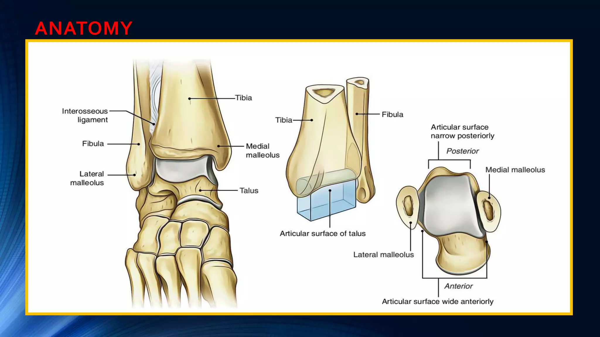
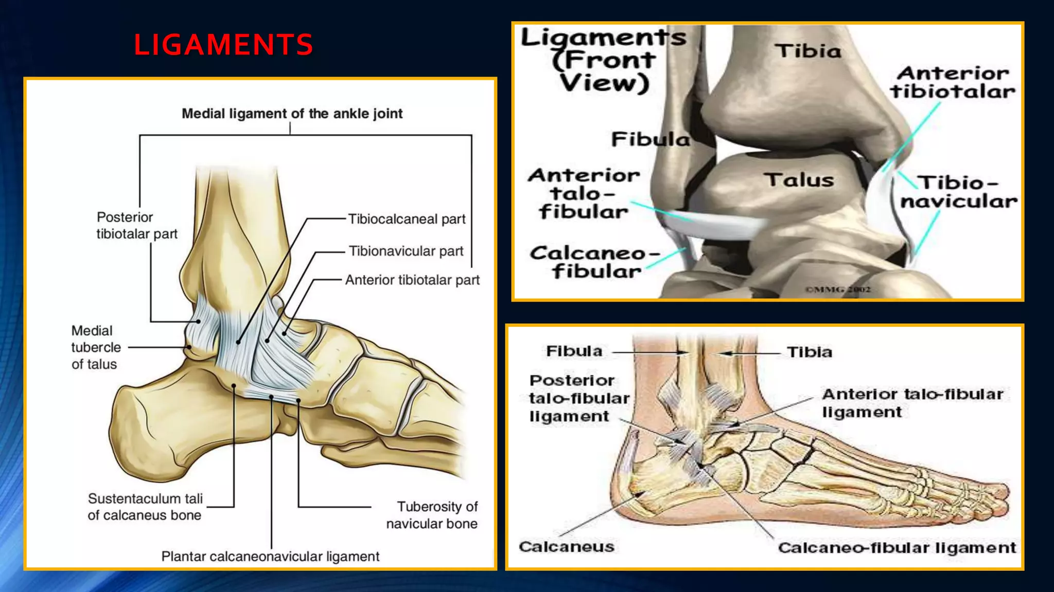
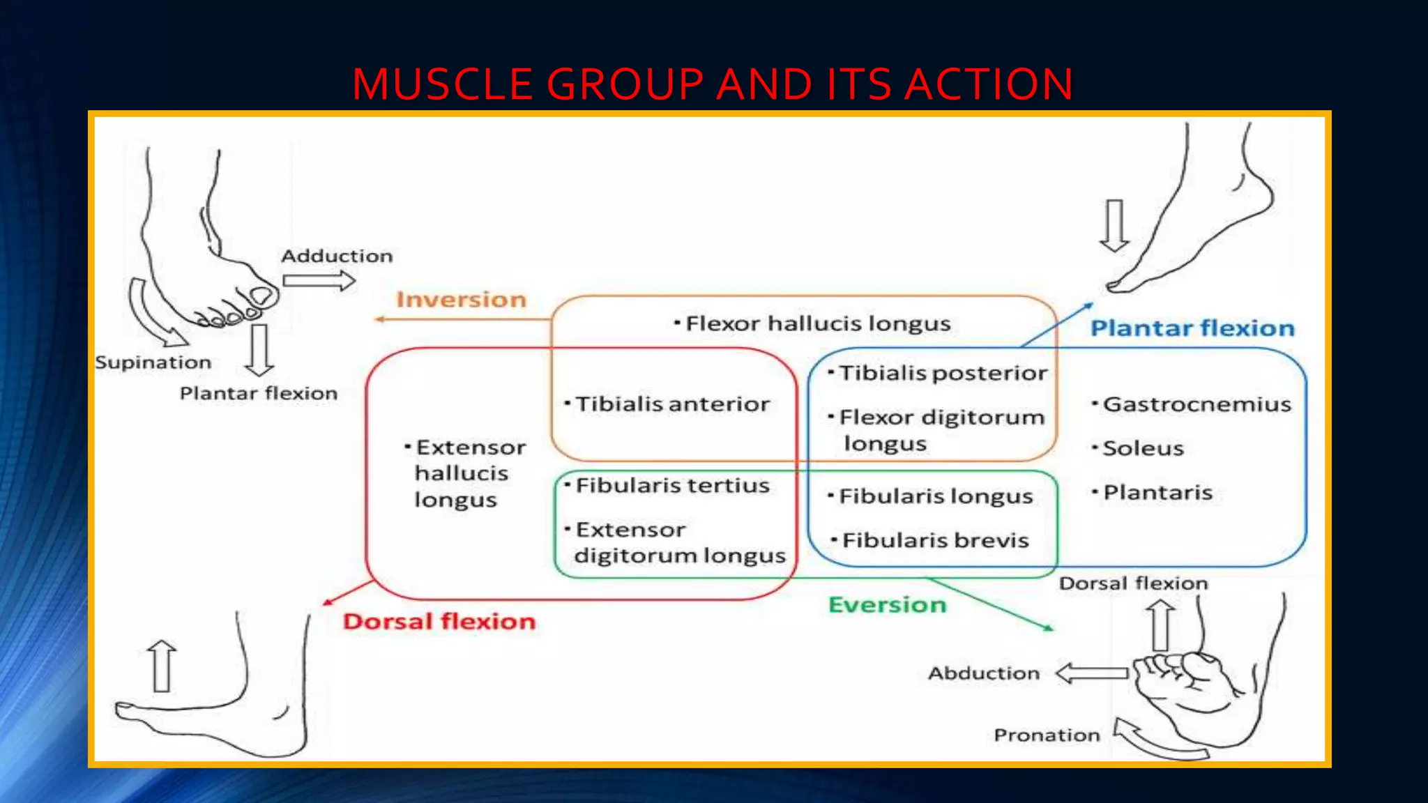
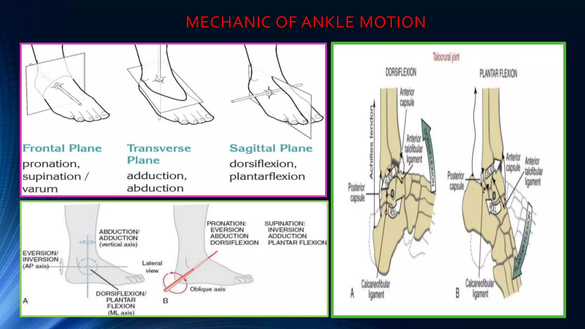
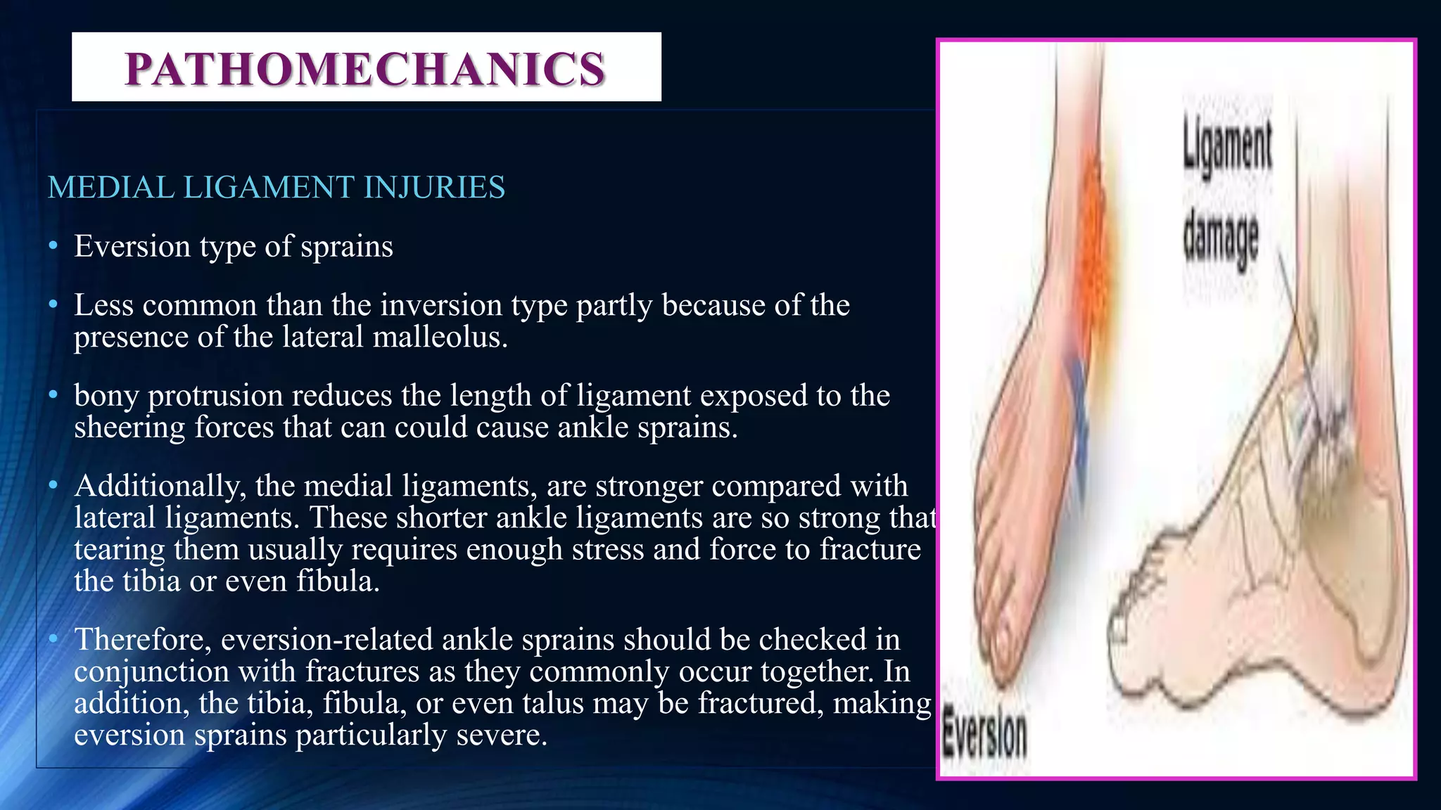
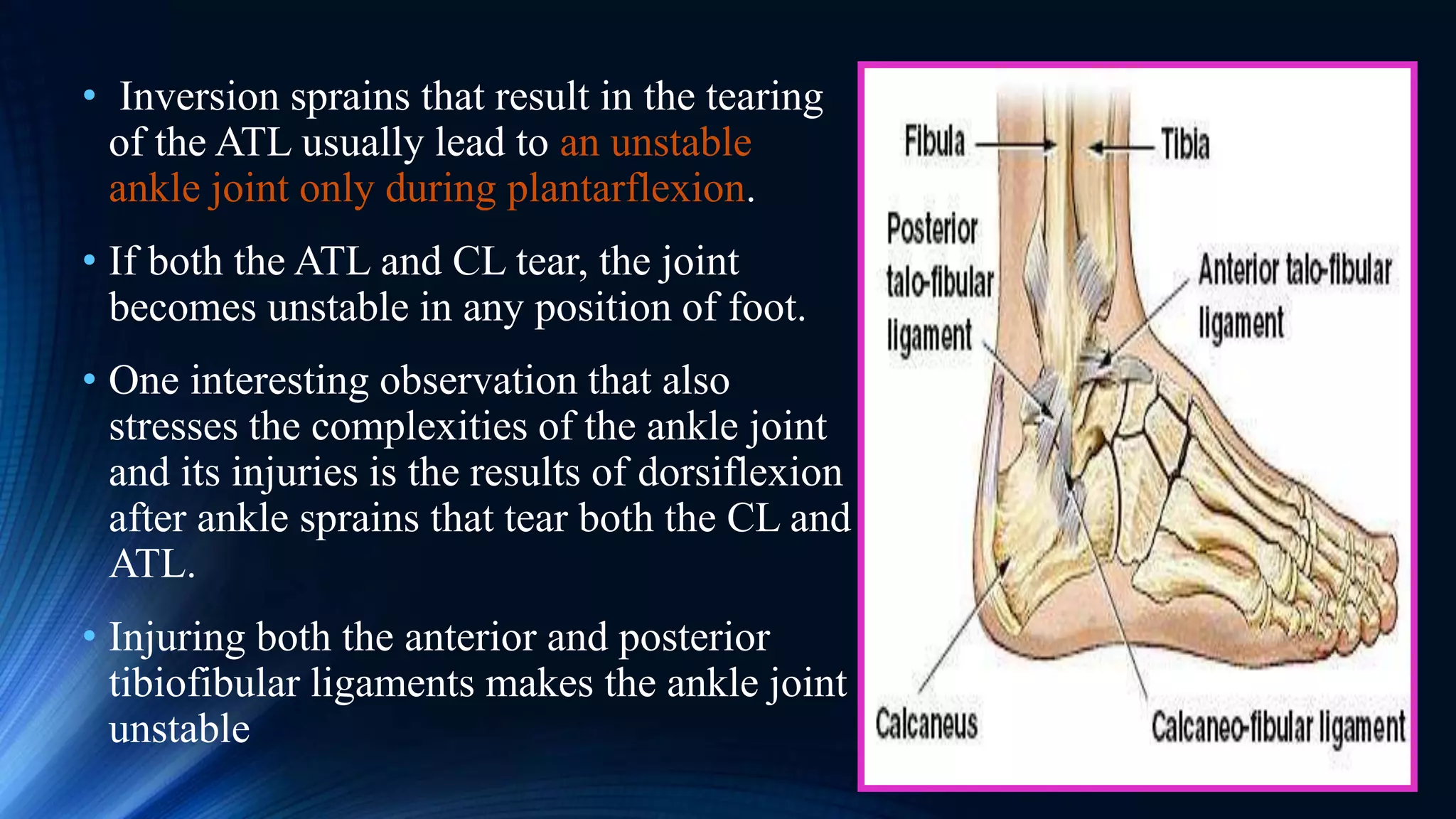
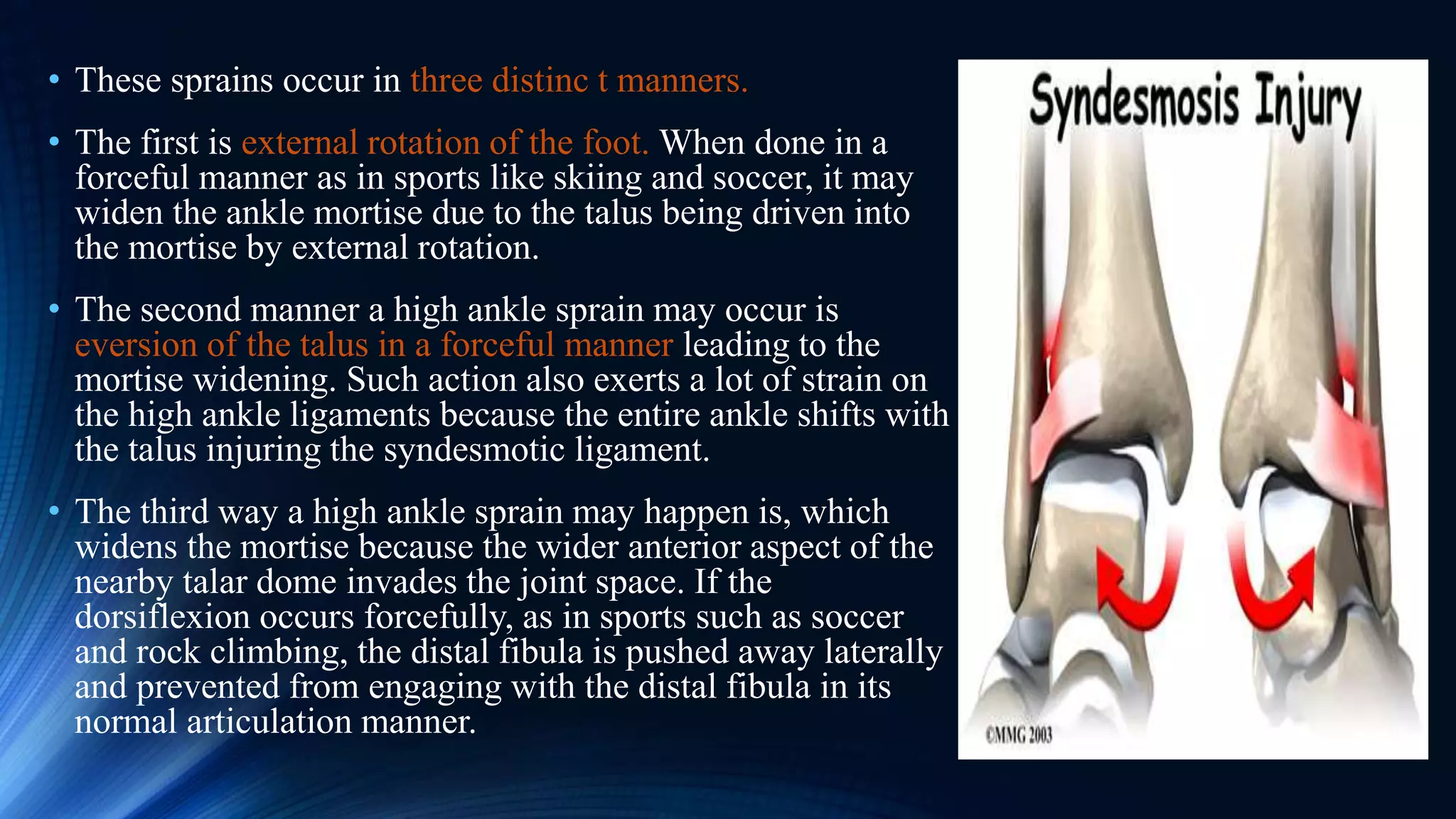
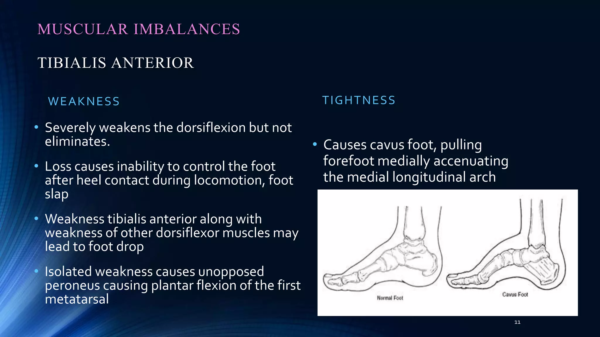
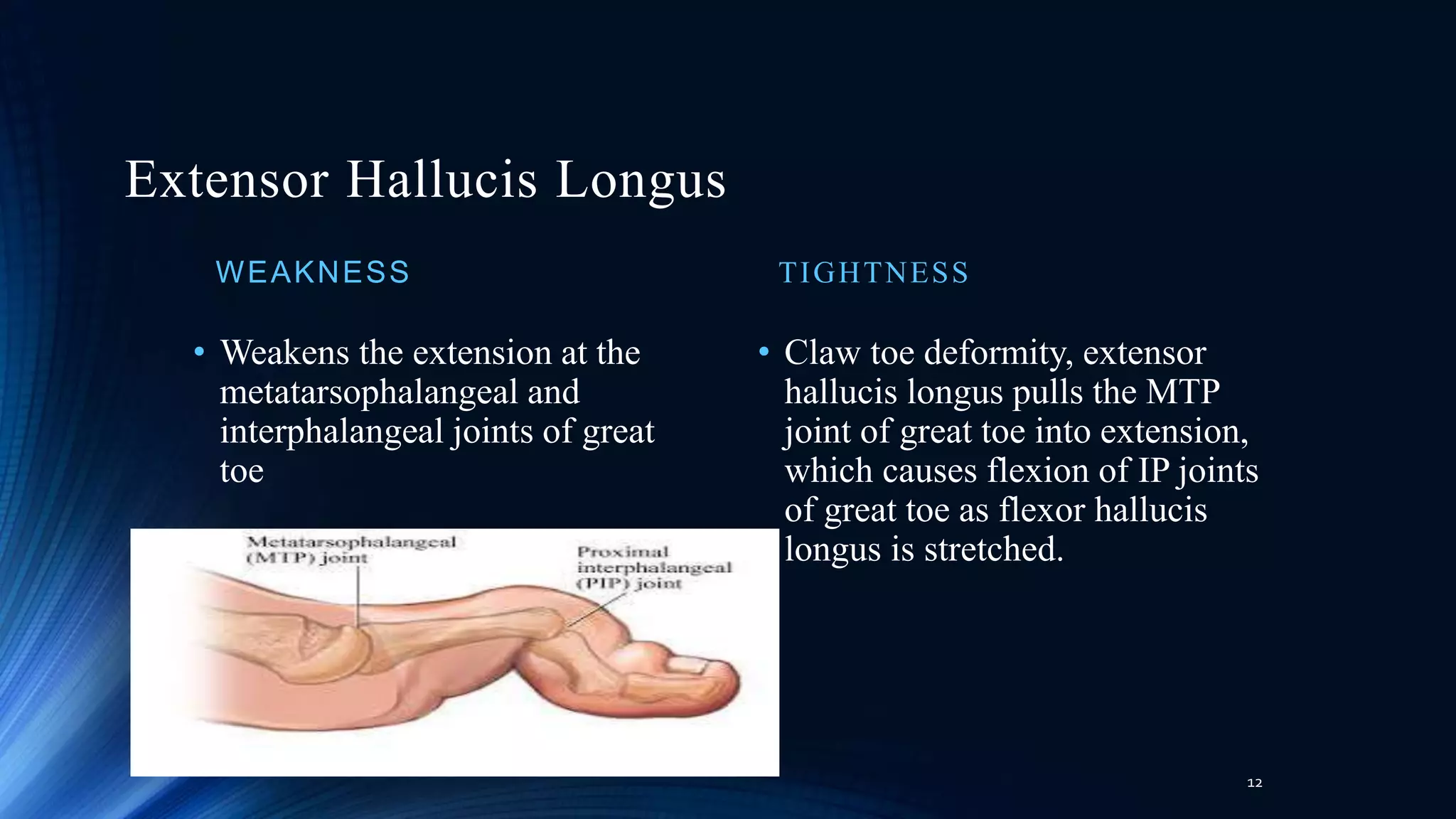
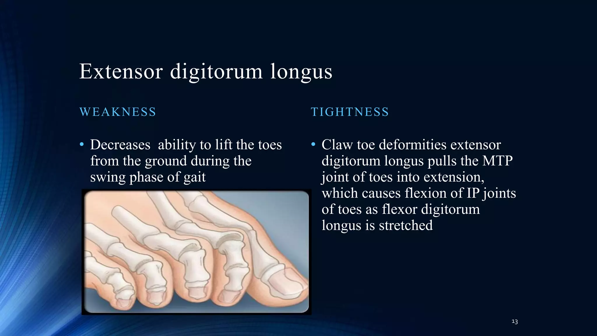
This document discusses the pathomechanics of ankle joint injuries. It begins with the anatomy and ligaments of the ankle joint. It then discusses the muscle groups around the ankle joint and their actions. Next, it explores the mechanics of ankle motion and different types of ankle injuries including lateral and medial ligament injuries, fractures, and muscular imbalances. It provides details on specific muscles like the tibialis anterior and their weaknesses or tightnesses. It concludes with discussing chronic ankle instability and recent literature on lateral ankle sprains and reinjury rates. In summary, the document provides an in-depth overview of ankle joint anatomy, mechanics, common injuries and their pathomechanics, as well as muscular factors.
Knee joint anatomy, biomechanics, pathomechanics and assessmentRadhika Chintamani
the knee complex complete anatomy, biomechanics, pathomechanics and its physical assessment in one single slideshow.a brief table given for easy understanding of what special test to be performed in which condition along with evidences of each special test.
small correction in slide number: 10
during flexion of tibia over femur in OKC; tibia glides and rolls posteriorly
during extension of tibia over femur in OKC: tibia glides and rolls anteriorly
Posture - a perquisite for functional abilities in daily life. Posture is a combination of anatomy and physiology with inherent application of bio-mechanics and kinematics. Sitting, standing, walking are all functional activities depending on the ability of the body to support that posture to carry out each activity. Injuries and pathologies either postural or structural can massively change the bio-mechanics of posture and thus affect functional abilities.
this is a slide show which gives in brief about anatomy and detailed description about biomechanics as well as pathomechanics of shoulder joint. various rhythms of shoulder complex are discussed as well along with the stability factors
A type of manual therapy in which the muscle or the joint is altered and placed in a position of comfort for certain duration after which the pain disappears completely or gets reduced. this slide show explains about the principles, mechanism and Phases of PRT
Brian Mulligan described novel concept of the simultaneous application of therapist applied accessory mobilizations and patient generated active movements
Includes detailed description of BIOMECHANICS & PATHOMECHANICS OF KNEE JOINT AND PATELLOFEMORAL JOINT with recent evidences . Hope you find it useful!!
This Presentation is about Mitchell relaxation technique also known a physiological relaxation technique Mitchell’s physiological relaxation technique is based on reciprocal inhibition and involves diaphragmatic breathing and a series of ordered isotonic contractions.
Muscle energy technique, a manual therapy technique with a long term history and 8 variations which can be used in various condition to treat muscle as well as joints. This slide show consists of detailed history, variations/types and summary of MET in detail.
SPORTS INJURIES OF ANKLE AND FOOT original.pptxMeghaPrakash9
sports injuries of ankle and foot is a seminar done by Ms. Megha ck on behalf of completing her master of physiotherapy program during the year 2020-2022
Research outcome measures related to ankle foot complex indications of de...Missions1
This presentation is about commonly used outcome measures of ankle foot complex. It also has information about delorme boot which a tool for progressive resisted exercise training
This is the Presentation on the topic "Pathomechanics of Knee Joint".
The presentation includes images and a clip for proper understanding. The sentences are framed in the way that you can learn it in a easy way.
Deformities observed with Pes cavus includes :
*clawing of the toes
*posterior hind foot deformity (described as an increased calcaneal angle)
*contracture of the plantar fascia
*cock-up deformity of the great toe
Following References were used to prepare this powerpoint presentation which makes the slides accurate and relaible for studying purpose; Therapeutic Exrercise – Carolyn Kisner
Orthopaedic Physical Assessment – Magee
Orthopaedic Medicine – L. Ombregt
Campbell’s Operative Orthopaedics
Slides includes following headings;
DEFINITION
TYPES
ORTHOPAEDIC ASSESSMENT
MEDICAL MANAGGEMENT
PHYSIOTHERAPY MANAGEMENT
SURGICAL MANAGEMENT
Anti ulcer drugs and their Advance pharmacology ||
Anti-ulcer drugs are medications used to prevent and treat ulcers in the stomach and upper part of the small intestine (duodenal ulcers). These ulcers are often caused by an imbalance between stomach acid and the mucosal lining, which protects the stomach lining.
||Scope: Overview of various classes of anti-ulcer drugs, their mechanisms of action, indications, side effects, and clinical considerations.
ARTIFICIAL INTELLIGENCE IN HEALTHCARE.pdfAnujkumaranit
Artificial intelligence (AI) refers to the simulation of human intelligence processes by machines, especially computer systems. It encompasses tasks such as learning, reasoning, problem-solving, perception, and language understanding. AI technologies are revolutionizing various fields, from healthcare to finance, by enabling machines to perform tasks that typically require human intelligence.
Ethanol (CH3CH2OH), or beverage alcohol, is a two-carbon alcohol
that is rapidly distributed in the body and brain. Ethanol alters many
neurochemical systems and has rewarding and addictive properties. It
is the oldest recreational drug and likely contributes to more morbidity,
mortality, and public health costs than all illicit drugs combined. The
5th edition of the Diagnostic and Statistical Manual of Mental Disorders
(DSM-5) integrates alcohol abuse and alcohol dependence into a single
disorder called alcohol use disorder (AUD), with mild, moderate,
and severe subclassifications (American Psychiatric Association, 2013).
In the DSM-5, all types of substance abuse and dependence have been
combined into a single substance use disorder (SUD) on a continuum
from mild to severe. A diagnosis of AUD requires that at least two of
the 11 DSM-5 behaviors be present within a 12-month period (mild
AUD: 2–3 criteria; moderate AUD: 4–5 criteria; severe AUD: 6–11 criteria).
The four main behavioral effects of AUD are impaired control over
drinking, negative social consequences, risky use, and altered physiological
effects (tolerance, withdrawal). This chapter presents an overview
of the prevalence and harmful consequences of AUD in the U.S.,
the systemic nature of the disease, neurocircuitry and stages of AUD,
comorbidities, fetal alcohol spectrum disorders, genetic risk factors, and
pharmacotherapies for AUD.
Tom Selleck Health: A Comprehensive Look at the Iconic Actor’s Wellness Journeygreendigital
Tom Selleck, an enduring figure in Hollywood. has captivated audiences for decades with his rugged charm, iconic moustache. and memorable roles in television and film. From his breakout role as Thomas Magnum in Magnum P.I. to his current portrayal of Frank Reagan in Blue Bloods. Selleck's career has spanned over 50 years. But beyond his professional achievements. fans have often been curious about Tom Selleck Health. especially as he has aged in the public eye.
Follow us on: Pinterest
Introduction
Many have been interested in Tom Selleck health. not only because of his enduring presence on screen but also because of the challenges. and lifestyle choices he has faced and made over the years. This article delves into the various aspects of Tom Selleck health. exploring his fitness regimen, diet, mental health. and the challenges he has encountered as he ages. We'll look at how he maintains his well-being. the health issues he has faced, and his approach to ageing .
Early Life and Career
Childhood and Athletic Beginnings
Tom Selleck was born on January 29, 1945, in Detroit, Michigan, and grew up in Sherman Oaks, California. From an early age, he was involved in sports, particularly basketball. which played a significant role in his physical development. His athletic pursuits continued into college. where he attended the University of Southern California (USC) on a basketball scholarship. This early involvement in sports laid a strong foundation for his physical health and disciplined lifestyle.
Transition to Acting
Selleck's transition from an athlete to an actor came with its physical demands. His first significant role in "Magnum P.I." required him to perform various stunts and maintain a fit appearance. This role, which he played from 1980 to 1988. necessitated a rigorous fitness routine to meet the show's demands. setting the stage for his long-term commitment to health and wellness.
Fitness Regimen
Workout Routine
Tom Selleck health and fitness regimen has evolved. adapting to his changing roles and age. During his "Magnum, P.I." days. Selleck's workouts were intense and focused on building and maintaining muscle mass. His routine included weightlifting, cardiovascular exercises. and specific training for the stunts he performed on the show.
Selleck adjusted his fitness routine as he aged to suit his body's needs. Today, his workouts focus on maintaining flexibility, strength, and cardiovascular health. He incorporates low-impact exercises such as swimming, walking, and light weightlifting. This balanced approach helps him stay fit without putting undue strain on his joints and muscles.
Importance of Flexibility and Mobility
In recent years, Selleck has emphasized the importance of flexibility and mobility in his fitness regimen. Understanding the natural decline in muscle mass and joint flexibility with age. he includes stretching and yoga in his routine. These practices help prevent injuries, improve posture, and maintain mobilit
Explore natural remedies for syphilis treatment in Singapore. Discover alternative therapies, herbal remedies, and lifestyle changes that may complement conventional treatments. Learn about holistic approaches to managing syphilis symptoms and supporting overall health.
The prostate is an exocrine gland of the male mammalian reproductive system
It is a walnut-sized gland that forms part of the male reproductive system and is located in front of the rectum and just below the urinary bladder
Function is to store and secrete a clear, slightly alkaline fluid that constitutes 10-30% of the volume of the seminal fluid that along with the spermatozoa, constitutes semen
A healthy human prostate measures (4cm-vertical, by 3cm-horizontal, 2cm ant-post ).
It surrounds the urethra just below the urinary bladder. It has anterior, median, posterior and two lateral lobes
It’s work is regulated by androgens which are responsible for male sex characteristics
Generalised disease of the prostate due to hormonal derangement which leads to non malignant enlargement of the gland (increase in the number of epithelial cells and stromal tissue)to cause compression of the urethra leading to symptoms (LUTS
These simplified slides by Dr. Sidra Arshad present an overview of the non-respiratory functions of the respiratory tract.
Learning objectives:
1. Enlist the non-respiratory functions of the respiratory tract
2. Briefly explain how these functions are carried out
3. Discuss the significance of dead space
4. Differentiate between minute ventilation and alveolar ventilation
5. Describe the cough and sneeze reflexes
Study Resources:
1. Chapter 39, Guyton and Hall Textbook of Medical Physiology, 14th edition
2. Chapter 34, Ganong’s Review of Medical Physiology, 26th edition
3. Chapter 17, Human Physiology by Lauralee Sherwood, 9th edition
4. Non-respiratory functions of the lungs https://academic.oup.com/bjaed/article/13/3/98/278874
MANAGEMENT OF ATRIOVENTRICULAR CONDUCTION BLOCK.pdfJim Jacob Roy
Cardiac conduction defects can occur due to various causes.
Atrioventricular conduction blocks ( AV blocks ) are classified into 3 types.
This document describes the acute management of AV block.
Couples presenting to the infertility clinic- Do they really have infertility...Sujoy Dasgupta
Dr Sujoy Dasgupta presented the study on "Couples presenting to the infertility clinic- Do they really have infertility? – The unexplored stories of non-consummation" in the 13th Congress of the Asia Pacific Initiative on Reproduction (ASPIRE 2024) at Manila on 24 May, 2024.
6. PATHOMECHANICS
MEDIAL LIGAMENT INJURIES
• Eversion type of sprains
• Less common than the inversion type partly because of the
presence of the lateral malleolus.
• bony protrusion reduces the length of ligament exposed to the
sheering forces that can could cause ankle sprains.
• Additionally, the medial ligaments, are stronger compared with
lateral ligaments. These shorter ankle ligaments are so strong that
tearing them usually requires enough stress and force to fracture
the tibia or even fibula.
• Therefore, eversion-related ankle sprains should be checked in
conjunction with fractures as they commonly occur together. In
addition, the tibia, fibula, or even talus may be fractured, making
eversion sprains particularly severe.
7. LATERAL LIGAMENTS INJURY
• Inversion type of sprains
• Most common type of ankle sprain
• Happens due to unstable landing after a jump or running/walking
on an uneven surface. It results from the plantarflexion of the foot
injuring the ATFL the ligament involved moves perpendicular to
the talus, exposing it to shear forces.
• A lot of force applied to the ATFL during the inversion sprain may
break it and affect the CFL. The effect occurs because the CFL is
the next ligament supposed to take the stress.
• The CFL can be injured if the inversion sprain is extreme enough.
8. • Inversion sprains that result in the tearing
of the ATL usually lead to an unstable
ankle joint only during plantarflexion.
• If both the ATL and CL tear, the joint
becomes unstable in any position of foot.
• One interesting observation that also
stresses the complexities of the ankle joint
and its injuries is the results of dorsiflexion
after ankle sprains that tear both the CL and
ATL.
• Injuring both the anterior and posterior
tibiofibular ligaments makes the ankle joint
unstable
9. `
• Some unique forms of ankle sprains involve
the syndesmotic ligaments that connect the
ankle joint to the bones forming the shin.
• This type of sprain is common among
footballers and result in persistent pain as
well as residual ankle dysfunction.
• Require almost twice as long to heal
compared with inversion and eversion
sprains. One reason for such lengthy healing
periods is the syndesmosis ligament, which is
hard to heal.
• Surgery is a common form of treatment for
cases where the high ankle sprain is serious
and the syndesmosis lIgaments are torn.
10. • These sprains occur in three distinc t manners.
• The first is external rotation of the foot. When done in a
forceful manner as in sports like skiing and soccer, it may
widen the ankle mortise due to the talus being driven into
the mortise by external rotation.
• The second manner a high ankle sprain may occur is
eversion of the talus in a forceful manner leading to the
mortise widening. Such action also exerts a lot of strain on
the high ankle ligaments because the entire ankle shifts with
the talus injuring the syndesmotic ligament.
• The third way a high ankle sprain may happen is, which
widens the mortise because the wider anterior aspect of the
nearby talar dome invades the joint space. If the
dorsiflexion occurs forcefully, as in sports such as soccer
and rock climbing, the distal fibula is pushed away laterally
and prevented from engaging with the distal fibula in its
normal articulation manner.
11. MUSCULAR IMBALANCES
TIBIALIS ANTERIOR
WEAKNESS TIGHTNESS
11
• Severely weakens the dorsiflexion but not
eliminates.
• Loss causes inability to control the foot
after heel contact during locomotion, foot
slap
• Weakness tibialis anterior along with
weakness of other dorsiflexor muscles may
lead to foot drop
• Isolated weakness causes unopposed
peroneus causing plantar flexion of the first
metatarsal
• Causes cavus foot, pulling
forefoot medially accenuating
the medial longitudinal arch
12. Extensor Hallucis Longus
WEAKNESS TIGHTNESS
12
• Weakens the extension at the
metatarsophalangeal and
interphalangeal joints of great
toe
• Claw toe deformity, extensor
hallucis longus pulls the MTP
joint of great toe into extension,
which causes flexion of IP joints
of great toe as flexor hallucis
longus is stretched.
13. Extensor digitorum longus
WEAKNESS TIGHTNESS
13
• Decreases ability to lift the toes
from the ground during the
swing phase of gait
• Claw toe deformities extensor
digitorum longus pulls the MTP
joint of toes into extension,
which causes flexion of IP joints
of toes as flexor digitorum
longus is stretched
14. Peroneus tertius
WEAKNESS TIGHTNESS
14
• Occurs in conjuction with
weakness of the extensor
digitorum longus and the other
dorsiflexor muscles.
• Occurs concomitantly with
extensor digitorum longus.
15. Gastrocnemius
WEAKNESS TIGHTNESS
15
• Decreased plantar flexion
strength
• Hampers an individual ability to
rise up on toes or climb hills or
ladders
• Decreases ROM of dorsiflexion
• Depends on position of knees
16. Soleus
WEAKNESS TIGHTNESS
16
• Plantar flexion strength is lost
• Impairs the leg control ability
as the body glides over the
stance foot
• Tightness of the soleus also restricts
dorsiflexion ROM
• Plantarflexion contracture is independent of
knee position.
• Restricts forward glide of the tibia, even
though momentum may continue the forward
progression of the thigh and trunk
• Tightness of the soleus is a risk factor for
genu recurvatum.
17. Deep Muscles Of The Posterior Compartment
Posterior Tibialis
WEAKNESS TIGHTNESS
17
• Impairs inversion strength.
• Impairs an individual’s ability to rise up on
the toes, even with intact plantarflexor
muscles, because the foot is unstable.
• Imbalance with the everter muscles, and
the foot tends to evert and abduct; that is, it
tends to pronate.
• Pulls the foot into inversion and
adduction of the forefoot and may
include slight plantarflexion,
producing a varus or an
equinovarus deformity of the foot.
• Such deformities are often found in
individuals with spasticity of the
posterior tibialis or with an
imbalance between the posterior
tibialis and the everters of the foot.
18. WEAKNESS
18
• Patients with posterior tibilalis tendon dysfunction (PTTD) exhibit increased
pronation at the hindfoot and forefoot, reflecting the muscle’s extensive role in
supporting most of the foot .
• PTTD is a primary cause of acquired flat feet and alters the normal movement
of the tarsal bones during weight bearing and gait .
• Factors associated with increased risk of PTTD are obesity, aging, hypertension,
diabetes, and vascular insufficiency within the tendon .
• A preexisting flat foot deformity also appears to be a risk factor for a rupture of
the posterior tibialis.
19. Flexor Digitorum Longus
WEAKNESS TIGHTNESS
19
• Produces weakness in toe flexion
MOSTLY at the distal interphalangeal
joints.
• Functionally, weakness of the flexor
digitorum longus produces difficulty in
stabilizing the foot and toes during stance
and is manifested by delayed or limited
heel rise as the body rolls over the foot.
• Tightness of the flexor digitorum longus
impairs extension ROM of the toes.
20. Flexor Hallucis Longus
WEAKNESS TIGHTNESS
20
• weakens flexion of the great .
• contributes to decreased plantar flexion
strength.
• Weakness may also contribute to slight
inversion weakness
• Limits extension of the joints of the toes
particularly when the ankle is dorsiflexed.
• Plantarflexing the ankle puts the muscle on
slack and allows more toe extension .
• Tightness of the flexor hallucis longus also is
implicated in a claw deformity of the great
toe, may also contribute to foot pain in the
medial longitudinal arch.
• Runners occasionally develop pain along the
flexor hallucis longus tendon as the result of
repeatedly stretching the contracting muscle
during the push off phase of running
21. MUSCLES OF THE LATERAL COMPARTMENT OF THE LEG
Peroneus Longus
WEAKNESS TIGHTNESS
21
• Weakness of the peroneus longus contributes
to weakness in eversion of the foot.
• the inverters, particularly the posterior
tibialis, pull the foot into inversion or
inversion with plantarflexion, and a varus, or
equinovarus, deformity
• Limit inversion ROM of the subtalar joint,
tightness is manifested primarily by a
plantarflexed first ray.
• In weight bearing the plantarflexed first ray
may produce excessive loading on the
metatarsal head of the great toe
• which can lead to pain and large callus
formation under the first metatarsal head
• Weight bearing in upright stance with a
plantarflexed first ray also produces a
supination moment on the foot.
22. Peroneus Brevis
WEAKNESS TIGHTNESS
22
• Weakness of the peroneus brevis decreases eversion
strength and contributes to an imbalance between
the inverter and everter muscles.
• increases the relative contribution of the inverters
and leads to a varus hindfoot deformity.
• tightness of the peroneus brevis may contribute
to valgus deformities of the foot.
• weakness of the posterior tibialis or
overactivity of the extensor digitorum longus
also are important contributors to valgus
deformities of the foot.
23. Fractures
23
Malleolar fracture and their classification
• Supination – Abduction injuries
Inversion force on the dorsiflexed ankle results in vertical or
oblique malleolar fracture and transverse avulsion type
fracture of distal fibula associated rupture of the lateral
collateral ligament.This is also associated with medial
displacement of talus.
• Supination-External rotation
This is the most common type of injury.This leads to typical
oblique fracture at distal fibula and either a transverse or
avulsion type of medial malleolus fracture and rupture of
the deltoid ligament.
24. 24
• Pronation-Abduction injuries
there will be deltoid ligament disruption and transverse fracture of the medial
malleolus with short oblique fracture of fibula at the level of syndesmosis with lateral
communiation fracture.
• Pronation-External rotation injuries
transverse fracture of the medial malleolus or rupture of the deltoid ligament may
occur.This can be associated with disruption of the tibiofibular ligament and fracture
of the posterior part of the distal articular surface of tibia, that is, posterior malleolar
fracture.
25. 25
• POTT’S FRACTURE
Described by percival Pott in 1765.
Combined abduction and external rotation violence
It includes rupture of the medial ligament/ fracture of medial malleolus, fracture
of lateral malleolus and lateral displacement of ankle.
27. 27
• Chronic ankle instability (CAI) is a term that is presently used to denote
the occurrence of repeated episodes of lateral ankle instability and the
presence of residual symptoms such as
• pain,
• swelling,
• ‘‘giving way,’’ and
• loss of motion
• Potential intrinsic risk factors for the development of ankle instability
due to, ligamentous stability,muscular strength,anatomic foot and ankle
alignment, postural sway, gait mechanics, and muscle reaction time..
28. 28
Two main types of instability can be distinguished:
• Mechanical instability related to anatomic abnormalities of the
ankle, usually related to ligament laxity.
• Functional instability related to posture defects or tendon and
muscle adjustment, usually related to a proprioceptive deficit.
29. 29
Mechanical instability
Bone instability:
• Unstable ankles can present a congruence defect with a wider talar dome and
reduced talar coverage as well as a more anterior position of the talus in
relation to the tibia on the loading.
• The lateral malleolus seems to be in a posterior position because of distension
or rupture of the anterior talofibular ligament, from medial rotation of the
talus (notably in pes cavus), or from malunion of the lateral malleolus.
30. 30
Mechanical ligament instability.
• Ankle instability does not result only from injury, constitutional hyperlaxity
also exists.These can be a stimulation defect of the joint mechanoreceptors
or proprioception dysfunction.
Mechanical joint instability
• The wider talus at the front explains that the deficit in dorsal flexion of the
ankle is a factor of instability.Therefore, anterior osteophytosis
(impingement exostosis) or anterior synovial hypertrophy (fibrous
impingement) are factors aggravating instability.
• This limitation in dorsi- flexion can also have a functional cause such as
retraction of the sural triceps or the gastrocnemial muscles or even a muscle
belly extended too far distally.
31. 31
Functional instability
• Functional muscular instability.
• The delay in muscle reactivity can be caused by a sometimes transitory
neurological deficit (paresis after sitting with the legs crossed) or a
mechanical muscle defect (muscle belly developed too distally,
tenosynovitis or luxation of the fibularis muscles).
• In a gait analysis, an increase in dorsal flexion of the first
metatarsophalangeal joint, an increase in ground contact time,
lateralization of pressure of the lateral edge of the midfoot and the
forefoot, and an increase in the pronosupination index
32. 32
Functional postural instability.
• Varus of the hindfoot is a cause of ligament reconstruction
failure .Varus results in excessive pressure on the lateral edge of
the foot and postural imbalance in monopedal weightbearing. It
is responsible for excessive tension of the fibularis muscles, with
most often a loss of the myotatic reflex.
• The associated equinus results in a contraction defect in the
extensor digitorum muscles or, excess flexion with clawing of
the lateral toes.
33. 33
• Clawing of the fifth toe or a callus under the head of the fourth
metatarsal is also a sign of ankle instability.
• Other morphostatic problems induce excessive pressure on the
lateral edge of the foot such as unequal length of the lower limbs in
which the shorter limb tends to position itself in varus-equinus, with
genu varum, adductus foot, or forefoot pronatus, causing an unstable
dynamic supination movement on weightbearing
34. RECENT ARTICLES
• Lateral and syndesmotic ankle sprain injuries: a narrative literature
review.
• Dubin JC1, Comeau D, McClelland RI, Dubin RA, Ferrel E Epub 2011 Jul 23.
• to review the literature that discusses normal anatomy and biomechanics
of the foot and ankle, mechanisms that may result in a lateral ankle sprain
or syndesmotic sprain
• Most ankle sprains respond favorably to nonsurgical treatment, such as
those offered by physical therapists, doctors of chiropractic, and
rehabilitation specialists.
An accurate diagnosis and prompt treatment can minimize an athlete's time
lost from sport and prevent future reinjury.
35. 2010 Jul 16;1:115-22.PUBMED
Ankle sprain: pathophysiology, predisposing
factors, and management strategies.
Hubbard TJ1, Wikstrom EA.
• review LAS pathophysiology, predisposing factors, and the current
evidence regarding therapeutic modalities and exercises used in the
treatment of LAS
• Recent evidence has shown the need for more stringent
immobilization to facilitate ligament healing and restoration of joint
stability and function after a LAS
36. Epub 2009 Jul 17PUBMED.
Reinjury after acute lateral ankle sprains in elite
track and field athletes.
Malliaropoulos N1, Ntessalen M, Papacostas
E, Longo UG, Maffulli N.
• The effect of a lateral ankle sprain on reinjury occurrence in the same
region.
Athletes with a grade I or II lateral ankle sprain are at higher risk of
experiencing a reinjury. Low-grade acute lateral ankle sprains result in
a higher risk of reinjury than high-grade acute lateral ankle sprains.
37. References:
37
• Application to Pathological Motion ;Gary L. Soderberg
• Kinesiology of the Musculoskeletal System: by Donald A. Neumann
• The Mechanics and Pathomechanics of Human Movement Carol A.
Oatis
• Joint Structure and Function : A Comprehensive Analysis, Pamela K.
Levangie Cynthia C. Norkin
• WEB SEARCH : pubmed, pedro, science direct