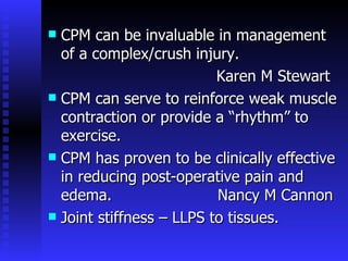
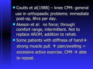
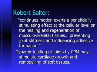
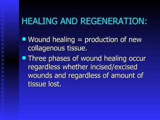
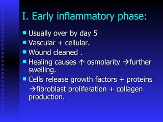
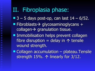
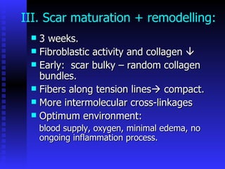
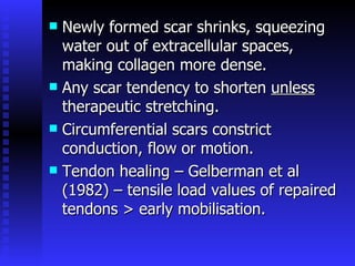
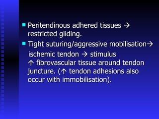
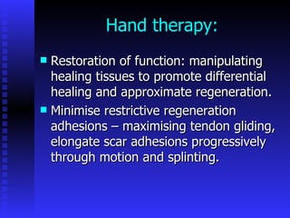
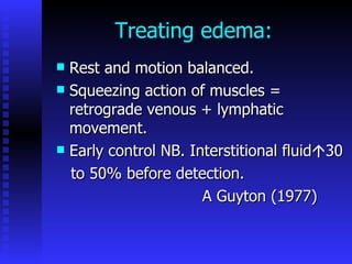
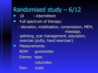
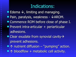
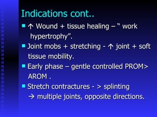
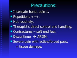
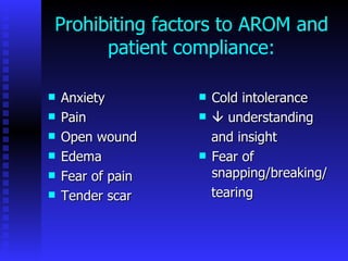
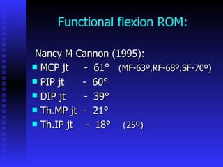
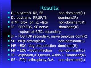
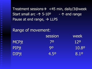
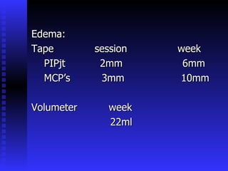
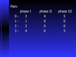
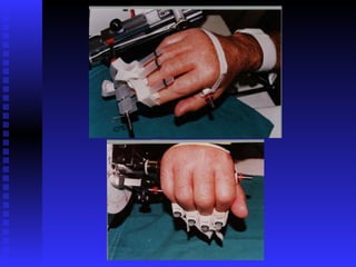
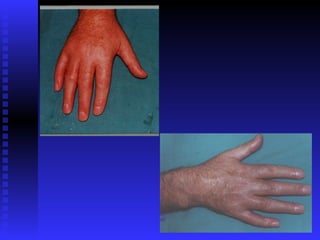
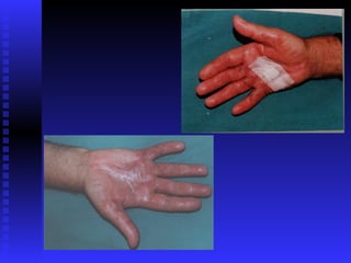
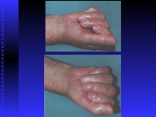
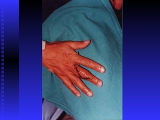
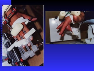
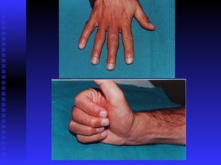
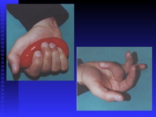
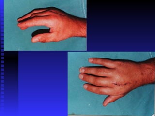
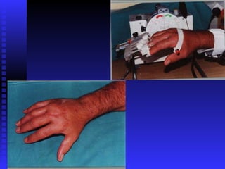
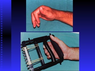
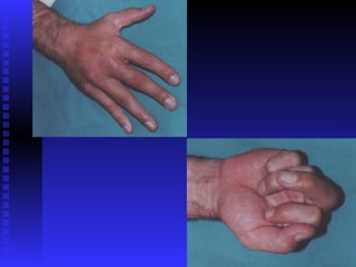
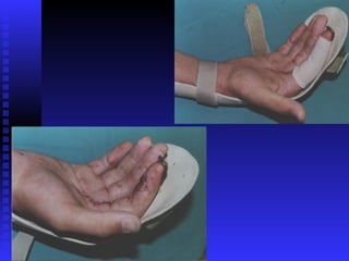
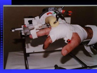
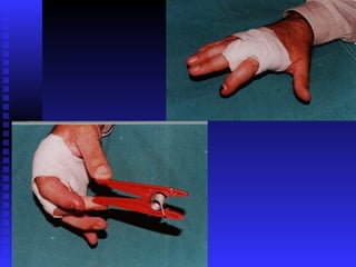
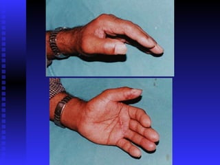
CONTINUOUS PASSIVE MOTION MACHINE AS AN INTEGRAL PART OF MANAGEMENT OF RANGE OF MOTION AND EDEMA IN HAND THERAPY Ms L Pringle Cape Town
“ Without passive motion there is no potential for active range..” J Colditz Active range of motion (AROM):  -contraction of muscle crossing a joint. Passive range of motion (PROM): -movement of a joint by an external  force.
Other definitions of PROM/CPM: Correct PROM: Gentle encouragement of tissues   max. available length. CPM: PROM continuously in a reciprocal fashion by a mechanical device which controls range, rate.  P La Stayo
CPM can be invaluable in management of a complex/crush injury.  Karen M Stewart CPM can serve to reinforce weak muscle contraction or provide a “rhythm” to exercise. CPM has proven to be clinically effective in reducing post-operative pain and edema.  Nancy M Cannon Joint stiffness – LLPS to tissues.
Coutts et al(1988) – knee CPM: general use in orthopaedic problems: immediate post-op, 8hrs per day. Akeson et al:  no force; through comfort range, intermittent. Not to replace AROM, addition to rehab. Some patients with stiffness of hand  strong muscle pull.    pain/swelling = excessive active exercise. CPM    able to repeat.
Robert Salter: “ continues motion exerts a beneficially stimulating effect at the cellular level on the healing and regeneration of musculo-skeletal tissues… preventing joint stiffness and influencing adhesive formation.” Dynamic loading of joints by CPM may stimulate cartilage growth and remodelling of soft tissues.
HEALING AND REGENERATION: Wound healing = production of new collagenous tissue. Three phases of wound healing occur regardless whether incised/excised wounds and regardless of amount of tissue lost.
I. Early inflammatory phase: Usually over by day 5 Vascular + cellular.  Wound cleaned . Healing causes    osmolarity   further swelling. Cells release growth factors + proteins  fibroblast proliferation + collagen production.
II.  Fibroplasia phase: 3 – 5 days post-op, can last 14 – 6/52. Fibroblasts   glycosaminoglycans + collagen   granulation tissue. Immobilisation helps prevent collagen fibre disruption = delay in    tensile wound strength. Collagen accumulation – plateau.Tensile strength 15%.    linearly for 3/12.
III. Scar maturation + remodelling: 3 weeks. Fibroblastic activity and collagen   Early:  scar bulky – random collagen bundles. Fibers along tension lines   compact. More intermolecular cross-linkages Optimum environment: blood supply, oxygen, minimal edema, no ongoing inflammation process.
Newly formed scar shrinks, squeezing water out of extracellular spaces, making collagen more dense. Any scar tendency to shorten  unless  therapeutic stretching. Circumferential scars constrict conduction, flow or motion. Tendon healing – Gelberman et al (1982) – tensile load values of repaired tendons > early mobilisation.
Peritendinous adhered tissues    restricted gliding. Tight suturing/aggressive mobilisation  ischemic tendon    stimulus    fibrovascular tissue around tendon juncture. (   tendon adhesions also occur with immobilisation).
Hand therapy: Restoration of function: manipulating healing tissues to promote differential healing and approximate regeneration. Minimise restrictive regeneration adhesions – maximising tendon gliding, elongate scar adhesions progressively through motion and splinting.
Treating edema:  Rest and motion balanced. Squeezing action of muscles = retrograde venous + lymphatic  movement. Early control NB. Interstitional fluid  30 to 50% before detection. A Guyton (1977)
Randomised study – 6/12 10  - intermittent Full spectrum of therapy: - elevation, mobilisation, compression, MEM,  massage, splinting, scar management, education, exercise (putty, hand exerciser). Measurements: ROM:  goniometer Edema:  tape. volumeter. Pain:  scale.
Pressure glove
Goniometer:  Tape:
Volumeter:
Pain: Scale: 0  =  no pain 1  =  all the time 2  =  with movement 3  =  at end range
Indications:  Edema   , limiting and managing. Pain, paralysis, weakness -   AROM. Commence ROM before close of phase I. Prevent intra-articular + periarticular  adhesions. Clear exudate from synovial cavity  prevent adhesions.    nutrient diffusion – “pumping” action.    bloodflow + metabolic cell activity.
Indications cont..    Wound + tissue healing – “ work  hypertrophy”. Joint mobs + stretching -    joint + soft tissue mobility. Early phase – gentle controlled PROM> AROM . Stretch contractures - > splinting    multiple joints, opposite directions.
Precautions: Insensate hand, pain 1. Repetitions +++. Not routinely. Therapist’s direct control and handling. Contractures – soft end feel.  Discontinue    AROM. Severe pain with active/forced pass. = tissue damage.
Prohibiting factors to AROM and patient compliance: Anxiety Pain Open wound Edema Fear of pain Tender scar Cold intolerance    understanding and insight Fear of snapping/breaking/ tearing
Functional flexion ROM: Nancy M Cannon (1995): MCP jt  -  61°  (MF-63º,RF-68º,SF-70º) PIP jt  -  60° DIP jt  -  39° Th.MP jt  -  21° Th.IP jt  -  18°  (25º)
Results: Du puytren’s  RF, SF  non-dominant(L) Du puytren’s  RF,SF,Th  dominant(R) # MF prox. ph. jt. –late  non-dominant(R) IF – FDP,FDS, SF-nerve  dominant(R) rupture at 6/52, secondary IF – FDS,FDP secondary, nerve tenolysis dom(R) SF –PIPjt arthroplasty  non-dominant(L) MF – EDC -dog bite,infection  dominant(R) MF – EDC –tooth,infection  non-dominant(L) IF – explosion,#’s,nerves,  skin ?  Dominant(R) RF – PIPjt arthroplasty,O.A.  non-dominant(L)
Treatment sessions    <45 min, daily/3@week Start small arc -   5-10º  -    end range Pause at end range,    LLPS Range of movement: session  week MCPjt  7º  12º PIPjt  9º  10.8º DIPjt  4.5º  8.1º
Edema: Tape  session  week PIPjt  2mm  6mm MCP’s  3mm  10mm Volumeter  week 22ml
Pain: phase I  phase II  phase III 0 -  3  4  5 1 -  1  0  0 2 -  2  0  0 3 -  4  6  5


Conclusion: The use of a CPM machine has proved a valuable adjunct to hand therapy, achieving goals such as the increase, maintenance and restoring of passive mobility, maintaining gliding, stimulating nutrient diffusion and decreasing edema and pain.

Hand Therapy - Continuous Passive Motion

  • 1.
    CONTINUOUS PASSIVE MOTIONMACHINE AS AN INTEGRAL PART OF MANAGEMENT OF RANGE OF MOTION AND EDEMA IN HAND THERAPY Ms L Pringle Cape Town
  • 2.
    “ Without passivemotion there is no potential for active range..” J Colditz Active range of motion (AROM): -contraction of muscle crossing a joint. Passive range of motion (PROM): -movement of a joint by an external force.
  • 3.
    Other definitions ofPROM/CPM: Correct PROM: Gentle encouragement of tissues  max. available length. CPM: PROM continuously in a reciprocal fashion by a mechanical device which controls range, rate. P La Stayo
  • 4.
    CPM can beinvaluable in management of a complex/crush injury. Karen M Stewart CPM can serve to reinforce weak muscle contraction or provide a “rhythm” to exercise. CPM has proven to be clinically effective in reducing post-operative pain and edema. Nancy M Cannon Joint stiffness – LLPS to tissues.
  • 5.
    Coutts et al(1988)– knee CPM: general use in orthopaedic problems: immediate post-op, 8hrs per day. Akeson et al: no force; through comfort range, intermittent. Not to replace AROM, addition to rehab. Some patients with stiffness of hand  strong muscle pull.  pain/swelling = excessive active exercise. CPM  able to repeat.
  • 6.
    Robert Salter: “continues motion exerts a beneficially stimulating effect at the cellular level on the healing and regeneration of musculo-skeletal tissues… preventing joint stiffness and influencing adhesive formation.” Dynamic loading of joints by CPM may stimulate cartilage growth and remodelling of soft tissues.
  • 7.
    HEALING AND REGENERATION:Wound healing = production of new collagenous tissue. Three phases of wound healing occur regardless whether incised/excised wounds and regardless of amount of tissue lost.
  • 8.
    I. Early inflammatoryphase: Usually over by day 5 Vascular + cellular. Wound cleaned . Healing causes  osmolarity  further swelling. Cells release growth factors + proteins  fibroblast proliferation + collagen production.
  • 9.
    II. Fibroplasiaphase: 3 – 5 days post-op, can last 14 – 6/52. Fibroblasts  glycosaminoglycans + collagen  granulation tissue. Immobilisation helps prevent collagen fibre disruption = delay in  tensile wound strength. Collagen accumulation – plateau.Tensile strength 15%.  linearly for 3/12.
  • 10.
    III. Scar maturation+ remodelling: 3 weeks. Fibroblastic activity and collagen  Early: scar bulky – random collagen bundles. Fibers along tension lines  compact. More intermolecular cross-linkages Optimum environment: blood supply, oxygen, minimal edema, no ongoing inflammation process.
  • 11.
    Newly formed scarshrinks, squeezing water out of extracellular spaces, making collagen more dense. Any scar tendency to shorten unless therapeutic stretching. Circumferential scars constrict conduction, flow or motion. Tendon healing – Gelberman et al (1982) – tensile load values of repaired tendons > early mobilisation.
  • 12.
    Peritendinous adhered tissues  restricted gliding. Tight suturing/aggressive mobilisation  ischemic tendon  stimulus  fibrovascular tissue around tendon juncture. (  tendon adhesions also occur with immobilisation).
  • 13.
    Hand therapy: Restorationof function: manipulating healing tissues to promote differential healing and approximate regeneration. Minimise restrictive regeneration adhesions – maximising tendon gliding, elongate scar adhesions progressively through motion and splinting.
  • 14.
    Treating edema: Rest and motion balanced. Squeezing action of muscles = retrograde venous + lymphatic movement. Early control NB. Interstitional fluid  30 to 50% before detection. A Guyton (1977)
  • 15.
    Randomised study –6/12 10 - intermittent Full spectrum of therapy: - elevation, mobilisation, compression, MEM, massage, splinting, scar management, education, exercise (putty, hand exerciser). Measurements: ROM: goniometer Edema: tape. volumeter. Pain: scale.
  • 16.
  • 17.
  • 18.
  • 19.
    Pain: Scale: 0 = no pain 1 = all the time 2 = with movement 3 = at end range
  • 20.
    Indications: Edema  , limiting and managing. Pain, paralysis, weakness -  AROM. Commence ROM before close of phase I. Prevent intra-articular + periarticular adhesions. Clear exudate from synovial cavity  prevent adhesions.  nutrient diffusion – “pumping” action.  bloodflow + metabolic cell activity.
  • 21.
    Indications cont..  Wound + tissue healing – “ work hypertrophy”. Joint mobs + stretching -  joint + soft tissue mobility. Early phase – gentle controlled PROM> AROM . Stretch contractures - > splinting  multiple joints, opposite directions.
  • 22.
    Precautions: Insensate hand,pain 1. Repetitions +++. Not routinely. Therapist’s direct control and handling. Contractures – soft end feel. Discontinue  AROM. Severe pain with active/forced pass. = tissue damage.
  • 23.
    Prohibiting factors toAROM and patient compliance: Anxiety Pain Open wound Edema Fear of pain Tender scar Cold intolerance  understanding and insight Fear of snapping/breaking/ tearing
  • 24.
    Functional flexion ROM:Nancy M Cannon (1995): MCP jt - 61° (MF-63º,RF-68º,SF-70º) PIP jt - 60° DIP jt - 39° Th.MP jt - 21° Th.IP jt - 18° (25º)
  • 25.
    Results: Du puytren’s RF, SF non-dominant(L) Du puytren’s RF,SF,Th dominant(R) # MF prox. ph. jt. –late non-dominant(R) IF – FDP,FDS, SF-nerve dominant(R) rupture at 6/52, secondary IF – FDS,FDP secondary, nerve tenolysis dom(R) SF –PIPjt arthroplasty non-dominant(L) MF – EDC -dog bite,infection dominant(R) MF – EDC –tooth,infection non-dominant(L) IF – explosion,#’s,nerves,  skin ? Dominant(R) RF – PIPjt arthroplasty,O.A. non-dominant(L)
  • 26.
    Treatment sessions  <45 min, daily/3@week Start small arc -  5-10º -  end range Pause at end range,  LLPS Range of movement: session week MCPjt 7º 12º PIPjt 9º 10.8º DIPjt 4.5º 8.1º
  • 27.
    Edema: Tape session week PIPjt 2mm 6mm MCP’s 3mm 10mm Volumeter week 22ml
  • 28.
    Pain: phase I phase II phase III 0 - 3 4 5 1 - 1 0 0 2 - 2 0 0 3 - 4 6 5
  • 29.
  • 30.
  • 31.
  • 32.
  • 33.
  • 34.
  • 35.
  • 36.
  • 37.
  • 38.
  • 39.
  • 40.
  • 41.
  • 42.
  • 43.
  • 44.
  • 45.
    Conclusion: The useof a CPM machine has proved a valuable adjunct to hand therapy, achieving goals such as the increase, maintenance and restoring of passive mobility, maintaining gliding, stimulating nutrient diffusion and decreasing edema and pain.