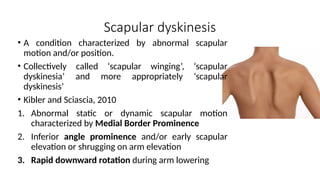
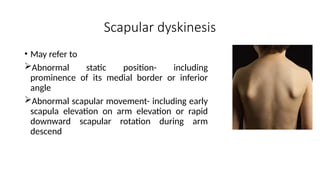
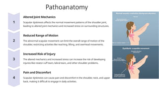
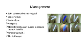
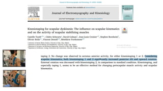
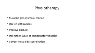
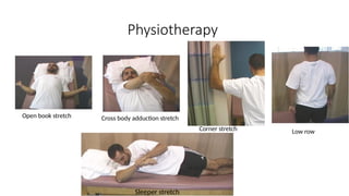
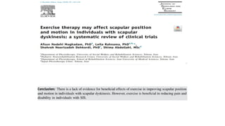
SCAPULAR DYSKINESIS
Presenter- Dr Binod Chaudhary
Moderator- Assoc. Prof. Dr Jhapindra Pokharel
HOD, Department of Orthopaedics
WRH, PoAHS
Contents
• Backgrounds
• Biomechanics
• Definition
• Etiology
• Diagnosis
• Management
Background
Scapula
• Large triangular bone located just lateral to
the vertebral column and posterior part of
the thoracic wall.
• Parts: body, neck, glenoid and coracoid
process
• Borders: medial, superior and inferior
• Surface: anterior and posterior
(supraspinatus and infraspinatus)
Scapulothoracic joint
• Interval between the scapula and underlying wall
of ribs
• Not a true joint
• Separated by thin layer of loose areolar tissue to
glide freely in all directions
• Any movement of the scapula on the thorax
results in movement of either the
acromioclavicular joint, the sternoclavicular joint
or both
Biomechanics
Resting position of scapula
• Anteriorly 10-20 degrees
• Internally rotated 30-45 degrees
• Upward tilt 3 degrees
4 biomechanical roles
• Center of rotation of humerus
• Anchor of humerus onto the thoracic wall
• Keeps acromion from obstructing movement
of humerus both into abduction and in flexion
• It is the means by which forces are
transmitted from core to the arm
Motion
• Primary motion: Elevation and Depression
Movement up and down along the rib cage
• Secondary motion:
Protraction and retraction
Upward and downward rotation
Internal and external rotation
Anterior and posterior tipping
Shoulder abduction
• The first 30-60 degrees of arm abduction
occur at glenohumeral joint, scapula
remaining fixed(setting phase)
• Further shoulder abduction the ratio of
glenohumeral motion to scapulothoracic
motion occurs at 2:1
• i.e for every 300
of abduction, 200
occurs at
glenohumeral and 100
occurs at scapula-
thoracic joint
• 17 different muscles attach to or originate
from scapula providing stability and
movement
• Muscles controlling glenohumeral joint
motion
Forward elevation: deltoid(anterior part), pectoralis major,
biceps, coracobrachialis
Abduction: deltoid(middle part), supraspinatus
External rotation: infraspinatus, teres minor, deltoid(posterior
part)’
Internal rotation: subscapularis, pectoralis major, latissimus
dorsi, deltoid
Adduction: pectoralis major, latissimus dorsi, teres major,
teres minor
Extension: deltoid(posterior part), latissimus dorsi, teres major
• Muscles controlling scapulothoracic
motion
Elevation: upper part of trapezius, rhomboids
Retraction: upper part of trapezius, rhomboids
Protraction: serratus anterior
Upward rotation: upper part of trapezius,
serratus anterior
Downward rotation: lower part of trapezius,
rhomboids
Scapular dyskinesis
• A condition characterized by abnormal scapular
motion and/or position.
• Collectively called ‘scapular winging’, ‘scapular
dyskinesia’ and more appropriately ‘scapular
dyskinesis’
• Kibler and Sciascia, 2010
1. Abnormal static or dynamic scapular motion
characterized by Medial Border Prominence
2. Inferior angle prominence and/or early scapular
elevation or shrugging on arm elevation
3. Rapid downward rotation during arm lowering
• May refer to
Abnormal static position- including
prominence of its medial border or inferior
angle
Abnormal scapular movement- including early
scapula elevation on arm elevation or rapid
downward scapular rotation during arm
descend
Scapular dyskinesis
• Scapulothoracic kinematics plays a key role in normal function of
upper extremity
• When rhythm is altered, there is changes in
Glenohumeral angulation
AC joint strain
Size of subacromial space
Activation of rotator cuff muscles
Loss of normal arm position and motion
Pathoanatomy
Altered Joint Mechanics
Scapular dyskinesis affects the normal movement patterns of the shoulder joint,
leading to altered joint mechanics and increased stress on surrounding structures.
Reduced Range of Motion
The abnormal scapular movement can limit the overall range of motion of the
shoulder, restricting activities like reaching, lifting, and overhead movements.
Increased Risk of Injury
The altered mechanics and increased stress can increase the risk of developing
injuries like rotator cuff tears, labral tears, and other shoulder problems.
Pain and Discomfort
Scapular dyskinesis can cause pain and discomfort in the shoulder, neck, and upper
back, making it difficult to engage in daily activities.
Etiology
Commonest causative mechanisms of dyskinesis have a soft tissue
components, involving either intrinsic muscle pathology, inflexibility or
inhibition of normal muscle activation
Etiology
• Primary, secondary or voluntary
Primary
• Due to abnormalities arising in the scapula-thoracic
articulation, periscapular muscles and nerves.
1. Neurological dysfunction: intrinsic or extrinsic nerve
lesion
2. Osseous: osteochondroma of scapula/ribs, fractures,
thoracic spine (scoliosis/kyphosis)
3. Soft tissues: congenital absence of muscles(SA, trapezius,
rhomboids), muscle avulsion, scapulothoracic bursitis
Secondary
• Arises as a response to abnormalities in the shoulder
1. Shoulder pain- reflex spasm of periscapular muscles
2. Glenohumeral stiffness- abnormal scapular motion in an attempt to
maintain overall motion
3. Deltoid fibrosis(congenital, secondary to intramuscular injections)
Voluntary
• Psychological issues
Risk factors
• Sports involving overhead activities
• Increase in throwing velocity
• Loose joint
• Previous or repeat shoulder injuries
• Poor physical condition (strength and
flexibility)
• Thoracic kyphosis/scoliosis
Postacchini R, Carbone S. Scapular dyskinesis Diagnosis and
treatment.OA Musculoskeletal Medicine 2013 Oct 18;1(2):20
SICK Scapula
• S: Scapular Malposition on rib cage
• I: Inferior Medial Scapular Winging
• C: Coracoid Tenderness
• K: Scapular DysKinesis
Burkhart SS, Morgan CD, Kibler WB. The disabled throwing shoulder: spectrum of pathology Part III: The SICK
scapula, scapular dyskinesis, the kinetic chain, and rehabilitation. Arthroscopy. 2003 Jul 1;19(6):641-61.
Types
• Type I: inferomedial scapular border prominence
• Type II: Medial border prominence
• Type III: Superomedial border prominence
• Type IV: normal scapular position and motion
Postacchini R, Carbone S. Scapular dyskinesis: diagnosis and treatment. OA Musculoskeletal Medicine. 2013 Oct
18;1(2):20.
Diagnosis Clinical Presentation
Symptom Description
Pain Aching, burning, or sharp pain in the
shoulder, neck, or upper back.
Weakness Difficulty lifting or reaching, especially
overhead, due to muscle weakness.
Scapular Winging Visible protrusion of the scapula from
the back, particularly during arm
movements.
Limited Range of Motion Restricted ability to move the shoulder
through its full range of motion.
Clicking or Popping Auditory sensations during shoulder
movements, indicating abnormal joint
Physical examination
• Assessment done in 3 stages
First stage: Assessment with arm at rest
Second stage: evaluation done while asking the subject to elevate
and then lower the arm in the sagittal and/or scapular plane
Third stage: observe the scapular motion while elevating and
lowering arm with some 2-3 kg weight in one hand and then in other
hand
• Bony prominences
• Stability of SC and AC joints
• Clavicle examination
• Coracoid position / tenderness
• Muscle bulk and symmetry
• Rule out presence of cervical pathology
• Assessment of vertebral curvature
• Patterns of Dyskinesis(Winging)
Physical examination
Winging
• Static (pseudo- winging) vs Dynamic (true winging)
• Medial vs lateral winging
• Clinical assessment is done by two methods
Four type method (4 types of dyskinesis)
Two type method (Yes/No)
Yes- indicates presence of one or more dyskinetic patterns
No- indicates a normal motion
• A 3D kinematic analysis by electromagnetic tracking can be also
performed to determine presence of dyskinesis and to establish
validity of two methods
Physical examination
Special tests
• Scapular assistance test(SAT)
• Scapular Retraction test (SRT)
• Lateral Scapular slide test
• Lennie test
• Scapular load test
• Rotator cuff integrity, labral stability and internal impingement should
be shought as they may represent the underlying cause of dyskinesis
• Glenohumeral stability test
Investigations
1)X-ray:- Complete shoulder series.
-usually unremarkable finding.
-can help identify clavicle or AC joint injury.
2)Nerve conduction study:- Long thoracic nerve , dorsal scapular nerve
or spinal accessory nerve.
3)Electromyography:- identify injury & the potential for recovery in
periscapular muscles.
• MRI :- Delineate muscle injury.
- identify inflammation or lesion that suggest
an alteration of
the scapular kinematics.
- MR arthrograms can identify associated
labral pathology.
• CT-Scan :- investigation of scapular position & soft
tissue to assess the
cause of symptoms.
Management
• Aim: To restore the scapular position and dynamics
• Goal: improve the kinematic chain at different levels from cervical and
thoracic spine to the shoulder
• Depend on underlying causes and clinical symptoms
Secondary to a shoulder pathology - address primary shoulder
disorder
Primary scapular dyskinesis -address scapula
• Both conservative and surgical
• Conservative:
Leave alone
Analgesia
Steroid injections of bursae in scapulo-
thoracic bursitis
Kinesio taping(KT)
Physiotherapy
Management
Physiotherapy
• Maintain glenohumeral motion
• Stretch stiff muscles
• Improve posture
• Strengthen weak or compensatory muscles
• Correct muscle dis-coordination
Physiotherapy
Open book stretch Cross body adduction stretch
Corner stretch Low row
Sleeper stretch
Physiotherapy
Inferior glide
Robbery exercise
Lawnmower exercise
Physiotherapy
Fencing Rhythmic stabilization
Scapular clock
Physiotherapy
Wall wash
Punches in multiple planes
Shoulder dump
Surgical
• Scapulothoracic compression test
• Nerve surgery
Neurolysis
Nerve grafting
Muscle transfer – to improve pain and function and
reduce winging
Serratus anterior palsy
Conservative treatment : 6-18 months
Scapular brace
Post trauma – poor prognosis
Operative
Nerve issues:
• Neurolysis of the long thoracic nerve – signs of nerve compression
• Supraclavicular decompression as the nerve traverses the scalene
• Nerve transfer – lateral branch of thoracodorsal nerve to the long
thoracic nerve
• Muscle avulsion – early repair of serratus anterior avulsion
Techniques
• Dynamic muscle transfer – split pectoralis
major
• Static stabilization uses fascial graft or sliing
• Scapulothoracic fusion – salvage operation,
FSHD
Trapezius palsy
• Less response to conservative
• Physical therapy, neck and core strengthening, transcutaneous nerve
stimulation, external support, analgesics
Surgery
• Neurolysis
• End-to-end suturing
• Nerve grafting
Eden-Lange muscle transfer
• Dynamic muscle transfer - transfer of levator scapulae
to the acromion and the rhomboid muscles to the
infraspinous fossa
Rhomboid palsy
• Majority conservatively
• Strengthening of the trapezius is the primary objective of physical
therapy
Surgery
• Neurolysis or decompression
• Repair of detached muscles
• Fascial sling operation
Osseous procedure
• Osteochondroma – resection
• Malunions of scapular fractures – osteotomy
• Scapulothoracic fusiobn – aims to improve pain but at the expense of
arm motion
Principles of management
• Management is focused on rehabilitation
• Asymptomatic subjects who occasionally play overhead sports:
if dyskinesis is mild: no need treatment
if clear cut alteration: rehabilitation
• Athletes ‘at risk’ must be treated before becoming symptomatic
• Symptomatic overhead athletes should initially avoid activitis
involving affected shoulder and start rehabilitation
• Return to sport at low level may be allowed when significant
improvement in tissue stretching is obtained
• Full return to competitive sports can be permitted after complete
resolution of scapulothoracic alterations, but rehabilitation should be
pursued at least for 4 to 6 months.
Rehabilitation Programs
• Aim :- Restore scapular muscular control & balance (scapular retraction ,
posterior tilt and ER).
• Serratus anterior & trapezius muscles act as scapular stabilizers.
• Objective :- is to balance the ratio between the three parts of trapezius,
that is UT/LT & UT/MT and activate serratus anterior.
• Rehabilitation is based on a proximal to distal regain of motion, control , &
strength
Take home message
• SD is not a diagnosis or an injury, instead it is an impairment of optimal
scapular motion and can be a risk factor for shoulder and elbow injuries
• History- primary, secondary or Voluntary
• Etiology – LTN palsy(traction), SAN (iatrogenic)
• Examine from back
• Medial or lateral winging(Trapezius or Rhomboid)
• Key muscles tested – S anterior, Trapezius, Rhomboid
• Majority improves non surgical 18 mths
• Scapular compression test – surgical intervention
References
• Azar FM, Canale ST, Beaty JH. Campbell's Operative Orthopaedics, E-Book. Elsevier Health Sciences;
2021
• Warwick D, Blom A, Whitehouse M. Apley and Solomon’s System of Orthopaedics and Trauma. CRC
Press; 2021 Dec 6.
• Miller MD, Thompson SR. DeLee & Drez's Orthopaedic Sports Medicine E-Book: 2-Volume Set.
Elsevier Health Sciences; 2018 Dec 20.
• Charalambous CP. The shoulder made easy. Springer; 2019 Feb 27.
• Kibler WB, Sciascia AD. Disorders of the Scapula and their role in shoulder injury. Gewerbestrasse,
Switzerland: Springer International Publishing. 2017:128-30.
• Kibler WB, Sciascia A. Current concepts: scapular dyskinesis. British journal of sports medicine. 2010
Apr 1;44(5):300-5.
• Burkhart SS, Morgan CD, Kibler WB. The disabled throwing shoulder: spectrum of pathology Part III:
The SICK scapula, scapular dyskinesis, the kinetic chain, and rehabilitation. Arthroscopy. 2003 Jul
1;19(6):641-61.
Thank you

SCAPULAR DYSKINESIS types and management.pptx

  • 1.
    SCAPULAR DYSKINESIS Presenter- DrBinod Chaudhary Moderator- Assoc. Prof. Dr Jhapindra Pokharel HOD, Department of Orthopaedics WRH, PoAHS
  • 2.
    Contents • Backgrounds • Biomechanics •Definition • Etiology • Diagnosis • Management
  • 3.
    Background Scapula • Large triangularbone located just lateral to the vertebral column and posterior part of the thoracic wall. • Parts: body, neck, glenoid and coracoid process • Borders: medial, superior and inferior • Surface: anterior and posterior (supraspinatus and infraspinatus)
  • 4.
    Scapulothoracic joint • Intervalbetween the scapula and underlying wall of ribs • Not a true joint • Separated by thin layer of loose areolar tissue to glide freely in all directions • Any movement of the scapula on the thorax results in movement of either the acromioclavicular joint, the sternoclavicular joint or both
  • 5.
    Biomechanics Resting position ofscapula • Anteriorly 10-20 degrees • Internally rotated 30-45 degrees • Upward tilt 3 degrees 4 biomechanical roles • Center of rotation of humerus • Anchor of humerus onto the thoracic wall • Keeps acromion from obstructing movement of humerus both into abduction and in flexion • It is the means by which forces are transmitted from core to the arm
  • 6.
    Motion • Primary motion:Elevation and Depression Movement up and down along the rib cage • Secondary motion: Protraction and retraction Upward and downward rotation Internal and external rotation Anterior and posterior tipping
  • 7.
    Shoulder abduction • Thefirst 30-60 degrees of arm abduction occur at glenohumeral joint, scapula remaining fixed(setting phase) • Further shoulder abduction the ratio of glenohumeral motion to scapulothoracic motion occurs at 2:1 • i.e for every 300 of abduction, 200 occurs at glenohumeral and 100 occurs at scapula- thoracic joint
  • 8.
    • 17 differentmuscles attach to or originate from scapula providing stability and movement • Muscles controlling glenohumeral joint motion Forward elevation: deltoid(anterior part), pectoralis major, biceps, coracobrachialis Abduction: deltoid(middle part), supraspinatus External rotation: infraspinatus, teres minor, deltoid(posterior part)’ Internal rotation: subscapularis, pectoralis major, latissimus dorsi, deltoid Adduction: pectoralis major, latissimus dorsi, teres major, teres minor Extension: deltoid(posterior part), latissimus dorsi, teres major
  • 9.
    • Muscles controllingscapulothoracic motion Elevation: upper part of trapezius, rhomboids Retraction: upper part of trapezius, rhomboids Protraction: serratus anterior Upward rotation: upper part of trapezius, serratus anterior Downward rotation: lower part of trapezius, rhomboids
  • 10.
    Scapular dyskinesis • Acondition characterized by abnormal scapular motion and/or position. • Collectively called ‘scapular winging’, ‘scapular dyskinesia’ and more appropriately ‘scapular dyskinesis’ • Kibler and Sciascia, 2010 1. Abnormal static or dynamic scapular motion characterized by Medial Border Prominence 2. Inferior angle prominence and/or early scapular elevation or shrugging on arm elevation 3. Rapid downward rotation during arm lowering
  • 11.
    • May referto Abnormal static position- including prominence of its medial border or inferior angle Abnormal scapular movement- including early scapula elevation on arm elevation or rapid downward scapular rotation during arm descend Scapular dyskinesis
  • 12.
    • Scapulothoracic kinematicsplays a key role in normal function of upper extremity • When rhythm is altered, there is changes in Glenohumeral angulation AC joint strain Size of subacromial space Activation of rotator cuff muscles Loss of normal arm position and motion
  • 13.
    Pathoanatomy Altered Joint Mechanics Scapulardyskinesis affects the normal movement patterns of the shoulder joint, leading to altered joint mechanics and increased stress on surrounding structures. Reduced Range of Motion The abnormal scapular movement can limit the overall range of motion of the shoulder, restricting activities like reaching, lifting, and overhead movements. Increased Risk of Injury The altered mechanics and increased stress can increase the risk of developing injuries like rotator cuff tears, labral tears, and other shoulder problems. Pain and Discomfort Scapular dyskinesis can cause pain and discomfort in the shoulder, neck, and upper back, making it difficult to engage in daily activities.
  • 14.
    Etiology Commonest causative mechanismsof dyskinesis have a soft tissue components, involving either intrinsic muscle pathology, inflexibility or inhibition of normal muscle activation
  • 15.
    Etiology • Primary, secondaryor voluntary Primary • Due to abnormalities arising in the scapula-thoracic articulation, periscapular muscles and nerves. 1. Neurological dysfunction: intrinsic or extrinsic nerve lesion 2. Osseous: osteochondroma of scapula/ribs, fractures, thoracic spine (scoliosis/kyphosis) 3. Soft tissues: congenital absence of muscles(SA, trapezius, rhomboids), muscle avulsion, scapulothoracic bursitis
  • 16.
    Secondary • Arises asa response to abnormalities in the shoulder 1. Shoulder pain- reflex spasm of periscapular muscles 2. Glenohumeral stiffness- abnormal scapular motion in an attempt to maintain overall motion 3. Deltoid fibrosis(congenital, secondary to intramuscular injections) Voluntary • Psychological issues
  • 17.
    Risk factors • Sportsinvolving overhead activities • Increase in throwing velocity • Loose joint • Previous or repeat shoulder injuries • Poor physical condition (strength and flexibility) • Thoracic kyphosis/scoliosis Postacchini R, Carbone S. Scapular dyskinesis Diagnosis and treatment.OA Musculoskeletal Medicine 2013 Oct 18;1(2):20
  • 18.
    SICK Scapula • S:Scapular Malposition on rib cage • I: Inferior Medial Scapular Winging • C: Coracoid Tenderness • K: Scapular DysKinesis Burkhart SS, Morgan CD, Kibler WB. The disabled throwing shoulder: spectrum of pathology Part III: The SICK scapula, scapular dyskinesis, the kinetic chain, and rehabilitation. Arthroscopy. 2003 Jul 1;19(6):641-61.
  • 19.
    Types • Type I:inferomedial scapular border prominence • Type II: Medial border prominence • Type III: Superomedial border prominence • Type IV: normal scapular position and motion Postacchini R, Carbone S. Scapular dyskinesis: diagnosis and treatment. OA Musculoskeletal Medicine. 2013 Oct 18;1(2):20.
  • 20.
    Diagnosis Clinical Presentation SymptomDescription Pain Aching, burning, or sharp pain in the shoulder, neck, or upper back. Weakness Difficulty lifting or reaching, especially overhead, due to muscle weakness. Scapular Winging Visible protrusion of the scapula from the back, particularly during arm movements. Limited Range of Motion Restricted ability to move the shoulder through its full range of motion. Clicking or Popping Auditory sensations during shoulder movements, indicating abnormal joint
  • 21.
    Physical examination • Assessmentdone in 3 stages First stage: Assessment with arm at rest Second stage: evaluation done while asking the subject to elevate and then lower the arm in the sagittal and/or scapular plane Third stage: observe the scapular motion while elevating and lowering arm with some 2-3 kg weight in one hand and then in other hand
  • 22.
    • Bony prominences •Stability of SC and AC joints • Clavicle examination • Coracoid position / tenderness • Muscle bulk and symmetry • Rule out presence of cervical pathology • Assessment of vertebral curvature • Patterns of Dyskinesis(Winging) Physical examination
  • 23.
    Winging • Static (pseudo-winging) vs Dynamic (true winging) • Medial vs lateral winging
  • 24.
    • Clinical assessmentis done by two methods Four type method (4 types of dyskinesis) Two type method (Yes/No) Yes- indicates presence of one or more dyskinetic patterns No- indicates a normal motion • A 3D kinematic analysis by electromagnetic tracking can be also performed to determine presence of dyskinesis and to establish validity of two methods Physical examination
  • 25.
    Physical examination Special tests •Scapular assistance test(SAT) • Scapular Retraction test (SRT) • Lateral Scapular slide test
  • 26.
    • Lennie test •Scapular load test • Rotator cuff integrity, labral stability and internal impingement should be shought as they may represent the underlying cause of dyskinesis • Glenohumeral stability test
  • 27.
    Investigations 1)X-ray:- Complete shoulderseries. -usually unremarkable finding. -can help identify clavicle or AC joint injury. 2)Nerve conduction study:- Long thoracic nerve , dorsal scapular nerve or spinal accessory nerve. 3)Electromyography:- identify injury & the potential for recovery in periscapular muscles.
  • 28.
    • MRI :-Delineate muscle injury. - identify inflammation or lesion that suggest an alteration of the scapular kinematics. - MR arthrograms can identify associated labral pathology. • CT-Scan :- investigation of scapular position & soft tissue to assess the cause of symptoms.
  • 29.
    Management • Aim: Torestore the scapular position and dynamics • Goal: improve the kinematic chain at different levels from cervical and thoracic spine to the shoulder • Depend on underlying causes and clinical symptoms Secondary to a shoulder pathology - address primary shoulder disorder Primary scapular dyskinesis -address scapula
  • 30.
    • Both conservativeand surgical • Conservative: Leave alone Analgesia Steroid injections of bursae in scapulo- thoracic bursitis Kinesio taping(KT) Physiotherapy Management
  • 32.
    Physiotherapy • Maintain glenohumeralmotion • Stretch stiff muscles • Improve posture • Strengthen weak or compensatory muscles • Correct muscle dis-coordination
  • 33.
    Physiotherapy Open book stretchCross body adduction stretch Corner stretch Low row Sleeper stretch
  • 34.
  • 35.
  • 36.
    Physiotherapy Wall wash Punches inmultiple planes Shoulder dump
  • 38.
    Surgical • Scapulothoracic compressiontest • Nerve surgery Neurolysis Nerve grafting Muscle transfer – to improve pain and function and reduce winging
  • 39.
    Serratus anterior palsy Conservativetreatment : 6-18 months Scapular brace Post trauma – poor prognosis
  • 40.
    Operative Nerve issues: • Neurolysisof the long thoracic nerve – signs of nerve compression • Supraclavicular decompression as the nerve traverses the scalene • Nerve transfer – lateral branch of thoracodorsal nerve to the long thoracic nerve • Muscle avulsion – early repair of serratus anterior avulsion
  • 41.
    Techniques • Dynamic muscletransfer – split pectoralis major • Static stabilization uses fascial graft or sliing • Scapulothoracic fusion – salvage operation, FSHD
  • 42.
    Trapezius palsy • Lessresponse to conservative • Physical therapy, neck and core strengthening, transcutaneous nerve stimulation, external support, analgesics
  • 43.
    Surgery • Neurolysis • End-to-endsuturing • Nerve grafting
  • 44.
    Eden-Lange muscle transfer •Dynamic muscle transfer - transfer of levator scapulae to the acromion and the rhomboid muscles to the infraspinous fossa
  • 45.
    Rhomboid palsy • Majorityconservatively • Strengthening of the trapezius is the primary objective of physical therapy
  • 46.
    Surgery • Neurolysis ordecompression • Repair of detached muscles • Fascial sling operation
  • 47.
    Osseous procedure • Osteochondroma– resection • Malunions of scapular fractures – osteotomy • Scapulothoracic fusiobn – aims to improve pain but at the expense of arm motion
  • 48.
    Principles of management •Management is focused on rehabilitation • Asymptomatic subjects who occasionally play overhead sports: if dyskinesis is mild: no need treatment if clear cut alteration: rehabilitation • Athletes ‘at risk’ must be treated before becoming symptomatic
  • 49.
    • Symptomatic overheadathletes should initially avoid activitis involving affected shoulder and start rehabilitation • Return to sport at low level may be allowed when significant improvement in tissue stretching is obtained • Full return to competitive sports can be permitted after complete resolution of scapulothoracic alterations, but rehabilitation should be pursued at least for 4 to 6 months.
  • 50.
    Rehabilitation Programs • Aim:- Restore scapular muscular control & balance (scapular retraction , posterior tilt and ER). • Serratus anterior & trapezius muscles act as scapular stabilizers. • Objective :- is to balance the ratio between the three parts of trapezius, that is UT/LT & UT/MT and activate serratus anterior. • Rehabilitation is based on a proximal to distal regain of motion, control , & strength
  • 51.
    Take home message •SD is not a diagnosis or an injury, instead it is an impairment of optimal scapular motion and can be a risk factor for shoulder and elbow injuries • History- primary, secondary or Voluntary • Etiology – LTN palsy(traction), SAN (iatrogenic) • Examine from back • Medial or lateral winging(Trapezius or Rhomboid) • Key muscles tested – S anterior, Trapezius, Rhomboid • Majority improves non surgical 18 mths • Scapular compression test – surgical intervention
  • 52.
    References • Azar FM,Canale ST, Beaty JH. Campbell's Operative Orthopaedics, E-Book. Elsevier Health Sciences; 2021 • Warwick D, Blom A, Whitehouse M. Apley and Solomon’s System of Orthopaedics and Trauma. CRC Press; 2021 Dec 6. • Miller MD, Thompson SR. DeLee & Drez's Orthopaedic Sports Medicine E-Book: 2-Volume Set. Elsevier Health Sciences; 2018 Dec 20. • Charalambous CP. The shoulder made easy. Springer; 2019 Feb 27. • Kibler WB, Sciascia AD. Disorders of the Scapula and their role in shoulder injury. Gewerbestrasse, Switzerland: Springer International Publishing. 2017:128-30. • Kibler WB, Sciascia A. Current concepts: scapular dyskinesis. British journal of sports medicine. 2010 Apr 1;44(5):300-5. • Burkhart SS, Morgan CD, Kibler WB. The disabled throwing shoulder: spectrum of pathology Part III: The SICK scapula, scapular dyskinesis, the kinetic chain, and rehabilitation. Arthroscopy. 2003 Jul 1;19(6):641-61.
  • 53.

Editor's Notes

  • #4 Sternoclavicular and acromioclavicular joints are interdependent with scapulothoracic joint
  • #5 Anterior surface of scapula is covered by subscapularis and corresponding part of the chest wall by SAglide over each other to provide movement
  • #11 Snapping scapula syndrome
  • #13  Athletes have increased risk of injuring : labrum, rotator cuff and capsule
  • #14 Decreased flexibility of short head of biceps muscles/pectoralis minor -Result in anterior tilt & protraction of scapula. Decreased in strength & activation of serratus anterior -Result in loss of posterior tilt & upward rotation of scapula
  • #15 1. Long thoracic nerve, spinal accessory nerve, dorsal scapular nerve
  • #16 In response to shoulder pain, the patient may adjust the glenohumeral motion, which then leads to an attempted compensation by scapula-thoracic motion. The scapula may try to compensate for limited movement in the glenohumeral joint by increasing scapula-thoracic motion, leading to scapular muscle fatigue and winging
  • #18 Burkhart et al recognized the importance of scapular dyskenesis in overhead athlets complaining of shoulder pain introduced the acronym SICK to indicate the clinical findings that are present in dyskinetic scapula S: asymmetric malposition of scapula that will appear as if one shoulder is lower than the other. I: contributes to loss of power & limited flexion & abduction of upper extremity (weak SA,TZ &Rh m/s). C: pectoralis minor tightens as the coracoid tilts inferiorly & shifts laterallyt away from the midline, and its insertion at the coracoid becomes very tender. K: alteration of the normal position or motion of the scapula during coupled scapulohumeral movements
  • #19 II: a/w SLAP lesions III: a/w decrease in the size of the AH space and potential rotator cuff injuries
  • #20 Medial winging- in SA palsy (long thoracic nerve). Lateral winging- in trapezius or rhomboids palsy (spinal accessory/dorsal scapular nerve).
  • #23 Static- present at rest, doesnot increase on forward elevation of the arm against resistance, due to structural abnormalities of scapula or ribs Dynamic- increases on forward flexion of the arm against resistance, due to muscle imbalance
  • #25 Slide*
  • #26 Lennie: a method for measuring the position of the scapulae and the strength of scapular stabilizers at different loads
  • #33 Open book stretch for anterior shoulder tightness Cross body adduction stretch for posterior shoulder tightness Corner stretch for anterior shoulder tightness Low row is an isometric exercise which helps strengthen the lower trapezius and serratus anterior muscle Sleeper stretch for posterior shoulder tightness
  • #34 Inferior glide: isometric; strengthen lower trapezius and serratus anterior Robbery exercise; placing the elbow in back pockets Lawnmower exercise; utilizes trunk rotation to help facilitate scapular retraction
  • #35 Fencing; stepping laterally while retracting the scapula against resistance Rhythmic stabilization Scapular clock; retraction, protraction and depression
  • #36 Wall wash; closed chained exercise; utilizes all kinetic chain segments Punches in multiple planes Shoulder dump
  • #37 Level IV evidence Sis: subacromial Impingement Syndrome
  • #38 Test for surgical stabilization
  • #41 Fasciocapulohumeral muscular dystrophy Humeral muscular dystrophies
  • #50 Discussed in physiotherapy section