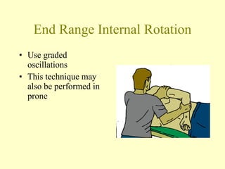
Shoulder Mobilization Case Study Proximal Humeral Fracture
History 61 year old male Fractured the greater tuberosity of the right shoulder eight weeks ago Partially tore the rotator cuff muscle of the same shoulder. Patient was immobilized in a sling for eight weeks.
Clinical Presentation Sever limitation of right shoulder motions Demonstrates a capsular pattern External rotation, abduction , medial rotation Complains on a dull constant ache within the shoulder at rest. Rating the resting pain as a  6/10 on the  pain scale.  Experiences sharp pains with any motion of the  shoulder . Pain is rated as a 8/10. X-rays and MRI indicates that the fracture is healed and the rotator cuff is partially healed.
Physical Therapy Referral Restore motion and normal strength to the right shoulder
Clinical Considerations Patient has moderate to sever pain with any movement. Shoulder restriction is due primarily to capsular and muscle shortening around the fracture site. Muscular strength of the right shoulder complex is weak due to the prolong immobilization.
Treatment Plan Modalities Mobilization techniques Strengthening exercises
Mobilization Joints to be mobilized Glenohumeral Sternoclaviclar Acromclavical Scapula Potential muscled that are shorten. Subscapularis Pectoral major & minor Infaspinatus & teres minor Lat Rhomboids  Serrtaus Upper mid and lower trap
Goal  Increase shoulder glenohumeral  motion without exacerbation of pain.
Concepts To Remember In The Glenohumeral Joint Osteokinematic  : There is 3 degrees of freedom Flexion/Extension, ABd /ADd, Internal/External Rot. Articulator surface anatomy Concave glenoid & convex humerus Loose pack position 20 degrees scapulohumeral abduction with 30 degrees elevation in the scapular plane.
Concepts To Remember In The Shoulder Complex Joint Accessory (C omponent ) Motions Arthokinematic movements that must occur in order for normal osteokinematic movement to take place  Eg. Inferior Glide Joint Play Motion Those accessory that can be produced passively at a joint but not actively. Eg.  Lateral Distraction
Physiological Movements Refer to Matiland CD
Shoulder Flexion Glenohumeral  Lateral distaction Inferior glide Posteior  glide Sternoclavicular Inferior gilde Anterior glide Scapula Distraction Upward rotation Elevation
Scapluar Plane Oscillations General technique Introductory Pain Lubication of tissues
Glenohumeral Lateral Distraction   Often one of the first technique to use Good for general capsular tightness Pain control
Inferior Glide In Loose Pack For restriction in flexion and abduction Used to decreased pain  with grade I & II  oscillation
Inferior Glide At 90ยบ of Abduction Increase mid-range flexion and abduction
Anterior Glide In Loose Pack The primary tissue affect by this technique is the anterior capsular region
Posterior Glide In Loose Pack Matiland Technique Indication for posterior capsular tightness Used in the early phases of the rehab to began   To increase internal rotation
Posterior Glide At 90ยบ Abduction Posterior Glide at 90 degrees abduction Increase flexion and internal rotation
Posterior Glide in Flexion Advance technique that gives a strong   localized stretch to posterior capsule
Sternoclavicluar Inferior Glide Used to improve component motion for shoulder flexion.
Anterior & Posterior Glide of AC Joint Assist in improving shoulder flexion Used to decreased joint pain in the AC joint
Scapula Mobilizations The purpose of these techniques is to increase range of motion in scapular: Superior glide Inferior glide  Medial rotation Lateral rotation
Advance Soft Tissue Stretching Latissmus Dorsi Patient supine  Therapist at the head of patient One hand grips medial side of patient hand just above elbow and move it into flexion while laterally rotating the shoulder The other hand and forearm stabilizes the lower thorax Using the grip begin to stretch into flex and lateral rotation
Advance Soft Tissue Stretching Pectoralis Major Patient supine Therapist using both hands grips the medial side of the patientโ€™s elbow and  flexs  and laterally rotate the arms Placing a stretch on the pectoral muscles
Subscapularis Stretch End Range
End Range Internal Rotation Use graded oscillations This technique may also be performed in prone

Shoulder Lecture

  • 1.
    Shoulder Mobilization CaseStudy Proximal Humeral Fracture
  • 2.
    History 61 yearold male Fractured the greater tuberosity of the right shoulder eight weeks ago Partially tore the rotator cuff muscle of the same shoulder. Patient was immobilized in a sling for eight weeks.
  • 3.
    Clinical Presentation Severlimitation of right shoulder motions Demonstrates a capsular pattern External rotation, abduction , medial rotation Complains on a dull constant ache within the shoulder at rest. Rating the resting pain as a 6/10 on the pain scale. Experiences sharp pains with any motion of the shoulder . Pain is rated as a 8/10. X-rays and MRI indicates that the fracture is healed and the rotator cuff is partially healed.
  • 4.
    Physical Therapy ReferralRestore motion and normal strength to the right shoulder
  • 5.
    Clinical Considerations Patienthas moderate to sever pain with any movement. Shoulder restriction is due primarily to capsular and muscle shortening around the fracture site. Muscular strength of the right shoulder complex is weak due to the prolong immobilization.
  • 6.
    Treatment Plan ModalitiesMobilization techniques Strengthening exercises
  • 7.
    Mobilization Joints tobe mobilized Glenohumeral Sternoclaviclar Acromclavical Scapula Potential muscled that are shorten. Subscapularis Pectoral major & minor Infaspinatus & teres minor Lat Rhomboids Serrtaus Upper mid and lower trap
  • 8.
    Goal Increaseshoulder glenohumeral motion without exacerbation of pain.
  • 9.
    Concepts To RememberIn The Glenohumeral Joint Osteokinematic : There is 3 degrees of freedom Flexion/Extension, ABd /ADd, Internal/External Rot. Articulator surface anatomy Concave glenoid & convex humerus Loose pack position 20 degrees scapulohumeral abduction with 30 degrees elevation in the scapular plane.
  • 10.
    Concepts To RememberIn The Shoulder Complex Joint Accessory (C omponent ) Motions Arthokinematic movements that must occur in order for normal osteokinematic movement to take place Eg. Inferior Glide Joint Play Motion Those accessory that can be produced passively at a joint but not actively. Eg. Lateral Distraction
  • 11.
  • 12.
    Shoulder Flexion Glenohumeral Lateral distaction Inferior glide Posteior glide Sternoclavicular Inferior gilde Anterior glide Scapula Distraction Upward rotation Elevation
  • 13.
    Scapluar Plane OscillationsGeneral technique Introductory Pain Lubication of tissues
  • 14.
    Glenohumeral Lateral Distraction Often one of the first technique to use Good for general capsular tightness Pain control
  • 15.
    Inferior Glide InLoose Pack For restriction in flexion and abduction Used to decreased pain with grade I & II oscillation
  • 16.
    Inferior Glide At90ยบ of Abduction Increase mid-range flexion and abduction
  • 17.
    Anterior Glide InLoose Pack The primary tissue affect by this technique is the anterior capsular region
  • 18.
    Posterior Glide InLoose Pack Matiland Technique Indication for posterior capsular tightness Used in the early phases of the rehab to began To increase internal rotation
  • 19.
    Posterior Glide At90ยบ Abduction Posterior Glide at 90 degrees abduction Increase flexion and internal rotation
  • 20.
    Posterior Glide inFlexion Advance technique that gives a strong localized stretch to posterior capsule
  • 21.
    Sternoclavicluar Inferior GlideUsed to improve component motion for shoulder flexion.
  • 22.
    Anterior & PosteriorGlide of AC Joint Assist in improving shoulder flexion Used to decreased joint pain in the AC joint
  • 23.
    Scapula Mobilizations Thepurpose of these techniques is to increase range of motion in scapular: Superior glide Inferior glide Medial rotation Lateral rotation
  • 24.
    Advance Soft TissueStretching Latissmus Dorsi Patient supine Therapist at the head of patient One hand grips medial side of patient hand just above elbow and move it into flexion while laterally rotating the shoulder The other hand and forearm stabilizes the lower thorax Using the grip begin to stretch into flex and lateral rotation
  • 25.
    Advance Soft TissueStretching Pectoralis Major Patient supine Therapist using both hands grips the medial side of the patientโ€™s elbow and flexs and laterally rotate the arms Placing a stretch on the pectoral muscles
  • 26.
  • 27.
    End Range InternalRotation Use graded oscillations This technique may also be performed in prone