This document summarizes a case report of conjoined twins diagnosed prenatally. Ultrasound and MRI found the twins to be thoraco-omphalopagus, joined at the chest and abdomen with a single heart and shared liver. The parents opted to terminate the pregnancy given the twins' poor prognosis. The document then reviews conjoined twins generally, including classification, embryology (they result from incomplete twinning after 14 days), diagnostic criteria with imaging modalities, associated anomalies, management approaches including delivery method, and challenges with separation surgery due to shared organs.
accurate assessment of gestational age by certain mensrtual data and clinical examination may have dating discrepancy with the ultrasound. this ppt critically addresses such issues
Fetal growth restriction (FGR), formerly called intrauterine growth restriction (IUGR), refers to a condition in which an unborn baby is smaller than it should be because it is not growing at a normal rate inside the womb.
Mild FGR usually doesn't cause long-term problems. In fact, most babies who have it catch up in height and weight by age 2. But severe FGR can seriously harm a baby before and after birth. The extent of the problems depends on the cause and how severe the growth restriction is. It also depends on what point in the pregnancy it starts.
USMLE GENERAL EMBRYOLOGY 017 Twins monozygotic monozygotic monozygotic.pdfAHMED ASHOUR
Twins are siblings born during the same pregnancy and are classified into different types based on how they develop.
The two primary types of twins are identical (monozygotic) twins and fraternal (dizygotic) twins.
The experience of having twins can vary, and each pregnancy is unique.
Expectant parents of twins should work closely with healthcare providers to ensure a healthy and well-managed pregnancy.
In this presentation we will discuss
First trimester US especially TVS is an integral part for confirmation of intrauterine pregnancy and to rule out ectopic pregnancy.
First trimester US helps us in suggesting conceptus viability.
First trimester US especially TVS is very efficient in approaching and evaluating the cause of vaginal bleeding.
Embryo implantation in the region of a previous caesarean section scar is a rare but potentially catastrophic complication of a previous cesarean birth.
micro teaching on communication m.sc nursing.pdfAnurag Sharma
Microteaching is a unique model of practice teaching. It is a viable instrument for the. desired change in the teaching behavior or the behavior potential which, in specified types of real. classroom situations, tends to facilitate the achievement of specified types of objectives.
Ozempic: Preoperative Management of Patients on GLP-1 Receptor Agonists Saeid Safari
Preoperative Management of Patients on GLP-1 Receptor Agonists like Ozempic and Semiglutide
ASA GUIDELINE
NYSORA Guideline
2 Case Reports of Gastric Ultrasound
Prix Galien International 2024 Forum ProgramLevi Shapiro
June 20, 2024, Prix Galien International and Jerusalem Ethics Forum in ROME. Detailed agenda including panels:
- ADVANCES IN CARDIOLOGY: A NEW PARADIGM IS COMING
- WOMEN’S HEALTH: FERTILITY PRESERVATION
- WHAT’S NEW IN THE TREATMENT OF INFECTIOUS,
ONCOLOGICAL AND INFLAMMATORY SKIN DISEASES?
- ARTIFICIAL INTELLIGENCE AND ETHICS
- GENE THERAPY
- BEYOND BORDERS: GLOBAL INITIATIVES FOR DEMOCRATIZING LIFE SCIENCE TECHNOLOGIES AND PROMOTING ACCESS TO HEALTHCARE
- ETHICAL CHALLENGES IN LIFE SCIENCES
- Prix Galien International Awards Ceremony
TEST BANK for Operations Management, 14th Edition by William J. Stevenson, Ve...kevinkariuki227
TEST BANK for Operations Management, 14th Edition by William J. Stevenson, Verified Chapters 1 - 19, Complete Newest Version.pdf
TEST BANK for Operations Management, 14th Edition by William J. Stevenson, Verified Chapters 1 - 19, Complete Newest Version.pdf
Ethanol (CH3CH2OH), or beverage alcohol, is a two-carbon alcohol
that is rapidly distributed in the body and brain. Ethanol alters many
neurochemical systems and has rewarding and addictive properties. It
is the oldest recreational drug and likely contributes to more morbidity,
mortality, and public health costs than all illicit drugs combined. The
5th edition of the Diagnostic and Statistical Manual of Mental Disorders
(DSM-5) integrates alcohol abuse and alcohol dependence into a single
disorder called alcohol use disorder (AUD), with mild, moderate,
and severe subclassifications (American Psychiatric Association, 2013).
In the DSM-5, all types of substance abuse and dependence have been
combined into a single substance use disorder (SUD) on a continuum
from mild to severe. A diagnosis of AUD requires that at least two of
the 11 DSM-5 behaviors be present within a 12-month period (mild
AUD: 2–3 criteria; moderate AUD: 4–5 criteria; severe AUD: 6–11 criteria).
The four main behavioral effects of AUD are impaired control over
drinking, negative social consequences, risky use, and altered physiological
effects (tolerance, withdrawal). This chapter presents an overview
of the prevalence and harmful consequences of AUD in the U.S.,
the systemic nature of the disease, neurocircuitry and stages of AUD,
comorbidities, fetal alcohol spectrum disorders, genetic risk factors, and
pharmacotherapies for AUD.
The prostate is an exocrine gland of the male mammalian reproductive system
It is a walnut-sized gland that forms part of the male reproductive system and is located in front of the rectum and just below the urinary bladder
Function is to store and secrete a clear, slightly alkaline fluid that constitutes 10-30% of the volume of the seminal fluid that along with the spermatozoa, constitutes semen
A healthy human prostate measures (4cm-vertical, by 3cm-horizontal, 2cm ant-post ).
It surrounds the urethra just below the urinary bladder. It has anterior, median, posterior and two lateral lobes
It’s work is regulated by androgens which are responsible for male sex characteristics
Generalised disease of the prostate due to hormonal derangement which leads to non malignant enlargement of the gland (increase in the number of epithelial cells and stromal tissue)to cause compression of the urethra leading to symptoms (LUTS
NVBDCP.pptx Nation vector borne disease control programSapna Thakur
NVBDCP was launched in 2003-2004 . Vector-Borne Disease: Disease that results from an infection transmitted to humans and other animals by blood-feeding arthropods, such as mosquitoes, ticks, and fleas. Examples of vector-borne diseases include Dengue fever, West Nile Virus, Lyme disease, and malaria.
263778731218 Abortion Clinic /Pills In Harare ,sisternakatoto
263778731218 Abortion Clinic /Pills In Harare ,ABORTION WOMEN’S CLINIC +27730423979 IN women clinic we believe that every woman should be able to make choices in her pregnancy. Our job is to provide compassionate care, safety,affordable and confidential services. That’s why we have won the trust from all generations of women all over the world. we use non surgical method(Abortion pills) to terminate…Dr.LISA +27730423979women Clinic is committed to providing the highest quality of obstetrical and gynecological care to women of all ages. Our dedicated staff aim to treat each patient and her health concerns with compassion and respect.Our dedicated group ABORTION WOMEN’S CLINIC +27730423979 IN women clinic we believe that every woman should be able to make choices in her pregnancy. Our job is to provide compassionate care, safety,affordable and confidential services. That’s why we have won the trust from all generations of women all over the world. we use non surgical method(Abortion pills) to terminate…Dr.LISA +27730423979women Clinic is committed to providing the highest quality of obstetrical and gynecological care to women of all ages. Our dedicated staff aim to treat each patient and her health concerns with compassion and respect.Our dedicated group of receptionists, nurses, and physicians have worked together as a teamof receptionists, nurses, and physicians have worked together as a team wwww.lisywomensclinic.co.za/
Pulmonary Thromboembolism - etilogy, types, medical- Surgical and nursing man...VarunMahajani
Disruption of blood supply to lung alveoli due to blockage of one or more pulmonary blood vessels is called as Pulmonary thromboembolism. In this presentation we will discuss its causes, types and its management in depth.
2. ABSTRACT
One of the most interesting congenital malformations is
a conjoined twin. Conjoined twins are a rare
occurrence. More commonly known as Siamese
twins,after First Siamese twins "exhibited" in America in
1829 ,Chang and Eng born in SIAM(Thailand)
•Current technology is providing a basis for earlier
diagnosis and a better prognosis.Magnetic resonance
imaging and CT scan provide excellent anatomic detail,
demonstrating organ position, shared viscera, and
vascular anatomy.
•We present a case of Thoraco-omphalopagus
Conjoined Twins.
3. Case report
22 year old women, primigravida,with 7 months of amenorrhea , 2nd degree
consanguineous marriage & no family history of twins. She had a previous antenatal
Ultrasound report at 16weeks which read as twin pregnancy. She presented to our
hospital at 31 weeks. Sonography was performed which showed following:
•Two fetuses with all 4 pair of limbs and two cranial cavities(dicephalus, tetrabrachius)
•The twins were joined at the thorax and upper abdomen.
•Single umbilical cord(5vessel), shared liver,omphalocoele, and only one fetal heart which
appeared normal.
•One fetus (acardia)had hydrocephalus with poor visualization of lungs and kyphoscoliotic
spine.
•To confirm our findings and get more details we did a fetal MRI.MRI confirmed our usg
findings.
•On the basis of these findings, the diagnosis of terata anacatadidyma, thoracoomphalopagus, conjoined twins was made, and the parents were informed about the
malformation and the twins' poor chance for survival. The parents decided to terminate
the pregnancy.Caesarean section was done and twins were delivered as breech.
•Twins had single male external genitalia, weighing 3 kg together. Placenta was single.
• An USG and CT w was done to further assess them; findings of antenatal scans were
confirmed.
9. INTRODUCTION
• Conjoined twins are rare, but the exact
prevalence is unknown. The estimated
prevalence in the literature varies widely from
1: 50,000 to 1: 200,000.
• There is a female predominance on the order
of 3:1
• The conjoined twinning process occurs when
the division of the embryonic disc occurs more
than 14 days after fertilization.
10. EMBRYOLOGY
• The twinning process occurs in the very early stages of
embryogenesis. Division after the 14th day of fertilization
results in an incomplete division, because once it becomes an
embryo, it is incapable of fusion. The later the division, the
more severe the effect.
• Duplicata incompleta is the most severe form of incomplete
division, because division is so late and incomplete, only a few
organ systems are duplicated. This makes surgical division
impossible.
• Many variations of conjoined twins are possible. It is
customary to name these large lateral fusions, which
incorporate multiple regions, on the basis of the anatomic
part that remains double and separate; for example,
dicephalus, two heads with fusion of the thorax and abdomen
11. •
•
•
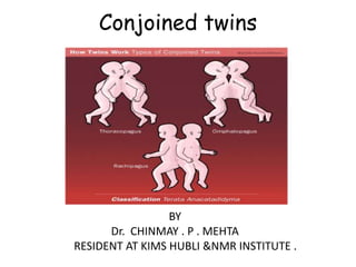
CLASSIFICATION
Inferior conjunction: lower body is single, or twin joined by some lower portion of
.
the body
Duplicata completa = terata catadidyma
Ischiopagus: joined by the lower portion of the coccyx & sacrum
Pygopagus: joined by the lateral and posterior aspect of the coccyx and sacrum
Middle conjunction: midbody fusion with separation of the upper and lower
portion of the body
• Duplicata completa = terata anacatadidyma
Thoracopagus: joined by the chest wall
Omphalopagus: joined between the umbilicus and the xyphoid process
Rachipagus: joined by the spine, above the sacrum
• Superior conjunction: upper body is single, or twin joined by some lower portion
of the body
• Duplicata completa = terata anadidyma
Syncephalus: joined by the face
Craniopagus: joined by the skull.
( Site of fusion + suffix "pagus“)
12.
13. USG DIAGNOSIS CRITERIA
• Demonstration of a continuous non seperated external skin
contour.
• Bifid appearance of the fetal pole in first trimester
• Conjoined body parts.
• Body parts of the twins are imaged on the same level and
in the same sonar plane.
• No change in the relative position of the twins to one
another and on successive scans.
• Monochorionic twinning-Single placental mass/ No intertwin membrane
• More than 3 vessels in a single umbilical cord.
• Complex multiple fetal anomalies.
14. THORACOPAGUS
• Thoracopagus twins are united face to face from the upper
thorax to the umbilicus with a common sternum, diaphragm,
and upper abdominal wall. Ninety percent of such twins have
a common pericardial sac, and there is always a degree of
cardiac fusion.
• The liver is invariably fused, and 25% of thoracopagus twins
share a biliary system. Initial liver assessment can be
performed with US. However, in twins joined anteriorly, there
is limited probe access; when viewed from the side, the
conjoined liver is oriented in an oblique plane to the axis of
the probe .
• A better appreciation of liver anatomy is gained from
multiplanar techniques, ideally MR imaging.
• It is important to evaluate bile excretion by using dynamic
biliary scintigraphy with Tc-99m HIDA
15. OMPHALOPAGUS
• Omphalopagus twins are joined ventrally in the
umbilical region, often including the lower
thorax.
• Liver fusion occurs in approximately 80% of cases.
As there is no mixing of blood in the cardiac
chambers, the liver can be well assessed by using
CT with intravenous injection of contrast material
into one twin.
• Gadolinium-enhanced MR imaging can also be
used in this context. As described in the section
on thoracopagus twins, biliary scintigraphy can be
helpful in determining biliary drainage
16. ROLE OF COLOR DOPPLER & 4D
• Color doppler is very useful in evaluation of
liver blood supply-Common portal vein
precludes separation & also to evaluate
number and orientation of hepatic veins.Also
very useful in craniopagus.
• 4D-Easier for parents to understand & helps
in counselling.Better surface views
• But 2D and Doppler better to determine
degree of organ sharing
17. ROLE OF MRI
• Pre-surgical planning
- Fetuses stable on placental support
- No sedation required
- Defines degree of organ sharing
- T2WI excellent for brain/renal/chest detail
- Tl WI for additional bowel and liver information
• Clarify anomalies
- Either fetus may have lethal anomaly in addition
to being conjoined.
- Information may influence management
Termination of pregnancy.
Requirement for emergent separation.
Mode of delivery
18. PROGNOSIS
• The prognosis for conjoined twins is generally
unfavorable, with approximately 40% of cases
stillborn. The worst prognoses concern
craniopagus twins and those with a sole cardiac
mass. Structural anomalies are frequently
found such as polyhydramnios (50%), cardiac
malformations, common omphaloceles, and
neural tube defects. Upon discovery of nonviable
conjoined twins, interruption of pregnancy
should therefore be recommended
19. MANAGEMENT
• In the case of potentially viable conjoined twins, after
24 weeks GA the choice between vaginal delivery
or prophylactic caesarian section should be made
based on maternal safety and neonatal
criteria. Caesarian section avoids dystocia, uterine
rupture, and fetal death in utero.
• Approximately six to ten cases of conjoined twins per
annum worldwide are treated surgically. The surgery is
most successful when commonality of fetal organs is
limited; surgical intervention often takes place around
one year of age
20.
21. CONCLUSION
• Diagnosis of conjoined twins is possible as early
as 8 weeks GA, but accurate evaluation of
common structures is not possible. The 12-week
scan allows clinicians to assess viability, and, in
the case of nonviability, to propose early medical
interruption of pregnancy, preventing
hysterotomy in the case of a delayed
termination. Early discovery of viable conjoined
twins permits assessment of the best route of
delivery and a planning for serial sonography and
fast MRI to plan separation surgery
22. REFERENCES
•
•
•
•
•
•
•
•
•
1. Barth RA, Filly RA, Goldberg JD: Conjoined twins: Prenatal diagnosis and
assessment of associated malformations. Radiology 177:201-07, 1990.
2. Jirous J, Radocha K, Hanas S: Dicephalus, tribrachius: Prenatal diagnosis and
management. Acta Obstet Gynecol Scand 66:79-81, 1987.
3. Hammond DI, Okun NB, Carpenter BF: Prenatal ultrasonographic diagnosis of
dicephalus conjoined twins. Can Association Radiol J 42:357-9: 1991.
4. Apuzzio JJ, Ganesh VV, Chervenak J: Prenatal diagnosis of dicephalous conjoined
twins in a triplet pregnancy. Am J Obstet Gynecol 159:1214-5, 1988.
5. Fitzgerald EJ, Toi A, Cochlin DL: Conjoined twins: Antanatal ultrasound diagnosis
and a review of the literature. Br J Radiol 11:94-6, 1983.
6. Chatterjee MS, Weiss RR, Verma UL: Prenatal diagnosis of conjoined twins.
Prenat Diagn 3:357-61, 1983.
7. Guttmacher AF, Nichols BL: Teratology of conjoined twins. Birth Defects 3:3-9,
1967.
8. Chan DPC: Thoracompholapagus diagnosed before delivery. Med J Aust, I,
pp480-3, 1976.
9. Romero R, Pilu GL, Jeanty P: Prenatal diagnosis of congenital anomalies.
Appleton-Lange, Norwalk, 1988, p 405.