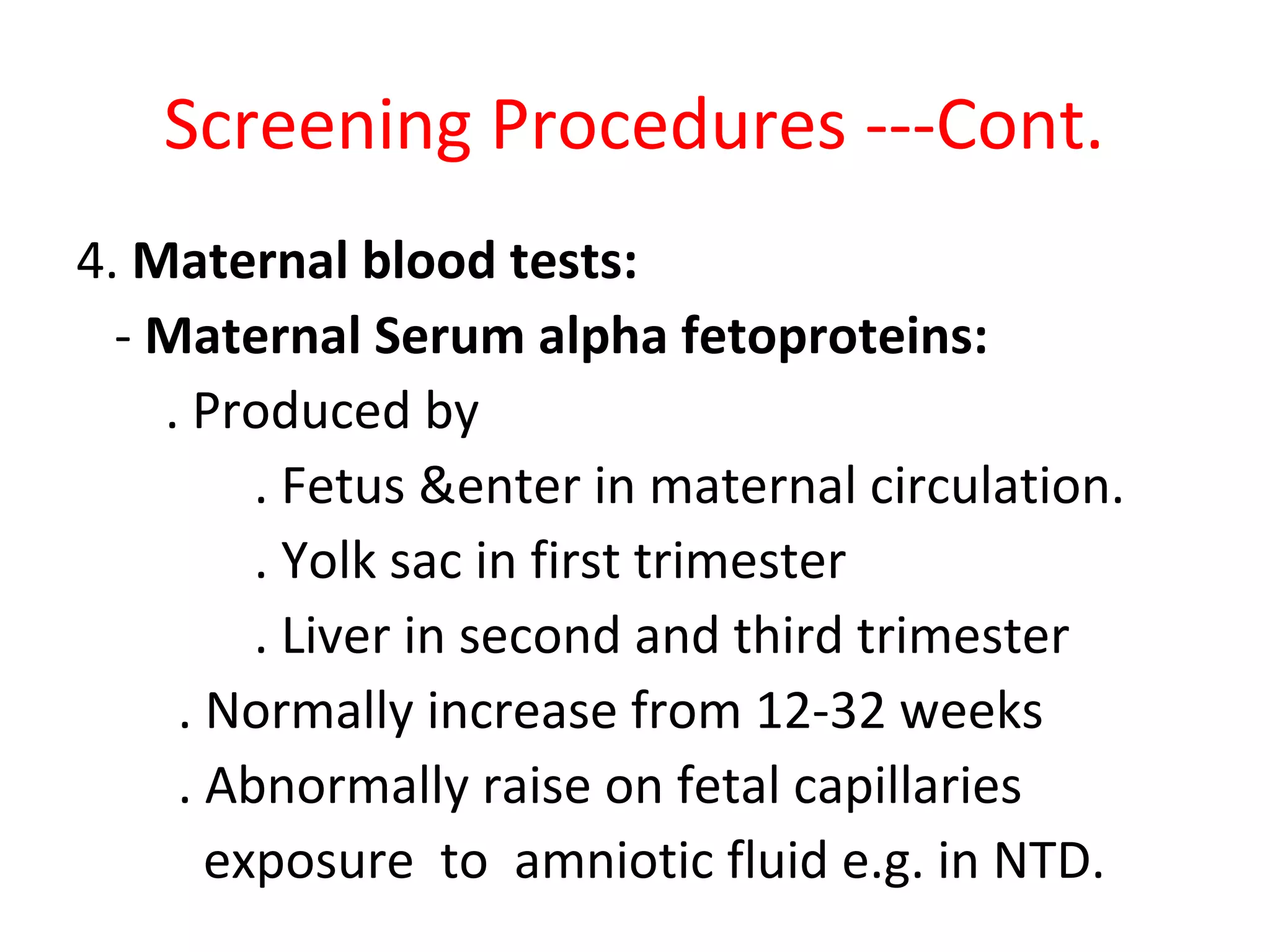
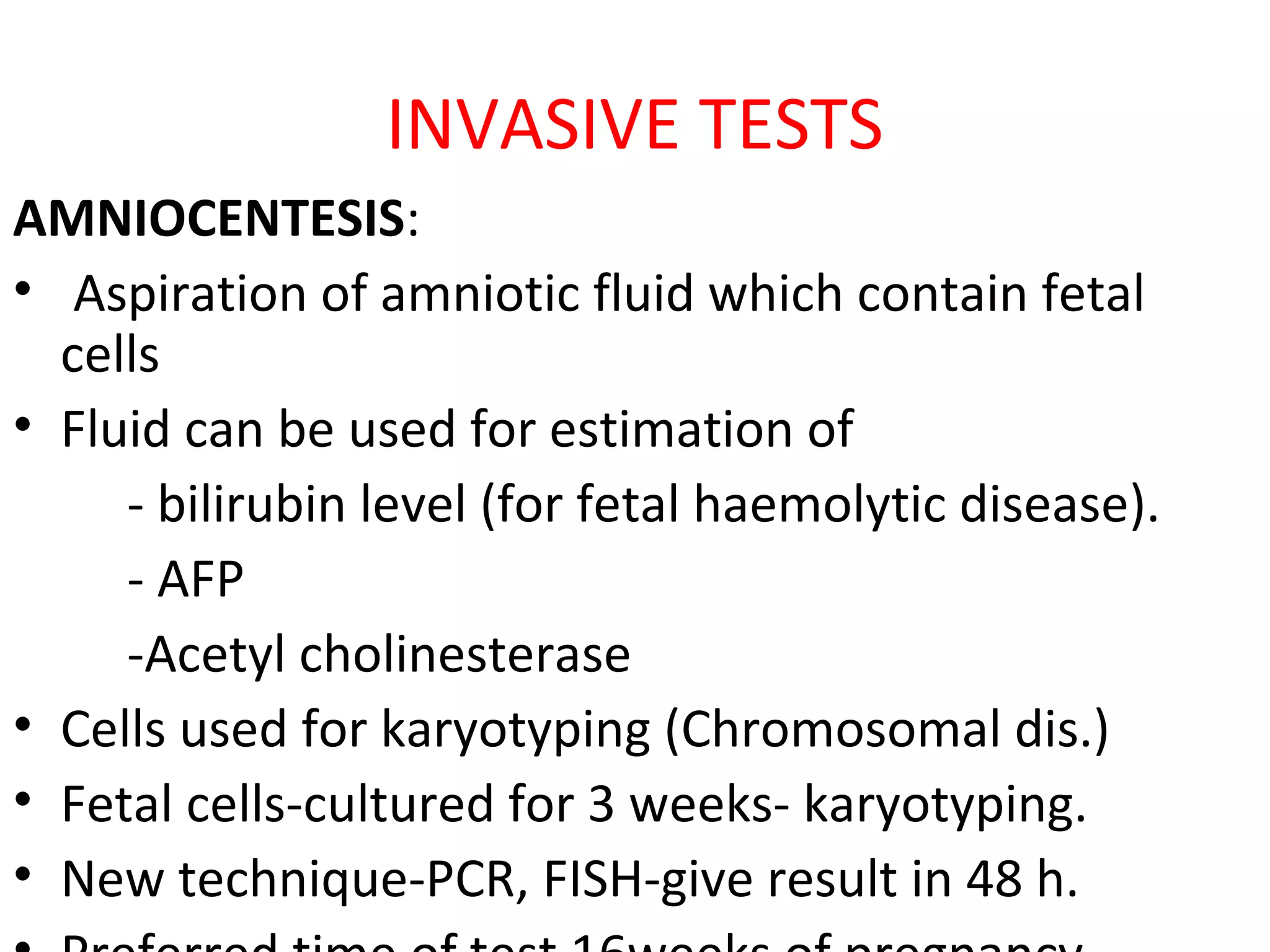
By
Dr. Jeetendra
Prenatal Diagnosis
• Over the last 4 decades, the genetic basis of
an increasing number of diseases is becoming
understood. At the same time, safe and
effective fetal diagnostic techniques are being
developed.
Prenatal Diagnosis of fetal
Abnormalities
• Benefits:
1.Malformation incompatible with life may be
terminated.
2.Certain abnormalities may be correctible in-utero.
3.-Provides opportunity to arrange corrective
measures before hand.
- offer a chance to be delivered at a place where
the required facilities are available.
4. Parents decision to continue pregnancy/ mentally
prepare to have a handicapped child.
Classification of Congenital
Abnormalities
1 Chromosomal Abnormalities: - Trisomy 21
(D.S)
- Trisomy 18 (E.S)
- Trisomy 13 (P.S)
2 Structural Abnormalities: - CNS
- CVS
- GIT
- Bone
- Renal system
3 Genetic Disorders: - Inborn error of metabolism
- Haemoglobinopathies
What Should We Do?
• Every pregnancy should be evaluated with the
most definite test.
• Practically & economically not feasible because
 expensive
 Invasive
Worldwide practice is to carry out
-Screening procedures
-Definite (diagnostic)tests for screening
positive cases
Screening Procedures
These are:
- Simple
- Cheap
- Least invasive
- safe
- Easily repeatable
Screening Procedures --- Cont.
1. History:
- Increasing maternal age
- Congenital anomalies in previous children
- F/Hx.
. Still birth
. Recurrent 1st
trimester abortion
. Cousin marriage
Screening Procedures ---Cont.
2. Features of current pregnancy:
- Drug intake(antiepileptics e.g. warfarin,
alcohol, smoking)
- Radiation exposure
- Maternal ch. diseases e.g.DM, cardiac, renal
- Uterine fundas large/ small for date
- Decrease fetal movements
- Fetal malpresentation
- Viral infection in early pregnancy
Screening Procedures --- Cont.
3. Ultrasonography:
- Screening tool in all trimesters
- At 10-14 weeks if fetal nuchal translucency
- > 2.5 mm- chromosomal anomalies
association
- At 18-20 weeks 75% fetal abnormalities
can
be diagnose
1st
Trimester Ultrasound
• NT Ultrasound
Cleft lip and palate
Ventriculomegaly
Posterior Urethral Valve
Multicystic Dysplastic kidney
Screening Procedures ---Cont.
4. Maternal blood tests:
- Maternal Serum alpha fetoproteins:
. Produced by
. Fetus &enter in maternal circulation.
. Yolk sac in first trimester
. Liver in second and third trimester
. Normally increase from 12-32 weeks
. Abnormally raise on fetal capillaries
exposure to amniotic fluid e.g. in NTD.
Maternal S. alpha fetoproteins --cont.
- Raised level in neural tube defect(NTD).
- Screen for NTD at 15-20 weeks if +ve confirm
with detailed USG.
- Also raised in following conditions:
. Miscalculated dates . Multiple pregnancies
. Threatened abortion . IUD
. Teratoma . Congenital nephrosis
. Ant. Abdominal wall defects
MSAFP
Triple Test
• - Used for Down Synd. Screening. It comprises
. AFP
. hCG
. uE3 (unconjugated oestriol )
- Best carried at 15-18 weeks. In DS AFP & uE3
are low while hCG is raised
- Triple test+ maternal age diagnose 60% DS
- In trisomy 18 all above components are low
Quadruple test
• Triple test+ Inhibin A estimation
• This test + maternal age detects 76% DS
Double Test
• Low pregnancy associated plasma proteins-A
(PAPP-A) level and raised serum Beta-hCG
during 1st
trimester
• Double test+ maternal age diagnose 60% DS.
↓ ↑ ↓
↓ ↑ ↓
↑ ↓ ↑
↓ ↓ ↓
↑
↑ ↓
? ↑ ↓
? ↓ ↓
↓ ↑ ↓
↓ ↓ ↓
Ν ↓↓↓
DIAGNOSTIC TESTS
• For high risk women on basis of screening tests
• An ideal test should be :
- Least invasive
- diagnose c. abnormality in early pregnancy.
- Minimally interfering developing pregnancy
• Diagnostic tests are also not risk free.
Counselling
• Organize an appointment
• Couple should be present
• Explain:
- Risk of occurance of c. abnormality
- All tests available, their procedure, cost,
diagnostic ability and benefits, possible risks
- Possible management plain
• If termination of pregnancy is unacceptable
diagnostic tests would be fruitless.
NON INVASIVE TESTS
• Ultrasonography:
• Diagnostic USG is different from screening USG,
- It takes longer time
- Dx. Wide range of c. anomalies
- Non invasive and diagnosis at spot possible
- But possible only at large gestational age
• Colour doppler further enhance the capability
especially for cardiac malformations and renal
agenesis.
Other Soft Signs
• short ears
• cerebellar hypoplasia
• cholecystomegaly
• Mild cerebral ventriculomegaly
• Hypoplasia of middle phalanx of 5th digit
• Increased Iliac angle
• Short frontal lobe
What are the 2T soft signs?
• Increased nuchal thickness
• short femur or humerus
• Pylectasis
• echogenic foci in heart
• Echogenic Bowel
• choroid plexus cysts
INVASIVE TESTS
AMNIOCENTESIS:
• Aspiration of amniotic fluid which contain fetal
cells
• Fluid can be used for estimation of
- bilirubin level (for fetal haemolytic disease).
- AFP
-Acetyl cholinesterase
• Cells used for karyotyping (Chromosomal dis.)
• Fetal cells-cultured for 3 weeks- karyotyping.
• New technique-PCR, FISH-give result in 48 h.
AMNIOCENTESIS---Cont.
• Procedure:
• Preliminary USG to confirm-duration of gestation,
-placental site,- adequacy of liqour (150-200 ml)
• Sterilize the abdomen
• 22 G spinal needle is used.
• About 20 cc amniotic fluid is withdrawn.
• Give Anti- D to all Rh-ve mothers.
• Ask rest for 30 min.& restrict movements for 48h
Amniocentesis
AMNIOCENTESIS---Cont.
• Limitations (difficulties)of procedure: if
- Anteriorly placed placenta
- Multiple pregnancy.
- Maternal obesity
- Oligohydramnios
• Risks:
- Pregnancy loss 1 % - Bleeding , Infection,
- Rupture of membrane - Preterm labour&IUD
- Leaking of Amniotic fluid
- Increase risk of RDS in newborn
CHORIONIC VILLUS SAMPLING
• Collection of fragments of placental tissue (chorionic
villi)- cells are examined for Dx. of C.Anomalies.
• Cytotrophoblastic (rapidly dividing) cells are used for
direct karyotyping- result available within 24-48 h.
• Chorionic villi are best source of DNA
• CVS can be performed at 10 weeks gestation.
• Indications:
1-DNA analysis for SCD,thallasemias, CF. hemophillias
2-Chromosomal abnormalities
3-Inborn error of metabolism
CHORIONIC VILLUS SAMPLING
• Procedure:
• Trans-abdominal approach preferred –under USG
guidance in supine position
• Trans-cervical approach is easy.
• In lithotomy position, sterilize area & Aspiration
catheter and biopsy forceps.
• Introduce through Cx. under USG into placental
tissue avoiding membrane rupture
• Risks: Pregnancy loss 2-6%
• Before 10 weeks- associated with limb deformities,
micrognathia, microglassia
FETAL BLOOD SAMPLING (FBS)
• Fetal blood- lymphocyte are rapidly cultured, results
within 48-72 hours.
• Indications: 1- Prenatal Dx. DNA available for
Cytogenetic studies In failed amniocentesis, and
mosaicism in chorion or amniotic fluid.
2-Fetal assessment: for red cell alloimmunization,
(Hb;Hc,TrF) Hydrops fetalis, viral infection, platelets
alloimmunization
• Unfortunately Associated with highest rate of fetal loss.
• Currently used for blood transfusion in-utero in fetal A.
FETAL BLOOD SAMPLING (FBS)
• Procedure : (cordocentesis):
• The sites for FBS are placental insertion of
umbilical cord, abdominal insertion of cord,
intrahepatic fetal vein and fetal heart.
• Suitable time is 20-28 weeks
• Risks:
- Bleeding from site of puncture
- Cord haematoma
- Fetal bradycardia
- Fetal death
EMBRYOSCOPY & FETOSCOPY
• Direct visualization of embryo and fetus.
• Limited field of vision.
• Provide information only about external fetal
structures .
NEW MOLECULAR ANALYTIC
TECHNIQUES
• Fetal cell obtained by CVS and Amniocentesis
can be used for prenatal Dx. For congenital
anomalies by following new techniques
1- Southern blotting:
Cleavage of chromosomal DNA at specific
sites and used for tests
2- PCR
3- FISH
Polymerase chain reaction (PCR)
• Amplify specific DNA and RNA fragments
• Once nucleotide sequence of a region of DNA
strand is known, complimentary
oligonucleotides & polymerase are added to
single strand DNA
• Repeat process 30 times to get adequate DNA
• PCR identify specific DNA sequence for gene
mutation & prenatal Dx. at an earlier stage
before an embryo transfer in IVF cycle.
FLOURESCENT IN SITU HYBRIDIZATION
• FISH allows detection & localization of specific
DNA sequence in interphase or metaphase.
• Advantage – results available in 24-48 h.
• Disadvantage – fail to detect big structural
rearrangements
• Identify 80% clinically relevant abnormalities,
helpful for early decision about further
management of affected pregnancies.
MANAGEMENT OF FETAL C. ANOMALIES
• It is a tedious task, requires skillful,
sympathetic & professional approach.
• Management options
- Termination of pregnancy
- In- utero management if possible
- Conservative management
POSTPARTUM MANAGEMENT OF C.A.
For better understanding of congenital anomalies
and its impact on future reproductive performance of
couple, following procedures are carried out on
affected babies/ abortusses:
1.Physical examination /postmortem
2.Fetal tissue(blood, skin, placenta) for karyotyping
3.Placenta and membrane for histopathology
4.Placental & baby swab for microbiology & virology.
5.Baby gram (x-rays of whole baby)
6.Baby photograph
THANKS

Prenatal diagnosis

  • 1.
  • 2.
    Prenatal Diagnosis • Overthe last 4 decades, the genetic basis of an increasing number of diseases is becoming understood. At the same time, safe and effective fetal diagnostic techniques are being developed.
  • 3.
    Prenatal Diagnosis offetal Abnormalities • Benefits: 1.Malformation incompatible with life may be terminated. 2.Certain abnormalities may be correctible in-utero. 3.-Provides opportunity to arrange corrective measures before hand. - offer a chance to be delivered at a place where the required facilities are available. 4. Parents decision to continue pregnancy/ mentally prepare to have a handicapped child.
  • 4.
    Classification of Congenital Abnormalities 1Chromosomal Abnormalities: - Trisomy 21 (D.S) - Trisomy 18 (E.S) - Trisomy 13 (P.S) 2 Structural Abnormalities: - CNS - CVS - GIT - Bone - Renal system 3 Genetic Disorders: - Inborn error of metabolism - Haemoglobinopathies
  • 5.
    What Should WeDo? • Every pregnancy should be evaluated with the most definite test. • Practically & economically not feasible because  expensive  Invasive Worldwide practice is to carry out -Screening procedures -Definite (diagnostic)tests for screening positive cases
  • 6.
    Screening Procedures These are: -Simple - Cheap - Least invasive - safe - Easily repeatable
  • 7.
    Screening Procedures ---Cont. 1. History: - Increasing maternal age - Congenital anomalies in previous children - F/Hx. . Still birth . Recurrent 1st trimester abortion . Cousin marriage
  • 8.
    Screening Procedures ---Cont. 2.Features of current pregnancy: - Drug intake(antiepileptics e.g. warfarin, alcohol, smoking) - Radiation exposure - Maternal ch. diseases e.g.DM, cardiac, renal - Uterine fundas large/ small for date - Decrease fetal movements - Fetal malpresentation - Viral infection in early pregnancy
  • 9.
    Screening Procedures ---Cont. 3. Ultrasonography: - Screening tool in all trimesters - At 10-14 weeks if fetal nuchal translucency - > 2.5 mm- chromosomal anomalies association - At 18-20 weeks 75% fetal abnormalities can be diagnose
  • 10.
  • 11.
  • 12.
  • 13.
  • 14.
  • 15.
  • 16.
    Screening Procedures ---Cont. 4.Maternal blood tests: - Maternal Serum alpha fetoproteins: . Produced by . Fetus &enter in maternal circulation. . Yolk sac in first trimester . Liver in second and third trimester . Normally increase from 12-32 weeks . Abnormally raise on fetal capillaries exposure to amniotic fluid e.g. in NTD.
  • 17.
    Maternal S. alphafetoproteins --cont. - Raised level in neural tube defect(NTD). - Screen for NTD at 15-20 weeks if +ve confirm with detailed USG. - Also raised in following conditions: . Miscalculated dates . Multiple pregnancies . Threatened abortion . IUD . Teratoma . Congenital nephrosis . Ant. Abdominal wall defects
  • 19.
  • 20.
    Triple Test • -Used for Down Synd. Screening. It comprises . AFP . hCG . uE3 (unconjugated oestriol ) - Best carried at 15-18 weeks. In DS AFP & uE3 are low while hCG is raised - Triple test+ maternal age diagnose 60% DS - In trisomy 18 all above components are low
  • 21.
    Quadruple test • Tripletest+ Inhibin A estimation • This test + maternal age detects 76% DS
  • 22.
    Double Test • Lowpregnancy associated plasma proteins-A (PAPP-A) level and raised serum Beta-hCG during 1st trimester • Double test+ maternal age diagnose 60% DS.
  • 25.
    ↓ ↑ ↓ ↓↑ ↓ ↑ ↓ ↑ ↓ ↓ ↓ ↑ ↑ ↓ ? ↑ ↓ ? ↓ ↓ ↓ ↑ ↓ ↓ ↓ ↓ Ν ↓↓↓
  • 26.
    DIAGNOSTIC TESTS • Forhigh risk women on basis of screening tests • An ideal test should be : - Least invasive - diagnose c. abnormality in early pregnancy. - Minimally interfering developing pregnancy • Diagnostic tests are also not risk free.
  • 27.
    Counselling • Organize anappointment • Couple should be present • Explain: - Risk of occurance of c. abnormality - All tests available, their procedure, cost, diagnostic ability and benefits, possible risks - Possible management plain • If termination of pregnancy is unacceptable diagnostic tests would be fruitless.
  • 28.
    NON INVASIVE TESTS •Ultrasonography: • Diagnostic USG is different from screening USG, - It takes longer time - Dx. Wide range of c. anomalies - Non invasive and diagnosis at spot possible - But possible only at large gestational age • Colour doppler further enhance the capability especially for cardiac malformations and renal agenesis.
  • 29.
    Other Soft Signs •short ears • cerebellar hypoplasia • cholecystomegaly • Mild cerebral ventriculomegaly • Hypoplasia of middle phalanx of 5th digit • Increased Iliac angle • Short frontal lobe
  • 30.
    What are the2T soft signs? • Increased nuchal thickness • short femur or humerus • Pylectasis • echogenic foci in heart • Echogenic Bowel • choroid plexus cysts
  • 31.
    INVASIVE TESTS AMNIOCENTESIS: • Aspirationof amniotic fluid which contain fetal cells • Fluid can be used for estimation of - bilirubin level (for fetal haemolytic disease). - AFP -Acetyl cholinesterase • Cells used for karyotyping (Chromosomal dis.) • Fetal cells-cultured for 3 weeks- karyotyping. • New technique-PCR, FISH-give result in 48 h.
  • 32.
    AMNIOCENTESIS---Cont. • Procedure: • PreliminaryUSG to confirm-duration of gestation, -placental site,- adequacy of liqour (150-200 ml) • Sterilize the abdomen • 22 G spinal needle is used. • About 20 cc amniotic fluid is withdrawn. • Give Anti- D to all Rh-ve mothers. • Ask rest for 30 min.& restrict movements for 48h
  • 33.
  • 34.
    AMNIOCENTESIS---Cont. • Limitations (difficulties)ofprocedure: if - Anteriorly placed placenta - Multiple pregnancy. - Maternal obesity - Oligohydramnios • Risks: - Pregnancy loss 1 % - Bleeding , Infection, - Rupture of membrane - Preterm labour&IUD - Leaking of Amniotic fluid - Increase risk of RDS in newborn
  • 35.
    CHORIONIC VILLUS SAMPLING •Collection of fragments of placental tissue (chorionic villi)- cells are examined for Dx. of C.Anomalies. • Cytotrophoblastic (rapidly dividing) cells are used for direct karyotyping- result available within 24-48 h. • Chorionic villi are best source of DNA • CVS can be performed at 10 weeks gestation. • Indications: 1-DNA analysis for SCD,thallasemias, CF. hemophillias 2-Chromosomal abnormalities 3-Inborn error of metabolism
  • 36.
    CHORIONIC VILLUS SAMPLING •Procedure: • Trans-abdominal approach preferred –under USG guidance in supine position • Trans-cervical approach is easy. • In lithotomy position, sterilize area & Aspiration catheter and biopsy forceps. • Introduce through Cx. under USG into placental tissue avoiding membrane rupture • Risks: Pregnancy loss 2-6% • Before 10 weeks- associated with limb deformities, micrognathia, microglassia
  • 39.
    FETAL BLOOD SAMPLING(FBS) • Fetal blood- lymphocyte are rapidly cultured, results within 48-72 hours. • Indications: 1- Prenatal Dx. DNA available for Cytogenetic studies In failed amniocentesis, and mosaicism in chorion or amniotic fluid. 2-Fetal assessment: for red cell alloimmunization, (Hb;Hc,TrF) Hydrops fetalis, viral infection, platelets alloimmunization • Unfortunately Associated with highest rate of fetal loss. • Currently used for blood transfusion in-utero in fetal A.
  • 40.
    FETAL BLOOD SAMPLING(FBS) • Procedure : (cordocentesis): • The sites for FBS are placental insertion of umbilical cord, abdominal insertion of cord, intrahepatic fetal vein and fetal heart. • Suitable time is 20-28 weeks • Risks: - Bleeding from site of puncture - Cord haematoma - Fetal bradycardia - Fetal death
  • 41.
    EMBRYOSCOPY & FETOSCOPY •Direct visualization of embryo and fetus. • Limited field of vision. • Provide information only about external fetal structures .
  • 42.
    NEW MOLECULAR ANALYTIC TECHNIQUES •Fetal cell obtained by CVS and Amniocentesis can be used for prenatal Dx. For congenital anomalies by following new techniques 1- Southern blotting: Cleavage of chromosomal DNA at specific sites and used for tests 2- PCR 3- FISH
  • 43.
    Polymerase chain reaction(PCR) • Amplify specific DNA and RNA fragments • Once nucleotide sequence of a region of DNA strand is known, complimentary oligonucleotides & polymerase are added to single strand DNA • Repeat process 30 times to get adequate DNA • PCR identify specific DNA sequence for gene mutation & prenatal Dx. at an earlier stage before an embryo transfer in IVF cycle.
  • 44.
    FLOURESCENT IN SITUHYBRIDIZATION • FISH allows detection & localization of specific DNA sequence in interphase or metaphase. • Advantage – results available in 24-48 h. • Disadvantage – fail to detect big structural rearrangements • Identify 80% clinically relevant abnormalities, helpful for early decision about further management of affected pregnancies.
  • 45.
    MANAGEMENT OF FETALC. ANOMALIES • It is a tedious task, requires skillful, sympathetic & professional approach. • Management options - Termination of pregnancy - In- utero management if possible - Conservative management
  • 46.
    POSTPARTUM MANAGEMENT OFC.A. For better understanding of congenital anomalies and its impact on future reproductive performance of couple, following procedures are carried out on affected babies/ abortusses: 1.Physical examination /postmortem 2.Fetal tissue(blood, skin, placenta) for karyotyping 3.Placenta and membrane for histopathology 4.Placental & baby swab for microbiology & virology. 5.Baby gram (x-rays of whole baby) 6.Baby photograph
  • 47.