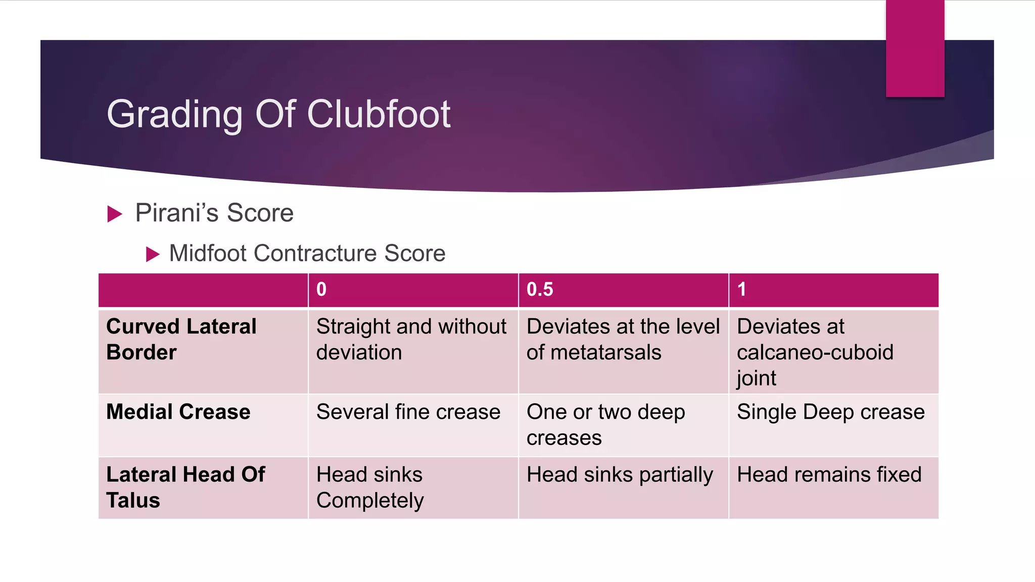
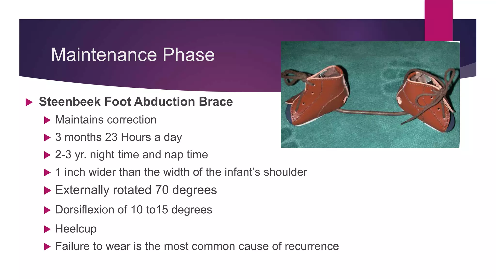
CONGENITAL TALIPES EQUINUS
VARUS
(CLUBFOOT)
BY: DR BIPUL BORTHAKUR
PROFESSOR
DEPT OF ORTHOPAEDICS, SMCH
Congenital Talipes Equino Varus
 It is derived from the Latin Term
 Talipes- talus ( Ankle) and pes (Foot)
 Equines -horse like
 Varus -inverted and adducted.
 It is also known as clubfoot because of its resemblance with
the club of golf
Deformities
• Cavus
• Adduction
• Varus
• Equinus
The four
basic
components
of clubfoot
are
• Internal rotation of tibia
• Medial subluxation of
the navicuiar on the
head of the talus
Other
Deformities
Include
 The incidence is approximately one in every 1000 live births.
 Bilateral deformities occur in 50% of patients.
 Most cases are sporadic.
 Families have been reported with clubfoot as an autosomal
dominant trait with incomplete penetrance.
Pathogenesis
Primary germ plasm defect in Talus
Continued plantar flexion and inversion
of Talus
Subsequent soft tissue changes in the
joints and musculotendinous complexes
Primary soft tissue abnormalities
within the neuromuscular units
Secondary Bony Changes
Types of Clubfoot
• Secondary Clubfoot
• Postural Clubfoot
• Metatarsus Adductus
• Idiopathic Clubfoot
According
to
Etiology
Secondary Clubfoot
• Neurogenic Clubfoot –
Associated with neurological
disorders such as Spina bifida,
Cerebral Palsy
• Syndromic Clubfoot – forms part
of a wider musculoskeletal
syndrome, such as arthrogryposis,
amniotic band syndrome
It is diagnosed
when clubfoot
forms part of
another health
condition
Postural Clubfoot
Very flexible clubfoot deformity
Can be reduced almost completely on passive
stretching
It is thought to result purely from posture in the womb
late in pregnancy
Metatarsus Adductus
It is not a true clubfoot
Diagnosed when child has forefoot adduction but
normal range of motion at ankle
In Pirani’s Scoring HFCS is zero and MFCS is higher
than zero
Types of Clubfoot
• Untreated Clubfoot
• Treated Clubfoot
• Resistant Clubfoot
• Recurrent Clubfoot
• Neglected Clubfoot
• Complex Clubfoot
According
to
Treatment
Stage
• When the affected child is under two
years of age and has had no treatment to
date
Untreated
Clubfoot
• When a child’s feet have corrected with
the Ponseti method and they have
completed the casting phase of treatment
Treated
Clubfoot
• When the child has previously
untreated clubfoot that doesnot
correct with Ponseti method
Resistant
Clubfoot
• When the child show signs of
recurrence in previously treated
clubfoot
Recurrent
Clubfoot
• Is a clubfoot in a child older than
two years who has little or no
treatment
Neglected
Clubfoot
• Is classified for any clubfoot that
has received any type of treatment
other than Ponseti method
Complex
Clubfoot
Pathoanatomy – Bony Changes
Talus-
• talar neck has been shown to be internally rotated relative
to the ankle mortise
• the talar body is externally rotated in the mortise
Navicular-
• navicular moves medially around the head of the talus
• The talonavicular joint is in an extreme position of inversion
Calcaneus
• The calcaneus rotates horizontally while pivoting on the
interosseous ligament, it slips beneath the head and neck
of the talus anterior to the ankle joint, and the calcaneal
tuberosity moves toward the fibular malleolus posteriorly
• The heel appears to be in varus because the calcaneus
rotates through the talocalcaneal joint in a coronal plane
and horizontally
Cuboid
• medially subluxated over calcaneal head
Forefoot
• The forefoot is adducted and supinated; severe
cases also have cavus
Tibia
• Smaller in size
• Internally rotated
Fibula
• Smaller in size
Soft Tissue Changes
 Muscle fibres are smaller in size
 Tibialis posterior, flexor digitorum longus (FDL), and flexor
hallucis longus (FHL) are contracted
 The calf is of a smaller size
 Atrophy of the leg muscles, especially the peroneal group
 Tendon sheaths: Thickening, especially of the tibialis
posterior and peroneal sheaths.
 Joint capsules: Contractures of posterior ankle, subtalar, and
talonavicular and calcaneocuboid capsules
 Ligaments: Contraction of calcaneofibular, talofibular, deltoid,
long and short plantar, spring, and bifurcate ligaments
 Fascia: The plantar fascial contracture contributes to the
cavus
Grading Of Clubfoot
 Pirani’s Score
 Midfoot Contracture Score
0 0.5 1
Curved Lateral
Border
Straight and without
deviation
Deviates at the level
of metatarsals
Deviates at
calcaneo-cuboid
joint
Medial Crease Several fine crease One or two deep
creases
Single Deep crease
Lateral Head Of
Talus
Head sinks
Completely
Head sinks partially Head remains fixed
 Hindfoot Contracture Score
0 0.5 1
Posterior Crease Several fine
Crease
Two or three
moderate crease
Single deep
crease
Rigidity Of
Equinus
<90 degrees =90 degrees >90 degrees
Empty Heel Calnaneus easy to
palpate
Palpated through
a layer of flesh
Very difficult to
palpate
GRADE TYPE SCORE REDUCIBILITY
I Benign 1-4 >90% soft-soft, resolving
II Moderate 5-9 >50% soft-stiff, reducible, partially resistant
III Severe 10-14 >50% stiff-soft, resistant, partially reducible
IV Very severe 15-20 <10% stiff-stiff, resistant
Treatment
 The aim of treatment is a normal, painfree and functional foot
 The initial treatment of clubfoot is non-operative
 The most widely accepted technique is that described by
Ignacio Ponseti.
Ponseti Method
 Consists of weekly serial manipulation and casting during the
first weeks of life
 It relies on the visco-elastic nature of the connective tissue to
produce plastic deformation through a process known as
stress relaxation.
 after about 1 minute of manipulation, toe-to-groin cast is
applied
 The casts should be applied in two stages:
 First, a short-leg cast to just below the knee
 Followed by extension above the knee when the plaster sets
 One week after application, the cast is removed and a new
cast is applied
 Cavus is corrected by supinating forefoot in proper alignment
with the hindfoot.
 Adduction corrected by abducting the entire foot under the
talus with the foot in slight supination
 Heel varus will be corrected when the entire foot is fully
abducted
 Equinus is corrected by dorsiflexing the foot (facilitated by
percutaneous tenotomy of Tendo-achillis)
Cavus Correction
 Correct method- Supination of forefoot
 Common Error : Pronation increases the cavus
Varus and Adduction Correction
 Correct- Abduct foot in supination while applying counter pressure against
the head of the talus
 Common Error: “Kite’s Error”- Abducting the forefoot while applying
counter-pressure at calcaneo-cuboid joint
Kite’s Error
Equinus Correction
 Dorsiflexion should be done in a fully abducted foot
 It is usually facilitated by percutaneous tenotomy
 The final cast is applied with the foot in the same maximally
abducted position and dorsiflexed 15 degrees
 Complete section of the tendon heals in three weeks
Maintenance Phase
 Steenbeek Foot Abduction Brace
 Maintains correction
 3 months 23 Hours a day
 2-3 yr. night time and nap time
 1 inch wider than the width of the infant’s shoulder
 Externally rotated 70 degrees
 Dorsiflexion of 10 to15 degrees
 Heelcup
 Failure to wear is the most common cause of recurrence
MANAGEMENT OF RECURRENCE
 Recurrence of the deformity is infrequent if the bracing
protocol is followed closely
 Early recurrences are best treated with repeat manipulation
and casting
 Achilles tendon lengthening may be necessary if dorsiflexion
is insufficient
 For dynamic metatarsal adduction transfer of the anterior
tibial tendon may be needed.
Surgical Management
 Surgery in clubfoot is indicated for deformities that do not
respond to conservative treatment by serial manipulation and
casting
 Must be tailored to the age of the child and to the deformity to
be corrected.
Timing Of Surgery
 When a plateau phase has been reached in non-operative
treatment.
 Turco (1971) recognized the best time period for surgical correction
to be one to two years of life
 Simons (1993) suggested that size and not age should be the
limiting factor - the foot should be at least 8 cm long.
Treatment By Age
 Less than 5 years : Correction can be obtained by soft tissue
procedures
 More than 5 years : Requires bony reshaping procedures
 More than 10 years : Triple arthrodesis if the foot is mature (
salvage procedures ).
Deformity Treatment
Metatarsus adductus >5 yrs: metatarsal osteotomy
Hind foot varus <2-3 yrs: modified McKay procedure
3-10 yrs: Dwyer osteotomy (isolated heel varus)
Dillwyn-Evans procedure (short medial column)
Lichtblau procedure (long lateral column)
10-12 yrs: triple arthrodesis
Equinus Achilles tendon lengthening plus posterior capsulotomy of
subtalar joint, ankle joint (mild-to-moderate deformity)
Lambrinudi procedure (severe deformity, skeletal immaturity)
All three
deformities
>10 yrs: triple arthrodesis
Gradual Distraction Procedures:
 Ilizarov Procedure: This is mostly reserved for recurrent and
resistant CTEVs. This is based on the principle that
continuous distraction of tissues resulted in neohistogenesis.
 JESS (Joshi's External Stabilising System) is a
modification of the llizarov procedure. It is based on the
principle of differential distraction of the soft tissues
Thank You

Congenital talipes equinus varus

  • 1.
    CONGENITAL TALIPES EQUINUS VARUS (CLUBFOOT) BY:DR BIPUL BORTHAKUR PROFESSOR DEPT OF ORTHOPAEDICS, SMCH
  • 2.
    Congenital Talipes EquinoVarus  It is derived from the Latin Term  Talipes- talus ( Ankle) and pes (Foot)  Equines -horse like  Varus -inverted and adducted.  It is also known as clubfoot because of its resemblance with the club of golf
  • 3.
    Deformities • Cavus • Adduction •Varus • Equinus The four basic components of clubfoot are
  • 5.
    • Internal rotationof tibia • Medial subluxation of the navicuiar on the head of the talus Other Deformities Include
  • 6.
     The incidenceis approximately one in every 1000 live births.  Bilateral deformities occur in 50% of patients.  Most cases are sporadic.  Families have been reported with clubfoot as an autosomal dominant trait with incomplete penetrance.
  • 7.
    Pathogenesis Primary germ plasmdefect in Talus Continued plantar flexion and inversion of Talus Subsequent soft tissue changes in the joints and musculotendinous complexes
  • 8.
    Primary soft tissueabnormalities within the neuromuscular units Secondary Bony Changes
  • 9.
    Types of Clubfoot •Secondary Clubfoot • Postural Clubfoot • Metatarsus Adductus • Idiopathic Clubfoot According to Etiology
  • 10.
    Secondary Clubfoot • NeurogenicClubfoot – Associated with neurological disorders such as Spina bifida, Cerebral Palsy • Syndromic Clubfoot – forms part of a wider musculoskeletal syndrome, such as arthrogryposis, amniotic band syndrome It is diagnosed when clubfoot forms part of another health condition
  • 11.
    Postural Clubfoot Very flexibleclubfoot deformity Can be reduced almost completely on passive stretching It is thought to result purely from posture in the womb late in pregnancy
  • 12.
    Metatarsus Adductus It isnot a true clubfoot Diagnosed when child has forefoot adduction but normal range of motion at ankle In Pirani’s Scoring HFCS is zero and MFCS is higher than zero
  • 13.
    Types of Clubfoot •Untreated Clubfoot • Treated Clubfoot • Resistant Clubfoot • Recurrent Clubfoot • Neglected Clubfoot • Complex Clubfoot According to Treatment Stage
  • 14.
    • When theaffected child is under two years of age and has had no treatment to date Untreated Clubfoot • When a child’s feet have corrected with the Ponseti method and they have completed the casting phase of treatment Treated Clubfoot
  • 15.
    • When thechild has previously untreated clubfoot that doesnot correct with Ponseti method Resistant Clubfoot • When the child show signs of recurrence in previously treated clubfoot Recurrent Clubfoot
  • 16.
    • Is aclubfoot in a child older than two years who has little or no treatment Neglected Clubfoot • Is classified for any clubfoot that has received any type of treatment other than Ponseti method Complex Clubfoot
  • 17.
    Pathoanatomy – BonyChanges Talus- • talar neck has been shown to be internally rotated relative to the ankle mortise • the talar body is externally rotated in the mortise Navicular- • navicular moves medially around the head of the talus • The talonavicular joint is in an extreme position of inversion
  • 18.
    Calcaneus • The calcaneusrotates horizontally while pivoting on the interosseous ligament, it slips beneath the head and neck of the talus anterior to the ankle joint, and the calcaneal tuberosity moves toward the fibular malleolus posteriorly • The heel appears to be in varus because the calcaneus rotates through the talocalcaneal joint in a coronal plane and horizontally
  • 19.
    Cuboid • medially subluxatedover calcaneal head Forefoot • The forefoot is adducted and supinated; severe cases also have cavus
  • 20.
    Tibia • Smaller insize • Internally rotated Fibula • Smaller in size
  • 21.
    Soft Tissue Changes Muscle fibres are smaller in size  Tibialis posterior, flexor digitorum longus (FDL), and flexor hallucis longus (FHL) are contracted  The calf is of a smaller size  Atrophy of the leg muscles, especially the peroneal group
  • 22.
     Tendon sheaths:Thickening, especially of the tibialis posterior and peroneal sheaths.  Joint capsules: Contractures of posterior ankle, subtalar, and talonavicular and calcaneocuboid capsules  Ligaments: Contraction of calcaneofibular, talofibular, deltoid, long and short plantar, spring, and bifurcate ligaments  Fascia: The plantar fascial contracture contributes to the cavus
  • 24.
    Grading Of Clubfoot Pirani’s Score  Midfoot Contracture Score 0 0.5 1 Curved Lateral Border Straight and without deviation Deviates at the level of metatarsals Deviates at calcaneo-cuboid joint Medial Crease Several fine crease One or two deep creases Single Deep crease Lateral Head Of Talus Head sinks Completely Head sinks partially Head remains fixed
  • 26.
     Hindfoot ContractureScore 0 0.5 1 Posterior Crease Several fine Crease Two or three moderate crease Single deep crease Rigidity Of Equinus <90 degrees =90 degrees >90 degrees Empty Heel Calnaneus easy to palpate Palpated through a layer of flesh Very difficult to palpate
  • 28.
    GRADE TYPE SCOREREDUCIBILITY I Benign 1-4 >90% soft-soft, resolving II Moderate 5-9 >50% soft-stiff, reducible, partially resistant III Severe 10-14 >50% stiff-soft, resistant, partially reducible IV Very severe 15-20 <10% stiff-stiff, resistant
  • 29.
    Treatment  The aimof treatment is a normal, painfree and functional foot  The initial treatment of clubfoot is non-operative  The most widely accepted technique is that described by Ignacio Ponseti.
  • 30.
    Ponseti Method  Consistsof weekly serial manipulation and casting during the first weeks of life  It relies on the visco-elastic nature of the connective tissue to produce plastic deformation through a process known as stress relaxation.  after about 1 minute of manipulation, toe-to-groin cast is applied
  • 31.
     The castsshould be applied in two stages:  First, a short-leg cast to just below the knee  Followed by extension above the knee when the plaster sets  One week after application, the cast is removed and a new cast is applied
  • 32.
     Cavus iscorrected by supinating forefoot in proper alignment with the hindfoot.  Adduction corrected by abducting the entire foot under the talus with the foot in slight supination  Heel varus will be corrected when the entire foot is fully abducted  Equinus is corrected by dorsiflexing the foot (facilitated by percutaneous tenotomy of Tendo-achillis)
  • 33.
    Cavus Correction  Correctmethod- Supination of forefoot  Common Error : Pronation increases the cavus
  • 34.
    Varus and AdductionCorrection  Correct- Abduct foot in supination while applying counter pressure against the head of the talus  Common Error: “Kite’s Error”- Abducting the forefoot while applying counter-pressure at calcaneo-cuboid joint
  • 35.
  • 36.
    Equinus Correction  Dorsiflexionshould be done in a fully abducted foot  It is usually facilitated by percutaneous tenotomy  The final cast is applied with the foot in the same maximally abducted position and dorsiflexed 15 degrees
  • 37.
     Complete sectionof the tendon heals in three weeks
  • 40.
    Maintenance Phase  SteenbeekFoot Abduction Brace  Maintains correction  3 months 23 Hours a day  2-3 yr. night time and nap time  1 inch wider than the width of the infant’s shoulder  Externally rotated 70 degrees  Dorsiflexion of 10 to15 degrees  Heelcup  Failure to wear is the most common cause of recurrence
  • 43.
    MANAGEMENT OF RECURRENCE Recurrence of the deformity is infrequent if the bracing protocol is followed closely  Early recurrences are best treated with repeat manipulation and casting  Achilles tendon lengthening may be necessary if dorsiflexion is insufficient  For dynamic metatarsal adduction transfer of the anterior tibial tendon may be needed.
  • 44.
    Surgical Management  Surgeryin clubfoot is indicated for deformities that do not respond to conservative treatment by serial manipulation and casting  Must be tailored to the age of the child and to the deformity to be corrected.
  • 45.
    Timing Of Surgery When a plateau phase has been reached in non-operative treatment.  Turco (1971) recognized the best time period for surgical correction to be one to two years of life  Simons (1993) suggested that size and not age should be the limiting factor - the foot should be at least 8 cm long.
  • 46.
    Treatment By Age Less than 5 years : Correction can be obtained by soft tissue procedures  More than 5 years : Requires bony reshaping procedures  More than 10 years : Triple arthrodesis if the foot is mature ( salvage procedures ).
  • 47.
    Deformity Treatment Metatarsus adductus>5 yrs: metatarsal osteotomy Hind foot varus <2-3 yrs: modified McKay procedure 3-10 yrs: Dwyer osteotomy (isolated heel varus) Dillwyn-Evans procedure (short medial column) Lichtblau procedure (long lateral column) 10-12 yrs: triple arthrodesis Equinus Achilles tendon lengthening plus posterior capsulotomy of subtalar joint, ankle joint (mild-to-moderate deformity) Lambrinudi procedure (severe deformity, skeletal immaturity) All three deformities >10 yrs: triple arthrodesis
  • 48.
    Gradual Distraction Procedures: Ilizarov Procedure: This is mostly reserved for recurrent and resistant CTEVs. This is based on the principle that continuous distraction of tissues resulted in neohistogenesis.  JESS (Joshi's External Stabilising System) is a modification of the llizarov procedure. It is based on the principle of differential distraction of the soft tissues
  • 49.