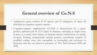
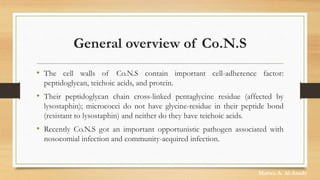
Coagulase negative staphylococci
Presentation
By
Marwa A. Al-Asady
General overview of Co.N.S
• Staphylococcus genus consists of 47 species and 23 subspecies, of these, 38
described as coagulase-negative species.
• Coagulase-negative staphylococci (Co.N.S) is characterized by a gram-
positive, spherical cell of (0.5-1.5µm) in diameter, occurring as single cocci,
in pairs, as a tetrad, short chains, or irregular cluster. Furthermore no-motile,
no-spore forming, encapsulated, catalase positive, cytochrome negative in
modified oxidase teste, also susceptible to lysostaphin and resistance to
bacitracin and they are grown in presence of 10% NaCl between 18℃ and
40℃.
Marwa A. Al-Asady
General overview of Co.N.S
• These groups of bacteria are facultative anaerobes so they can grow in the presence
or absence of oxygen.
• Besides, they are reduced nitrate to nitrite, fermenting carbohydrate and producing
pigments when growing on media that vary from white to deep yellow. Colonies
appearance round, smooth, raised and glistening on solid media.
• Called coagulase-negative staphylococci because they lack coagulase is a surface
protein, which can convert fibrinogen to fibrin and resulting in clot formation in
plasma.
Marwa A. Al-Asady
General overview of Co.N.S
• The cell walls of Co.N.S contain important cell-adherence factor:
peptidoglycan, teichoic acids, and protein.
• Their peptidoglycan chain cross-linked pentaglycine residue (affected by
lysostaphin); micrococci do not have glycine-residue in their peptide bond
(resistant to lysostaphin) and neither do they have teichoic acids.
• Recently Co.N.S got an important opportunistic pathogen associated with
nosocomial infection and community-acquired infection.
Marwa A. Al-Asady
General overview of Co.N.S
• They can adhere to medicinal instruments and surfaces through slime layer which
has a mucopolysaccharide arrangement, therefore they can simply colonize and
spread with-in the hospital environment. The slime feature also assistances in
pathogenicity by protective them from phagocytosis, chemotaxis, and anti-microbial
agents.
• Co.N.S are the most commonly isolated organism related with clinical infection and
communal bacteria residents of the skin, and mucous membranes humans.
• They are causes a variety of infections in immune-compromised individuals and
people with implanted medicinal devices.
Marwa A. Al-Asady
General overview of Co.N.S
• The incidence of sepsis infections in neonates caused by Co.N.S is still very
high and preventing and treating disease remain difficult in nosocomial
patient and cause a significant number of death.
• Co.N.S isolated from hospital environments are sometimes resistant to
various anti-microbial agents.
• About 80%–90% of Co.N.S isolates related with hospital infections are
methicillin-resistant (MR-Co.N.S).
Marwa A. Al-Asady
Co.N.S Species
• Co.N.S include many species:-
• S. epidermidis, S. caprae, S. sacchrolyticus, S. capitis(ssp.capitis ,ssp.urealyticus)
• S. haemolyticus, S. devriesie, S. jeffensis,
• S. hominis (ssp.hominis, ssp.novobisepticus),
• S. petrasii (ssp.croceilyticus, ssp.petrasii)S.lugdunensis
• S. warneri , S. pasteuri
• S. Saprophyticus (ssp. saprophyticus, ssp. bovis) S. equorum (ssp. equorum,
ssp.linens), S.xylosus
• S. Succinus (ssp.succinus, ssp.casei), S .gallinarum S. pettenkoferi, S. massiliensis
Co.N.S Species
• S. conhii (ssp.conhii , ssp.urealyticus),
• S. nepalensis S. klossi , S .arletta
• S. sciuri(ssp.sciuri, ssp.carnaticus, ssp.rodentium)
• S. fleurettii, S. lentus, S. stepanovicii, S. vitulinus, S .simulans, S. carnosus (ssp.utilis,
ssp.carnosus),
• S. codimenti, S. piscifermentans, S. muscae, S. microti, S.rosteri
• S. auricularis
Marwa A. Al-Asady
Pathogenesis of Co.N.S
• The pathogenesis of Co.N.S infection typically depends on their ability to
produce biofilms on polymer.
• Formation of biofilms around microorganisms protected them from the
host’s immune defense mechanisms lead to cause chronic infection, extra
resistant to various disinfectants, anti-microbial treatment than their
planktonic cell and from environment variables factors make them difficult
to eradicate.
Marwa A. Al-Asady
Virulence factors of Co.N.S
• To start an infection, bacteria need possess a group of molecules named
virulence factors that are critical for its invasion of the host, its persistence
within the host, and the initiation of the clinical symptoms.
• Co.N.S contain various virulence factors that promote colonization host
tissue, immune-evasion, immune-stimulation, develops antibiotic resistance,
also methicillin-resistant.
Marwa A. Al-Asady
Virulence factors of Co.N.S
• The virulence factor that produce and dangerous to host tissue, include
toxins, and degradative exoenzymes: metallo-protease with elastase activity,
cysteine protease, serine, protease, lipase, fatty-acid modifying enzymes
(FAME) and δ-toxin also can form biofilm is an important one.
Furthermore, produces lantibiotics are bacteriocins such as epidermin, which
are active to inhibit other gram-positive bacteria.
Marwa A. Al-Asady
Infection caused by Co.N.S
• Skin infection
• Endocarditis
• Endophthalmitis
• Bloodstream infections
• Foreign body-related infections
• Urinary tract infection
• Other infections (cerebrospinal shunt infections, meningitis, ventriculitis).
Marwa A. Al-Asady
Mechanism of antimicrobial resistance
• Antimicrobial resistance may be acquired through mutation and selection of
resistant bacterial strains or horizontal transfer of resistance genes from
other bacteria of the same or different species.
• The common famous resistance mechanisms in Co.N.S are the production
of enzymes that inactivate or destroy the antibiotics (e.g., stated by the genes
ermA, ermB, ermC, Blaz, and aac-apD), active deletion of antibiotics from the
cell (e.g., efflux-mechanisms pumps), and decrease of the antibiotic binding
affinity to the drug.
Marwa A. Al-Asady
Mechanism of antimicrobial resistance
• A large and theatrical increase in the number of resistant strains has
detected, in particular to penicillin, oxacillin, methicillin, clindamycin,
erythromycin, ciprofloxacin, and gentamicin.
• Resistance to β-lactams, that is, MR-Co.N.S (methicillin-resistant Co.N.S) is
determined by the presence of mecA gene, which encodes other penicillin
binding protein (PBP-2a) and is inserted into a mobile-gene element (MGE),
called the staphylococcal cassette chromosomal mec (SCCmec).
Marwa A. Al-Asady
Mechanism of antimicrobial resistance
• The mecA gene, which encodes a PBP-2a, with reduced affinity for
methicillin compared with the attractions of other PBP. In addition to
methicillin resistance, Co.N.S strains have acquired resistance to several other
antibiotics, including rapamycin, fluoroquinolones, gentamycin, tetracycline,
erythromycin, chloramphenicol, clindamycin and sulphonamides
Marwa A. Al-Asady
Thank you for lessening
Any Question

Coagulase negative staphylococci

  • 1.
  • 2.
    General overview ofCo.N.S • Staphylococcus genus consists of 47 species and 23 subspecies, of these, 38 described as coagulase-negative species. • Coagulase-negative staphylococci (Co.N.S) is characterized by a gram- positive, spherical cell of (0.5-1.5µm) in diameter, occurring as single cocci, in pairs, as a tetrad, short chains, or irregular cluster. Furthermore no-motile, no-spore forming, encapsulated, catalase positive, cytochrome negative in modified oxidase teste, also susceptible to lysostaphin and resistance to bacitracin and they are grown in presence of 10% NaCl between 18℃ and 40℃. Marwa A. Al-Asady
  • 3.
    General overview ofCo.N.S • These groups of bacteria are facultative anaerobes so they can grow in the presence or absence of oxygen. • Besides, they are reduced nitrate to nitrite, fermenting carbohydrate and producing pigments when growing on media that vary from white to deep yellow. Colonies appearance round, smooth, raised and glistening on solid media. • Called coagulase-negative staphylococci because they lack coagulase is a surface protein, which can convert fibrinogen to fibrin and resulting in clot formation in plasma. Marwa A. Al-Asady
  • 4.
    General overview ofCo.N.S • The cell walls of Co.N.S contain important cell-adherence factor: peptidoglycan, teichoic acids, and protein. • Their peptidoglycan chain cross-linked pentaglycine residue (affected by lysostaphin); micrococci do not have glycine-residue in their peptide bond (resistant to lysostaphin) and neither do they have teichoic acids. • Recently Co.N.S got an important opportunistic pathogen associated with nosocomial infection and community-acquired infection. Marwa A. Al-Asady
  • 5.
    General overview ofCo.N.S • They can adhere to medicinal instruments and surfaces through slime layer which has a mucopolysaccharide arrangement, therefore they can simply colonize and spread with-in the hospital environment. The slime feature also assistances in pathogenicity by protective them from phagocytosis, chemotaxis, and anti-microbial agents. • Co.N.S are the most commonly isolated organism related with clinical infection and communal bacteria residents of the skin, and mucous membranes humans. • They are causes a variety of infections in immune-compromised individuals and people with implanted medicinal devices. Marwa A. Al-Asady
  • 6.
    General overview ofCo.N.S • The incidence of sepsis infections in neonates caused by Co.N.S is still very high and preventing and treating disease remain difficult in nosocomial patient and cause a significant number of death. • Co.N.S isolated from hospital environments are sometimes resistant to various anti-microbial agents. • About 80%–90% of Co.N.S isolates related with hospital infections are methicillin-resistant (MR-Co.N.S). Marwa A. Al-Asady
  • 7.
    Co.N.S Species • Co.N.Sinclude many species:- • S. epidermidis, S. caprae, S. sacchrolyticus, S. capitis(ssp.capitis ,ssp.urealyticus) • S. haemolyticus, S. devriesie, S. jeffensis, • S. hominis (ssp.hominis, ssp.novobisepticus), • S. petrasii (ssp.croceilyticus, ssp.petrasii)S.lugdunensis • S. warneri , S. pasteuri • S. Saprophyticus (ssp. saprophyticus, ssp. bovis) S. equorum (ssp. equorum, ssp.linens), S.xylosus • S. Succinus (ssp.succinus, ssp.casei), S .gallinarum S. pettenkoferi, S. massiliensis
  • 8.
    Co.N.S Species • S.conhii (ssp.conhii , ssp.urealyticus), • S. nepalensis S. klossi , S .arletta • S. sciuri(ssp.sciuri, ssp.carnaticus, ssp.rodentium) • S. fleurettii, S. lentus, S. stepanovicii, S. vitulinus, S .simulans, S. carnosus (ssp.utilis, ssp.carnosus), • S. codimenti, S. piscifermentans, S. muscae, S. microti, S.rosteri • S. auricularis Marwa A. Al-Asady
  • 9.
    Pathogenesis of Co.N.S •The pathogenesis of Co.N.S infection typically depends on their ability to produce biofilms on polymer. • Formation of biofilms around microorganisms protected them from the host’s immune defense mechanisms lead to cause chronic infection, extra resistant to various disinfectants, anti-microbial treatment than their planktonic cell and from environment variables factors make them difficult to eradicate. Marwa A. Al-Asady
  • 10.
    Virulence factors ofCo.N.S • To start an infection, bacteria need possess a group of molecules named virulence factors that are critical for its invasion of the host, its persistence within the host, and the initiation of the clinical symptoms. • Co.N.S contain various virulence factors that promote colonization host tissue, immune-evasion, immune-stimulation, develops antibiotic resistance, also methicillin-resistant. Marwa A. Al-Asady
  • 11.
    Virulence factors ofCo.N.S • The virulence factor that produce and dangerous to host tissue, include toxins, and degradative exoenzymes: metallo-protease with elastase activity, cysteine protease, serine, protease, lipase, fatty-acid modifying enzymes (FAME) and δ-toxin also can form biofilm is an important one. Furthermore, produces lantibiotics are bacteriocins such as epidermin, which are active to inhibit other gram-positive bacteria. Marwa A. Al-Asady
  • 12.
    Infection caused byCo.N.S • Skin infection • Endocarditis • Endophthalmitis • Bloodstream infections • Foreign body-related infections • Urinary tract infection • Other infections (cerebrospinal shunt infections, meningitis, ventriculitis). Marwa A. Al-Asady
  • 13.
    Mechanism of antimicrobialresistance • Antimicrobial resistance may be acquired through mutation and selection of resistant bacterial strains or horizontal transfer of resistance genes from other bacteria of the same or different species. • The common famous resistance mechanisms in Co.N.S are the production of enzymes that inactivate or destroy the antibiotics (e.g., stated by the genes ermA, ermB, ermC, Blaz, and aac-apD), active deletion of antibiotics from the cell (e.g., efflux-mechanisms pumps), and decrease of the antibiotic binding affinity to the drug. Marwa A. Al-Asady
  • 14.
    Mechanism of antimicrobialresistance • A large and theatrical increase in the number of resistant strains has detected, in particular to penicillin, oxacillin, methicillin, clindamycin, erythromycin, ciprofloxacin, and gentamicin. • Resistance to β-lactams, that is, MR-Co.N.S (methicillin-resistant Co.N.S) is determined by the presence of mecA gene, which encodes other penicillin binding protein (PBP-2a) and is inserted into a mobile-gene element (MGE), called the staphylococcal cassette chromosomal mec (SCCmec). Marwa A. Al-Asady
  • 15.
    Mechanism of antimicrobialresistance • The mecA gene, which encodes a PBP-2a, with reduced affinity for methicillin compared with the attractions of other PBP. In addition to methicillin resistance, Co.N.S strains have acquired resistance to several other antibiotics, including rapamycin, fluoroquinolones, gentamycin, tetracycline, erythromycin, chloramphenicol, clindamycin and sulphonamides Marwa A. Al-Asady
  • 16.
    Thank you forlessening Any Question