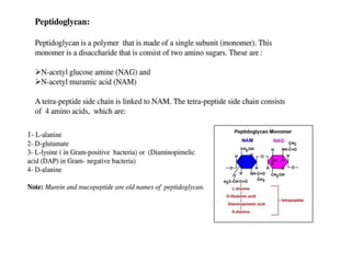
This document defines and describes several important bacteria. It discusses their classification (e.g. Gram-positive/negative), morphology, role in disease, and treatment approaches. Key bacteria mentioned include Clostridium difficile, Staphylococcus aureus (including MRSA), Streptococcus pneumoniae, Escherichia coli, Helicobacter pylori, Mycobacterium tuberculosis, and Neisseria gonorrhoeae. The document provides details on their pathogenic mechanisms, antibiotic resistance patterns, and recommended treatment regimens.
1. BATERIAL CELL WALL
Antibiotic. This term was originally coined to refer to only those
compounds produced by micro-organisms and capable of inhibiting
bacterial growth (Waksman & Woodruff 1940). However the term
“antibiotic” is now commonly used to refer to any drug (natural or
synthetic) that is used for treating bacterial infections.
Bactericidal. The property associated with an antibiotic that causes
the death of bacteria. As distinguished from “bacteriostatic”. Cell
wall synthesis inhibitors are bactericidal against susceptible growing
(dividing) bacteria.
Bacteriostatic. The property of inhibiting bacterial growth, usually
without killing them. Most (but not all) protein synthesis inhibitors
are bacteriostatic (e.g. tetracyclines). This drug effect makes bacteria
more vulnerable to destruction by the immune system.
Biofilm. An adherent extracellular matrix consisting of microbes (e.g.
bacteria), polysaccharides, proteins and DNA. Biofilms are
ubiquitous, and are typically found on solid substrates, and can cause
bacteria to be more resistant to the effects of antibiotics.
2. Peptidoglycan. A complex interwoven matrix that surrounds most
bacterial cells, and consists of a covalently linked macromolecule.
The term derives from the structural components of the cell wall
which are peptides (peptido-) and sugars (glycan). Other synonyms
are murein and mucopeptide.
Penicillin Binding Protein (PBP). The binding sites for penicillin (and
cephalosporin) antibiotics associated with the active site of
transpeptidase enzymes in the cell wall. These transpeptidases are
responsible for creating covalent cross-links between adjacent
glycan chains in the cell wall of growing bacteria. Antibiotic-induced
inhibition of cell wall transpeptidases interferes with the formation
of covalent bonds between peptide chains, and makes the growing
bacterial cell susceptible to rupture from a high intracellular osmotic
pressure.
Pseudomembranous colitis. Inflammation of the colon associated
with an overgrowth of gram-positive C. difficile, usually caused by
the use of broad spectrum antibiotics.
3. Obligative aerobe. A bacteria that requires oxygen in the
environment to survive, because they require oxygen to
make ATP. Examples: Mycobacterium tuberculosis.
Obligate anaerobe. These bacteria do not require oxygen,
and are typically killed by normal atmospheric levels of
oxygen (20.95%). These bacteria produce insufficient
amounts of the enzymes required to detoxify oxygen free
radicals & hydrogen peroxide (e.g. superoxide dismutase &
catalase). Note: the toxic effects of superoxide are used by
cells of the immune system to kill invading
microorganisms). Examples: Actinomyces, Bacterioides,
Clostridium (although some species of Clostridium can
survive in the form of tough dormant “endospores” in the
presence of oxygen).
4. Facultative anaerobe. A bacteria that makes ATP by oxidative
phosphorylation if oxygen is present, but can switch to anaerobic
respiration (fermentation) to make energy if oxygen is absent.
Examples include: species of Staphylococcus, Streptococcus,
Escherichia coli & Listeria. They are the most versatile type of
bacteria - they can live with or without oxygen.
Gram-negative. A bacteria that does not absorb the crystal violet
stain used in the Gram stain process. These bacteria have thin
layer of peptidoglycan, as well as an outer membrane that
prevents them from retaining the crystal violet stain used in
Gram's method. Use of a counterstain (e.g. safranin) stains such
cells pink or red.
Gram-positive. A bacteria that retains the color of the crystal
violet stain in the Gram stain. These cells have a cell wall
containing a thick layer of peptidoglycan, which interacts with the
stain, resulting in a purple/violet appearance under a microscope.
5. GRAM STAING
The process of Gram staining was developed by a
Danish bacteriologist (Hans Christian Gram) who
found that bacteria could be differentiated into
two large groups based upon different physical
properties of their cell walls. Gram-positive
bacteria which have a thick peptidoglycan wall
retain the crystal violet dye (staining them violet or
purple), while all other bacteria (e.g. those having
a thin peptidoglycan wall covered by an outer
membrane) can be stained pink using a
counterstain (safranin or fuchsine) added after the
crystal violet dye
6.
7.
8.
9.
10. Gram Positive
Clostridium botulinum
a heterogeneous group of eight strains of gram-positive, rod-shaped, spore-forming,
obligate anaerobic bacteria responsible for producing a type of food poisoning called
“botulism”, associated with a rare but potentially life-threatening neuro-paralytic
syndrome. different strains of these bacteria produce one of eight different types of
neurotoxins (botulinum toxin A-H), the most active of which produce an irreversible
disruption of acetylcholine release by that presynaptic nerve terminals. Return of
synaptic function requires growth of new presynaptic terminals and subsequent
formation of a new synapses, a process that requires approximately six months
(Pegram & Stone, 2016).
Clostridium difficile
an obligate anaerobic, spore-forming Gram positive rod found in the intestine (normally in
small numbers) broad spectrum antibiotics (e.g. clindamycin or ampicillin/amoxicillin,
cephalosporins) that reduce the normal population of competing flora can allow
overgrowth of C. difficile which releases two cytotoxins (toxins A&B) that bind to and kill
host cells, producing bowel necrosis and a severe colitis
infections can spread from patient to patient in hospital settings by spores adhering to
hands, clothes or equipment in a hospital setting (Lamont, 2016)
the name “difficile” reflects the difficulty in isolating and growing the bacteria using routine
media initial treatment typically involves therapy with oral metronidazole or vancomycin
(Kelly & Lamont, 2015)
11. Staphylococcus aureus (MRSA or ORSA)
frequently found in the nose, respiratory tract, and on the skin
a common cause of minor skin infections (pimple, boils),
respiratory infections (sinusitis), and food poisoning. can also
cause life-threatening diseases including meningitis,
pneumonia, endocarditis, toxic shock syndrome, bacteremia &
sepsis strains of antibiotic resistant S. aureus including
“methicillin-resistant S. aureus” (MRSA) (now referred to as
“oxacillin-resistant S. aureus”: ORSA) are a worldwide problem
in medicine. MRSA is a common cause for hospital-acquired
infections (CDC:MRSA in Healthcare Settings) susceptibility
testing should guide the selection of an appropriate antibiotic.
Empiric therapy has typically included oral therapy with
clindamycin, trimethoprim-sulfamethoxazole, or a long-acting
tetracycline (e.g. doxycycline). Linezolid, vancomycin and
daptomycin are being used for more resistant strains (Lowy,
2016).
12. Streptococcus pneumoniae
a gram-positive, alpha-hemolytic, facultative anaerobe.
the most common cause for community-acquired bacterial
pneumonia
can also cause bacteremia (especially in splenectomized patients),
meningitis, conjunctivitis, and other respiratory related infections
(otitis media, sinusitis)
drug-resistance is a growing concern, with the degree of resistance
varying by region. In 2013, 30% of S. pneumoniae causing severe
infections were fully resistant to one or more relevant antibiotics.
(CDC Threat Report, 2013).
Penicillin-susceptible strains can be treated with a beta-lactam
antibiotic, but empiric treatment of community acquired pneumonia
(CAP) also involves coverage of other causative bacteria, some of
which are insensitive to beta-lactam antibiotics (e.g. mycoplasma).
Hence doxycycline or a macrolide are considered drugs of first choice
for empiric treatment of CAP (for patients with no other
comorbidities, or recent use of these antibiotics). Hospitalized
patients are typically treated with drug combinations of beta-lactams,
13. Streptococcus pyogenes
a gram-positive, beta-hemolytic, facultative anaerobe that typically appear
as chains of cocci shaped bacteria (Stevens & Bryant, 2015).
can cause a broad range of infections, but most commonly originate in the
throat (strep throat, pharyngitis) or skin (impetigo) most strains remain
sensitive to penicillin (e.g. Pen G) or clindamycin; some strains are resistant
to macrolides, tetracyclines & clindamycin (Stevens, 2016).
Gram Negative
Treponema pallidum (syphilis)
a spriochaete bacterium that has both a cytoplasmic and outer membrane
these bacteria are 10-13 μm long & 0.15 μm in width, making them too slender to
be visualized by direct microscopy
based upon the structure of their cell wall, they are considered gram-negative, but
technically they are also too thin to be effectively Gram stained (Hicks & Clement,
2016).
Penicillin (parenteral Pen G) is the treatment of choice (doxycycline or tetracycline)
are used in patients with penicillin allergy; for patients who are pregnant,
desensitization followed by Pen G is recommended
14. Acinetobacter baumannii
an opportunistic pathogen, most commonly affecting patients with compromised
immune systems, most commonly associated with infections in tropical environments,
wars and natural disasters, or hospital outbreaks in temperate climates (Kanafani & Kanj,
2014)becoming increasingly important as a hospital-derived infection
it can survive on artificial surfaces for extended time periods due to its ability to form
biofilms Multidrug-resistant A. baumannii has been a major factor complicating the
treatment and rehabilitation of injured soldiers from Iraq & Afghanistan, with the source
of infection most likely being hospital facilities to which injured soldiers are exposed to
during care of their injuries
When A. baumannii is introduced to a hospital by a colonized patient, it can survive on
surfaces, resist desiccation, and infect new patients
In 2009, A. baumannii was responsible for 19% of ventilator-associated pneumonia cases
in Europe (Koulenti et al, 2009)
Susceptible strains may be treated with broad-spectrum 3rd or 4th generation
cephalosporins, a combination beta-lactam & beta-lactam inhibitor or a carbapenem.
Polymyxins, minocycline, tigecycline, or combination therapy are the main treatments
currently used for more resistant strains (Kanafani & Kanj, 2016)
15. Escherichia coli
as its name indicates, this bacteria is part of the normal intestinal flora
many strains are harmless, but five strains have been identified that can
cause diarrheal illness (e.g. travelor's diarrhea, bloody diarrhea), and food
poisoning
Infections commonly occur from eating contaminated food, including
undercooked hamburger, unpasteurized milk, mayonnaise, unwashed fruit
and vegetables, or even swallowing pool water contaminated with human
waste (Southwick 2008; NLM:MedlinePlus/Ecoli)
Severe infections can also result in kidney failure or death (most commonly
in patients with a compromised immune system)(Southwick 2008; Wanke,
2015)
Treatment must be guided by in vitro susceptibility test results. Most strains
are resistant to beta-lactam antibiotics.
Azithromycin is effective for treatment of travelers diarrhea. Other agents
including fluoroquinolones, and rifaximin are being used as well (Wanke,
2015b). Treatment of food poisoning (which can be caused by several types
of bacteria and viruses) is typically supportive (e.g. fluids, rest), and does
not involve use of antibiotics (Acheson, 2015).
16. Haemophilus influenzae
an aerobic (facultative anaerobe) gram-negative rod that
colonizes the respiratory tract & is transmitted by airborne
secretions (coughing & sneezing) (Yeh, 2015).
historically was mistakenly as a cause of influenza until the flu
virus was discovered in 1933. a common cause for community
acquired pneumonia.
some strains are resistant to penicillins due to production of
beta-lactamases and modification of its penicillin-binding
proteins alternative agents for treatment of infection include
amoxicillin, 2nd or 3rd generation cephalosporins, macrolides,
fluoroquinolones, tetracyclines and aminoglycosides (Yeh, 2015).
17. Helicobacter pylori
H. pylori is a spiral shaped gram-negative bacterium
it contains two to seven unipolar sheathed flagella that enhance its mobility through
viscous solutions. The bacteria produces the enzyme urease to hydrolyze urea in the
gastric lumen to form ammonia that helps neutralize gastric acid and form a protective
cloud around the organism, enabling it to penetrate the gastric mucus layer. Urease
makes up at least 5 percent of the bacteria's total protein weight, and the detection of
bacterial urease activity forms the clinical basis for several invasive and noninvasive
tests to diagnose infection (Crowe, 2016)
H. pylori is “microaerophilic”, as it needs oxygen to survive (it cannot ferment or
respire anaerobically), but it is killed by higher (atmospheric) concentrations of oxygen.
It is the most common chronic bacterial infection in humans & causes chronic gastritis
& most peptic ulcers.
Treatment regimens involve taking several medications for 14 days, as no single drug is
effective against H. pylori. Most treatments include a proton pump inhibitor (to
decrease stomach acidity, allowing damaged ulcerated tissue to heal) and two or three
antibiotics, including metronidazole, clarithromycin, tetracycline (doxycycline) or
amoxicillin (Crowe, 2016b).
18. Mycobacterium tuberculosis (TB)
Mycobacterium tuberculosis is the 2nd most common infectious cause of death in
adults worldwide (HIV is the most common) (Riley, 2015).
Mycobacterium tuberculosis is an aerobic, slow growing nonmotile bacillus that
has a waxy lipid-rich outer wall that contains high concentrations of mycolic acid.
This lipid rich outer cell wall accounts for its many unique clinical characteristics,
including its ability to resist killing by macrophages & PMNs, and to survive within
the body for several years.
The waxy cell wall of M. tuberculosis does not take up Gram stain. Hence it is not
seen on Gram stain. Heating is needed to partially melt the outer cell wall to
permit the penetration & binding of the red dye fuchsin with binds with high
affinity & is resistant to acid-alcohol decolorization. Hence M. tuberculosis is
referred to as an Acid Fast Bacterium. The waxy cell wall limits the bacterial access
to nutrients, which may explain its very slow rate of growth, which is about 1/20th
the rate of most other bacteria.
Tuberculosis is one of the oldest diseases known to man, and a major cause of
death worldwide. It primarily affects the lungs, although other organ systems are
involved in up to 30% of cases. If caused by drug-sensitive strains and properly
treated, it is curable. If left untreated, it causes death in roughly 50% of patients
within 5 years.
Treatment typically involves isoniazid or rifampin (for prevention/prophylaxis), and
various drug combinations (RI, RIPE or RESPI) for susceptible & resistant forms (R:
rifampin; E: ethambutol; S: streptomycin; P: pyrazinamide; I: isoniazid) (Sterling,
2016).
19. Mycoplasma pneumoniae
a very small bacterium (0.1-0.2 μm in length) that lacks a peptidoglycan cell wall,
which makes it insensitive to all cell wall inhibitors (beta-lactams & vancomycin).
these bacteria are considered the smallest self-replicating organisms (Baum, 2016)
a common cause for atypical (“walking”) pneumonia; (Chlamydia pneumoniae and
viruses are other causes for atypical pneumonia)
treatment typically involves use of tetracyclines (e.g. doxycycline), or macrolides (e.g
azithromycin or clarithromycin) (Baum, 2016).
Neisseria gonorrhoeae
gram-negative coccus responsible for causing gonorrhea, the 2nd most commonly
reported communicable disease in the US (Ghanem, 2016).
They are facultatively intracellular and typically appear in pairs (diplococci), in the
shape of coffee beans
a major cause of urethritis in men and cervicitis in women; cervicitis can result in
pelvic inflammatory disease (PID), infertility, ectopic pregnancy, and chronic pelvic
pain (Ghanem, 2016).
antibiotic resistance is an evolving challenge. Drug combinations are typically used
(e.g.ceftriaxone + azithromycin or doxycycline) (Swygard et al, 2016).
Sexual partners should also be treated.
20. Neisseria meningitidis (also called meningococcus)
one of several bacteria that can cause meningitis (it can also cause life-
threatening septicemia)
a round shaped bacteria that tends to cluster in pairs (diplococcus)
~10% of adults are asymptomatic carriers of N meningitidis in their
nasopharynx (CDC:meningococcal)
epidemics tend to occur in crowded environments, such as dorms
susceptible to treatment with 3rd generation cephalosporins (Apicella,
2015)
Pseudomonas aeruginosa
a common Gram-negative, rod-shaped bacteria known for being a multi
drug resistant (MDR) pathogen
a common cause of illness in immunocompromised patients, and typically
affects the urinary tract, airway, wounds, and can cause blood infections
a common colonizer of medical devices (e.g. catheters), and can be
spread by equipment that is not properly cleaned
it is aerobic, and at times a facultative anaerobe
is commonly isolated from the droppings of the domestic cockroach,
which can be pervasive in households as well as hospital settings
21.
22.
23.
24.
25.
26.
27.
28. Peptidoglycan:
•Peptidoglycan is porous cross linked polymer which is responsible for strength of
cell wall.
•Peptidoglycan is composed of three components.
1.Glycan backbone
2.Tetra-peptide side chain ( chain of 4 amino acids) linked to NAM
3.Peptide cross linkage
•Glycan backbone is the repeated unit of N-acetyl muramic acid (NAM) and N-
acetyl glycosamine (NAG) linked by β-glycosidic bond.
•The glycan backbone are cross linked by tetra-peptide linkage. The tetra-peptide
are only found in NAM.
•More than 100 peptidoglycan are known with the diversity focused on the
chemistry of peptide cross linkage and interbridge.
•Although the peptidoglycan chemistry vary from organism to organism the glycan
backbone ie NAG-NAM is same in all species of bacteria.
29. The aminoacids found in tetra-peptide are-
L-alanine: 1st position in both gm+ve and gm-ve bacteria
D-glutamic acid: 2nd position
D-aminopimelic acid/ L-lysine: 3rd position (variation occurs)
D-alanine: 4th position
30. In gram negative bacteria, peptide cross linkage occur between Diaminopamilic
acid (3rd position) of one glycan back bone and D-alanine of adjacent glycan
back bone.
In gram positive bacteria, peptide cross linkage occur by peptide interbridge. The
type and number of aminoacids in interbridge vary among bacterial species.
2. Teichoic acid:
Teichoic acid is water soluble polymer of glycerol or ribitol phosphate.
It is present in gram positive bacteria.
It constitutes about 50% of dry weight of cell wall.
It is the major surface antigen of gram positive bacteria.
Outer membrane
Outer membrane
Outer membrane in cell wall of gram negative bacteria
•It is an additional layer present in gram negative bacteria.
•It is composed of lipid bilayer, protein and lipopolysaccharide (LPS) layer
Function of outer membrane:
•Structure component of gram-ve cell wall
•LPS is an endotoxin produced by gram –ve bacteria
•Lipid-A is antigenic
4. LPS
•LPS is attached to outer membrane by hydrophobic bond. LPS is synthesized in cytoplasmic
membrane and transported to outer membrane.
•LPS is composed of lipid-A and polysaccharide.
•Lipid-A: it is phosphorylated glucosamine disaccharide.
•Polysaccharide: it consists of core-polysaccharide and O-polysaccharide
31.
32. Diagram of the typical cell wall and peptidoglycan structure of
bacteria, including the endolysin cleavage sites. The
peptidoglycan is composed of repeating sugar units,
Nacetylglucosamine (GlcNAc) and N-acetylmuramic acid
(MurNAc), which are cross-linked via an interpeptide bridge
between the meso-diaminopimelic acid (m-DAP) and D-alanine
(D-Ala) residues of adjacent tetrapeptide chains. The chains also
contain L-alanine (L-Ala) and D-glutamic acid (D-Glu). Gram-
negative bacteria contain an outer membrane (OM) structure
not present in Gram-positive bacteria. Both contain an inner
membrane (IM) structure. The cleaved bonds and major
classifications of endolysin are indicated: (1) N-acetylmuramoyl-
L-alanine amidase; (2-4) various endopeptidases; (5) N-acetyl-β-
D-glucosaminidase; (6) N-acetyl-β-D-muramidase (lysozyme).
33. Basic structure of the bacterial cell wall peptidoglycan. The
possible enzymatic activities of PLEs and the bonds they cleave
are indicated. Typically, PLEs carry one or two catalytic domains
displaying one of the indicated enzymatic activities. m-DAP is
found in the peptide chains of the peptidoglycan of most Gram-
negative bacteria, Bacillus spp. and Listeria spp., which present
also direct m-DAP-D-Ala bonding between adjacent stem
peptides. In most Gram-positive bacteria, mDAP is replaced by
L-Lys. Cross-linking between this residue and D-Ala of a
neighbor peptide chain usually occurs by an interpeptide bridge
of variable amino acidic composition (X). Despite some
variation observed among isolates of the same bacterial
species, examples of X bridges are (Gly)5 found in
Staphylococcus aureus, L-Ala-L-Ala in Enterococcus faecalis and
Streptococcus pyogenes, D-Asp in E. faecium, and L-Ser-L-Ala in
S. pneumoniae. The D-Ala residue in light blue may be lost after
peptidoglycan maturation.
34.
35.
36.
37.
38.
39.
40. A) The cell wall of Gram-positive bacteria.
• It is thick monolayered and accounts about 10 to 50% dry weight of cell.
•Gram-positive cell wall is mainly composed of two important components.
1. Peptidoglycan.
2. Teichoic acid.
1. Peptidoglycan
Components of peptidoglycan of Gram-positive cell.
41. Components of Peptidoglycan of the Gram-positive cell wall.
•Peptidoglycan is an important component of the cell wall.
•It is also called as glycopeptide and mucopeptide.
•Peptidoglycan is present almost in all bacteria except Halococcus
and Halobacter.
•The three main components of peptidoglycan are
a ) Glycan backbone strand.
b ) Tetrapeptide side chain.
c ) Cross bridges.
43. Peptidoglycan structure of Gram-positive cell wall
a ) Glycan backbone strand.
•Glycan backbone strand contains a long chain of amino sugars these sugars
are N-acetylmuramic acid and N-acetyl glucosamine.
•These sugars are linked to each other by Beta-1,4 linkage.
•It is the unbranched chain.
b ) Tetrapeptide side chain
•Tetrapeptide side chain consists of four amino acids in a particular sequence.
•The sequence is L-alanine, D-glutamic acid, L-lysin, D-alanine.
•Each N-acetylmuramic acid of glycan strand is linked to four amino acid by the
peptide bond.
•It is called as tetrapeptide side chain.
c ) Cross-linkage
When two adjacent tetrapeptide side chain is linked to each other by a glycan
molecule called as cross-linkage.
The carboxyl group of fourth amino aid D-alanine of the side chain is linked to amino
group third amino acid L-lysin of second tetrapeptide side chain.
The linkage of carboxyl group and the amino group is formed by five glycan
molecules so called as a cross bridge.
44. 2. Teichoic acid
•Teichoic acid is the second most important component of the Gram-positive
bacterial cell wall.
•Teichoic acid is a water-soluble polymer.
•It has repeating sugars.
•Depending on types of repeating sugars teichoic acid is of two types.
•They are Glycerol teichoic acid and Ribitol teichoic acid.
45. Components of teichoic acid of Gram-positive cell wall
1.Glycerol teichoic acid
•It is a long chain of glycerol 3 phosphate molecule.
•These glycerol 3 phosphate are linked by phosphodiester bonds.
2. Ribitol teichoic acid.
•It is a long chain of ribitol 5 phosphate molecule.
These are also linked by phosphodiester bonds
Cell wall of Gram negative bacteria
Cell wall of Gram negative bacteria is more rigid and
thick then Gram postive bacteria.
It is made up of two layers
1. Peptidoglycan
2. Outer cell wall layer
46.
47. Component of peptidoglycan of Gram negative cell wall
1. Peptidoglycan
•It is present in between outer wall layer and cell membrane.
•The structure is similar as gram positive cell wall.
•There are some minor differences in the Gram positive and Gram negative peptidoglycan
structure.
•The differences are as follows:-
1.The third amino acid of tetrapeptide side chainin Gram positive cell wall is L-lysin
which is replaced by Mesodiaminopimilic acid in Gram negative cell wall.
2. All N-acetymuramic acid molecules are not linked to tetrapeptide side chain.
3. Cross bridges are not made up of glycin molecule they are made up of peptide bond
48. Structure of peptidoglcan of Gram-Ve cell
•So due to all these above factors and mainly because of peptide bond cross linkage the network is lose.
•These lose network in peptidoglycan fails to give rigidity to the cell wall.
•In Gram negative cell wall rigidity of a cell is due to outer wall layer.
49. 2.Outer wall layer
•Outer wall layer is present outer side of peptidoglycan layer.
•Outer wall layer is similar to cell membrane.
•It is made up of four components.
Components of outer wall layer of Gram negative cell
1. Phospholipids
2. Lipopolysaccharides
3. Proteins
4.Lipoproteins
50.
51. Components of outer wall layer of Gram negative bacteria
1. Phospholipids
•Phospholipids are present in outer side of outer wall layer.
•It has a polar hydrophilic head and hydrophilic tail.
•It is a bilipid layer.
2. Lipopolysaccharides
•It is also present in outer side of outer wall layer.
•It is made up of lipid A, R-core, and “O” side chain.
•Lipid A molecule is made up of lipid so it is hydrophobic in nature.
•R core and “O” side chain is made up of sugar so it is hydrophobic
in nature.
3. Protein
•These protiens are structural protiens embeded in phospholipid
bilayer.
4. Lipoproteins
•Lipoproteins are present in inner side of outer cell wall near
peptidoglycan.