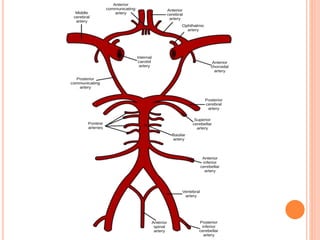
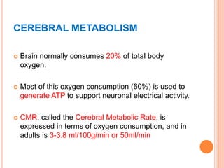
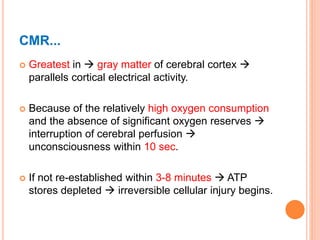
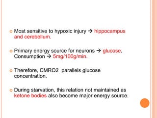
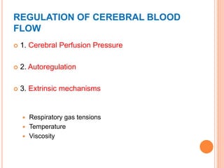
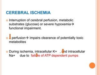
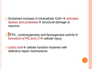
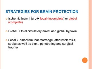
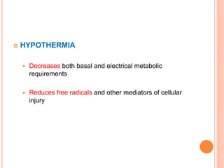
CEREBRAL PHYSIOLOGY
Speaker: Dr. Parul Sharma
Moderator: Dr. Virendra
CONTENTS
 Cerebral Circulation
 Cerebral Metabolism
 Cerebral Blood Flow
 Regulation of Cerebral Blood Flow
 Cerebral Perfusion Pressure
 Blood Brain Barrier
 Cerebrospinal Fluid
 Intracranial Pressure
 Cerebral Ischemia
 Strategies for Brain Protection
CEREBRAL CIRCULATION
 Arterial supply to brain  paired right and left
Internal Carotid Artery anterior circulation,
 And , paired right and left vertebral arteries
posterior circulation.
 3 paired arteries that originate from the circle of
Willis perfuse the brain anterior, middle and
posterior cerebral arteries.
 Posterior communicating arteries and the anterior
communicating artery complete the loop
 Anterior and posterior circulation equally.
CEREBRAL METABOLISM
 Brain normally consumes 20% of total body
oxygen.
 Most of this oxygen consumption (60%) is used to
generate ATP to support neuronal electrical activity.
 CMR, called the Cerebral Metabolic Rate, is
expressed in terms of oxygen consumption, and in
adults is 3-3.8 ml/100g/min or 50ml/min
CMR...
 Greatest in  gray matter of cerebral cortex 
parallels cortical electrical activity.
 Because of the relatively high oxygen consumption
and the absence of significant oxygen reserves 
interruption of cerebral perfusion 
unconsciousness within 10 sec.
 If not re-established within 3-8 minutes  ATP
stores depleted  irreversible cellular injury begins.
 Most sensitive to hypoxic injury  hippocampus
and cerebellum.
 Primary energy source for neurons  glucose.
Consumption  5mg/100g/min.
 Therefore, CMRO2 parallels glucose
concentration.
 During starvation, this relation not maintained as
ketone bodies also become major energy source.
CEREBRAL BLOOD FLOW
 Variety of methods available to measure
1) Positron emission tomography
2) Xenon enhanced CT
3) Single photon emission CT
4) CT perfusion scans
 Best described by Hagen Poiseuille equation for
laminar flow  direct relationship between flow,
CPP and calibre of vessels.
 CBF averages 50ml/100g/min.
 Grey matter  80ml/100g/min
 White matter  20ml/100g/min
 Total CBF  750ml/min i.e. 15-20% of cardiac
output
 Flow rates below  20-25ml/100g/min  cerebral
impairment  slowing on EEG
 15-20ml/100g/min  flat or isoelectric line on EEG
 <10ml/100g/min  irreversible brain damage.
REGULATION OF CEREBRAL BLOOD
FLOW
 1. Cerebral Perfusion Pressure
 2. Autoregulation
 3. Extrinsic mechanisms
 Respiratory gas tensions
 Temperature
 Viscosity
RESPIRATORY GAS TENSIONS
 Particularly PaCO2
 BF changes 1-2ml/100g/min/mmHg change in
PaCO2. immediate secondary to changes in
pH of CSF and cerebral tissue
 Acute changes in PaCO2, not HCO3- affects
CBF acute metabolic acidosis has little effect on
CBF H+ ions cant cross BBB
 Marked hyperventilation shifts ODC left  may
result in cerebral impairment even in normal
individuals
 Only marked changes in PaO2 alter CBF.
TEMPERATURE
 CBF changes 5-7%/1 degree change in
temperature
 Hypothermia both CMR and CBF
 Between 17-37 degree celcius, Q10 for humans
approx. 2 for every 10 degree increase in
temperature, CMR doubles.
 Conversely, CMR by 50% if temperature of brain
falls by 10%, and another 50% if temperature falls
from 27 to 17 degree Celsius
 At 20 degrees isoelectric EEG
 Hyperthermia above 42 degrees neuronal cell
injury
VISCOSITY
 Most important determinant hematocrit
 hematocrit viscosity  improves CBF
 But, reduction in hematocrit decreases oxygen
carrying capacity impair oxygen delivery
 Polycythemia  CBF
 Optimal cerebral oxygen delivery  hematocrit of
30%
CEREBRAL PERFUSION PRESSURE
 Difference in the pressures between the
arterial and venous circulation  dictates
the blood flow to the organ.
 CPP = MAP- ICP/CVP (whichever if greater)
 80-100mm Hg (or 70-90mm Hg)
 Because ICP is normally <15 mm Hg (5-15 mmHg)
 CPP primarily dependent  MAP
 The cerebrovascular resistance (CVR) is the
hindrance to the CBF predominantly by the calibre
of the vessels.
 Moderate to severe increases in ICP 
compromise CPP and CBF, even with normal MAP.
 CPP <50mm Hg slowing on EEG
 CPP 40-25mm Hg flat EEG
 Sustained pressure <25mm Hg irreversible brain
damage
 Cerebral vasculature rapidly adapts to changes in
CPP  10-60 sec
 in CPP  cerebral vasodilation
 in CPP  cerebral vasoconstriction
 CBF nearly constant between MAP 60-160mm Hg.
 Above these pressures  disrupts BBB  cerebral
edema and haemorrhage
 This curve is shifted to right in patients with chronic
arterial HTN.
 Directly proportional to PaCO2 between 20-80mm
Hg.
 Ions do not, but CO2 readily crosses BBB and
directly affects CBF.
 Intense sympathetic stimulation
vasoconstriction limits CBF
 Following brain injury or stroke vasospasm
BLOOD BRAIN BARRIER
 Cerebral vessels unique endothelial cells are
nearly fused
 The paucity of pores  BBB
 Lipid barrier allows passage of lipid soluble
substances, restricting those with high molecular
weight and ionized substances.
 Therefore, movement governed by size, lipid
solubility, and degree of protein binding in blood.
 Freely permeable CO2, O2, lipid soluble
molecules
 Poorly permeable  most ions, proteins, large
substances like mannitol.
 Acute hypertonicity  movement of water out of
brain parenchyma
 Acute hypotonicity water into the brain
 BBB disrupted by  severe HTN, tumours, trauma,
stroke, infection, marked hypercapnia, hypoxia, and
sustained seizures
CEREBROSPINAL FLUID
 Found in ventricles, cisterns, subarachnoid space
surrounding the brain and spinal cord.
 Function protect CNS against trauma as brain
floats in CSF (bouyancy)
 Formation  choroid plexus, minimal amount by
fluid leaking into perivascular spaces surrounding
cerebral vessels.
 Normal total CSF production 0.3-0.4 ml/min OR
21ml/hr OR 500ml/day
 Yet total CSF is only  150 ml
 Absorbed  arachnoid granulations over cerebral
hemispheres
 Active secretion of sodium in the choroid plexus
 Resulting fluid hypotonic, with lower potassium,
bicarbonate and glucose concentration
 Absorption translocation from arachnoid
granulations to ceberal venous sinuses.
INTRACRANIAL PRESSURE
 Cranial vault  rigid  fixed total volume of brain
(80%), blood (12%), and CSF (8%).
 Any increase in one component equivalent
decrease in another to prevent rise in ICP. 
Monroe Kellie Hypothesis / Doctrine
 Normal values 5-15mm Hg
 Major compensatory mechanisms for in ICP
 Initial displacement of CSF into spinal compartment
 absorption
o Sustained increase in ICP herniation
CEREBRAL ISCHEMIA
 Interruption of cerebral perfusion, metabolic
substrates (glucose) or severe hypoxemia
functional impairment.
 perfusion impairs clearance of potentially toxic
metabolites
 During ischemia, intracellular K+ , and intracellular
Na+ due to failure of ATP dependent pumps
 Sustained increase in intracellular Ca2+ activates
lipases and proteases structural damage to
neurons
 FFA , cyclooxygenase and lipoxygenase activity
formation of PG and LT cellular injury
 Lactic acid cellular function impaired with
defective repair mechanisms
STRATEGIES FOR BRAIN PROTECTION
 Ischemic brain injury focal (incomplete) or global
(complete)
 Global total circulatory arrest and global hypoxia
 Focal embolism, haemorrhage, atherosclerosis,
stroke as well as blunt, penetrating and surgical
trauma
 HYPOTHERMIA
 Decreases both basal and electrical metabolic
requirements
 Reduces free radicals and other mediators of cellular
injury
 OTHER GENERAL MEASURES
 Maintaining satisfactory CPP
 Oxygen carrying capacity
 Normal arterial oxygen tension
 Hyperglycemia avoided
 Normocarbia
Thank you 

Cns physiology

  • 1.
    CEREBRAL PHYSIOLOGY Speaker: Dr.Parul Sharma Moderator: Dr. Virendra
  • 2.
    CONTENTS  Cerebral Circulation Cerebral Metabolism  Cerebral Blood Flow  Regulation of Cerebral Blood Flow  Cerebral Perfusion Pressure  Blood Brain Barrier  Cerebrospinal Fluid  Intracranial Pressure  Cerebral Ischemia  Strategies for Brain Protection
  • 3.
    CEREBRAL CIRCULATION  Arterialsupply to brain  paired right and left Internal Carotid Artery anterior circulation,  And , paired right and left vertebral arteries posterior circulation.  3 paired arteries that originate from the circle of Willis perfuse the brain anterior, middle and posterior cerebral arteries.
  • 4.
     Posterior communicatingarteries and the anterior communicating artery complete the loop  Anterior and posterior circulation equally.
  • 6.
    CEREBRAL METABOLISM  Brainnormally consumes 20% of total body oxygen.  Most of this oxygen consumption (60%) is used to generate ATP to support neuronal electrical activity.  CMR, called the Cerebral Metabolic Rate, is expressed in terms of oxygen consumption, and in adults is 3-3.8 ml/100g/min or 50ml/min
  • 7.
    CMR...  Greatest in gray matter of cerebral cortex  parallels cortical electrical activity.  Because of the relatively high oxygen consumption and the absence of significant oxygen reserves  interruption of cerebral perfusion  unconsciousness within 10 sec.  If not re-established within 3-8 minutes  ATP stores depleted  irreversible cellular injury begins.
  • 8.
     Most sensitiveto hypoxic injury  hippocampus and cerebellum.  Primary energy source for neurons  glucose. Consumption  5mg/100g/min.  Therefore, CMRO2 parallels glucose concentration.  During starvation, this relation not maintained as ketone bodies also become major energy source.
  • 9.
    CEREBRAL BLOOD FLOW Variety of methods available to measure 1) Positron emission tomography 2) Xenon enhanced CT 3) Single photon emission CT 4) CT perfusion scans
  • 10.
     Best describedby Hagen Poiseuille equation for laminar flow  direct relationship between flow, CPP and calibre of vessels.
  • 11.
     CBF averages50ml/100g/min.  Grey matter  80ml/100g/min  White matter  20ml/100g/min  Total CBF  750ml/min i.e. 15-20% of cardiac output  Flow rates below  20-25ml/100g/min  cerebral impairment  slowing on EEG
  • 12.
     15-20ml/100g/min flat or isoelectric line on EEG  <10ml/100g/min  irreversible brain damage.
  • 13.
    REGULATION OF CEREBRALBLOOD FLOW  1. Cerebral Perfusion Pressure  2. Autoregulation  3. Extrinsic mechanisms  Respiratory gas tensions  Temperature  Viscosity
  • 14.
    RESPIRATORY GAS TENSIONS Particularly PaCO2  BF changes 1-2ml/100g/min/mmHg change in PaCO2. immediate secondary to changes in pH of CSF and cerebral tissue  Acute changes in PaCO2, not HCO3- affects CBF acute metabolic acidosis has little effect on CBF H+ ions cant cross BBB
  • 15.
     Marked hyperventilationshifts ODC left  may result in cerebral impairment even in normal individuals  Only marked changes in PaO2 alter CBF.
  • 16.
    TEMPERATURE  CBF changes5-7%/1 degree change in temperature  Hypothermia both CMR and CBF  Between 17-37 degree celcius, Q10 for humans approx. 2 for every 10 degree increase in temperature, CMR doubles.
  • 17.
     Conversely, CMRby 50% if temperature of brain falls by 10%, and another 50% if temperature falls from 27 to 17 degree Celsius  At 20 degrees isoelectric EEG  Hyperthermia above 42 degrees neuronal cell injury
  • 18.
    VISCOSITY  Most importantdeterminant hematocrit  hematocrit viscosity  improves CBF  But, reduction in hematocrit decreases oxygen carrying capacity impair oxygen delivery  Polycythemia  CBF
  • 19.
     Optimal cerebraloxygen delivery  hematocrit of 30%
  • 21.
    CEREBRAL PERFUSION PRESSURE Difference in the pressures between the arterial and venous circulation  dictates the blood flow to the organ.  CPP = MAP- ICP/CVP (whichever if greater)  80-100mm Hg (or 70-90mm Hg)
  • 22.
     Because ICPis normally <15 mm Hg (5-15 mmHg)  CPP primarily dependent  MAP  The cerebrovascular resistance (CVR) is the hindrance to the CBF predominantly by the calibre of the vessels.
  • 23.
     Moderate tosevere increases in ICP  compromise CPP and CBF, even with normal MAP.  CPP <50mm Hg slowing on EEG  CPP 40-25mm Hg flat EEG  Sustained pressure <25mm Hg irreversible brain damage
  • 24.
     Cerebral vasculaturerapidly adapts to changes in CPP  10-60 sec  in CPP  cerebral vasodilation  in CPP  cerebral vasoconstriction  CBF nearly constant between MAP 60-160mm Hg.  Above these pressures  disrupts BBB  cerebral edema and haemorrhage
  • 26.
     This curveis shifted to right in patients with chronic arterial HTN.  Directly proportional to PaCO2 between 20-80mm Hg.  Ions do not, but CO2 readily crosses BBB and directly affects CBF.
  • 27.
     Intense sympatheticstimulation vasoconstriction limits CBF  Following brain injury or stroke vasospasm
  • 28.
    BLOOD BRAIN BARRIER Cerebral vessels unique endothelial cells are nearly fused  The paucity of pores  BBB  Lipid barrier allows passage of lipid soluble substances, restricting those with high molecular weight and ionized substances.  Therefore, movement governed by size, lipid solubility, and degree of protein binding in blood.
  • 29.
     Freely permeableCO2, O2, lipid soluble molecules  Poorly permeable  most ions, proteins, large substances like mannitol.  Acute hypertonicity  movement of water out of brain parenchyma  Acute hypotonicity water into the brain
  • 30.
     BBB disruptedby  severe HTN, tumours, trauma, stroke, infection, marked hypercapnia, hypoxia, and sustained seizures
  • 31.
    CEREBROSPINAL FLUID  Foundin ventricles, cisterns, subarachnoid space surrounding the brain and spinal cord.  Function protect CNS against trauma as brain floats in CSF (bouyancy)  Formation  choroid plexus, minimal amount by fluid leaking into perivascular spaces surrounding cerebral vessels.
  • 32.
     Normal totalCSF production 0.3-0.4 ml/min OR 21ml/hr OR 500ml/day  Yet total CSF is only  150 ml
  • 34.
     Absorbed arachnoid granulations over cerebral hemispheres  Active secretion of sodium in the choroid plexus  Resulting fluid hypotonic, with lower potassium, bicarbonate and glucose concentration  Absorption translocation from arachnoid granulations to ceberal venous sinuses.
  • 36.
    INTRACRANIAL PRESSURE  Cranialvault  rigid  fixed total volume of brain (80%), blood (12%), and CSF (8%).  Any increase in one component equivalent decrease in another to prevent rise in ICP.  Monroe Kellie Hypothesis / Doctrine  Normal values 5-15mm Hg
  • 37.
     Major compensatorymechanisms for in ICP  Initial displacement of CSF into spinal compartment  absorption o Sustained increase in ICP herniation
  • 38.
    CEREBRAL ISCHEMIA  Interruptionof cerebral perfusion, metabolic substrates (glucose) or severe hypoxemia functional impairment.  perfusion impairs clearance of potentially toxic metabolites  During ischemia, intracellular K+ , and intracellular Na+ due to failure of ATP dependent pumps
  • 39.
     Sustained increasein intracellular Ca2+ activates lipases and proteases structural damage to neurons  FFA , cyclooxygenase and lipoxygenase activity formation of PG and LT cellular injury  Lactic acid cellular function impaired with defective repair mechanisms
  • 40.
    STRATEGIES FOR BRAINPROTECTION  Ischemic brain injury focal (incomplete) or global (complete)  Global total circulatory arrest and global hypoxia  Focal embolism, haemorrhage, atherosclerosis, stroke as well as blunt, penetrating and surgical trauma
  • 41.
     HYPOTHERMIA  Decreasesboth basal and electrical metabolic requirements  Reduces free radicals and other mediators of cellular injury
  • 42.
     OTHER GENERALMEASURES  Maintaining satisfactory CPP  Oxygen carrying capacity  Normal arterial oxygen tension  Hyperglycemia avoided  Normocarbia
  • 43.