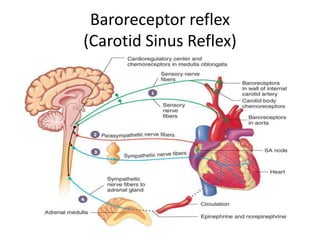
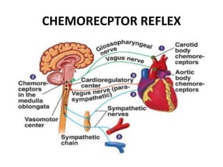
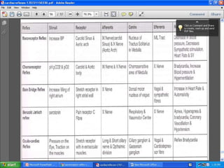
Cardiac Reflexes
Introduction
• Fast acting reflex loops between the heart and
CNS that contribute to regulation of cardiac
function and the maintenance of physiologic
Haemostasis.
• Specific cardiac receptors elicit the physiologic
responses by various pathways.
• Cardiac receptors are linked to CNS by
myelinated or unmyelinated afferent fibers
that travel along the vagus nerve.
• Cardiac receptors are in the atria,
ventricles,pericardium, and coronary arteries.
• Extracardiac receptors are located in the great
vessels and carotid artery.
• Sympathetic and parasympathetic nerve input is
processed in the CNS.
• After central processing, efferent fibers to the
heart or the systemic circulation will provoke a
particular reaction.
• The response of cardiovascular system to efferent
stimulation varies with age and duration of the
underlying condition.
Reflexes
• 1. Baroreceptor Reflex
• 2. Chemoreceptor Reflex
• 3. Brain Bridge Reflex
• 4. Bezold-Jarisch Reflex
• 5. Valsalva Maneuver
• 6. Cushing Reflex
• 7. Occulocardiac Reflex
Baroreceptor reflex
(Carotid Sinus Reflex)
• The best known of nervous mechanisms for
arterial pressure control (baroreceptor reflex)
• Baroreceptors are stretch receptors found in
the carotid body, aortic body and the wall of
all large arteries of the neck and thorax.
• Respond progressively at 50-170 mm Hg.
• Respond more to a rapidly changing pressure
than stationary pressure
• Changes in arterial blood pressure are
monitored by stretch receptors located in the
carotid sinus and aortic arch.
• The nucleus solitarius, located in the
cardiovascular center of the medulla, receives
the impulse from these stretch receptors
through afferent glossopharyngeal and vagus
nerves.
• The response from depressor system includes
decreased sympathetic activity, leading to
decrease in cardiac contractility, heart rate
and vascular tone.
• In addition, activation of the parasympathetic
system further decreases the heart rate,
myocardial contractility.
• Reverse effects are elicited with the onset of
hypotension
CHEMORECPTOR REFLEX
• Chemosensitive cells are located in the carotid
bodies and the aortic body.
• These cell responds to changes in pH status
and blood O2 tension.
• At an arterial partial O2 pressure of less than
50mmof Hg or in condition of acidosis, the
chemoreceptors send their impulses along the
sinus nerve of Henring and vagus nerve to
chemosensitive area of medulla.
• This area responds by stimulating the
respiratory centers and there by increasing the
ventilatory drive.
• In addition, activation of the parasympathetic
system, leads to a reduction in heart rate,and
myocardial contractility
• In the case, of persistant hypoxia, the CNS will
be directly stimulated, with a resultant
increase in sympathetic activity.
Brain Bridge Reflex
• It is elicited by stretch receptors located in the
right atrial wall and the cavo atrial junction
• An increase in the right sided filling pressure
sends vagal afferent signals to the cardiovascular
center in the medulla
• These afferent signals inhibit the
parasympathetic system activity, thereby
increasing the heart rate.
• Acceleration of heart rate also results from a
direct effect on the SA node by the stretching
the atrium.
BEZOLD- JARISCH REFLEX
• It responds to ventricular stimuli sensed by
the chemoreceptors and mechanoreceptors
within the left ventricular wall by inducing the
triad of
– Hypotension
– Bradycardia
– Coronary artery dilatation
• The activated receptors communicate along
unmyelinated vagal afferent type C fibers
• It produce increase in parasympathetic tone
• Because it invokes bradycardia, the Bezold
jarisch reflex is thought of as a
cardioprotective Reflex
• The reflex has been implicated in physiologic
response to a range of cardiovascular conditions
such as Myocardial ischemia, thrombolysis,or
revascularisation and syncope
ValsalvaManeuver
• Forced Expiration against a closed glottis
produces-
– Increased Intrathoracic pressure
– Increased central venous pressure
– Decreased Venous return
• Cardiac output and blood pressure will be
decreased after the valsalva maneuver
• This decrease will be sensed by baroreceptors
and will reflexively result in an increase in
heart rate and myocardial contractility
through sympathetic stimulation
• When the glottis open, venous return
increases and causes the heart to respond by
vigorous contraction and an increase in blood
Pressure.
• This increase in arterial blood pressure will in turn, be
sensed by baroreceptors, thereby stimulating the
parasympathetic efferent pathway to the heart.
CUSHING REFLEX
• The cushing reflex is a result of cerebral
ischemia caused by intracranial pressure
• Cerebral ischemia at the medullary vasomotor
center induces initial activation of the sympathetic
nervous system
• This activation will leads to increase in heart
rate, arterial blood pressure, and myocardial
contractility in an effort to improve cerebral
perfusion
OCULOCARDIAC REFLEX
• This pressure is provoked by pressure applied
to the globe of the eye
• Stretch receptors are located in the
extraocular muscles
• Once activated, stretch receptors will send
afferent signals through short and long ciliary
nerves.
• This ciliary nerves will merge with ophthalmic
division of trigeminal nerve at the ciliary
ganglion.
• The trigeminal nerves will carry the impulse to
gasserian ganglion,thereby resulting in
increased parasympathetic tone and
subsequent BRADYCARDIA
Cardiac reflexes

Cardiac reflexes

  • 1.
  • 2.
    Introduction • Fast actingreflex loops between the heart and CNS that contribute to regulation of cardiac function and the maintenance of physiologic Haemostasis. • Specific cardiac receptors elicit the physiologic responses by various pathways.
  • 3.
    • Cardiac receptorsare linked to CNS by myelinated or unmyelinated afferent fibers that travel along the vagus nerve. • Cardiac receptors are in the atria, ventricles,pericardium, and coronary arteries. • Extracardiac receptors are located in the great vessels and carotid artery.
  • 4.
    • Sympathetic andparasympathetic nerve input is processed in the CNS. • After central processing, efferent fibers to the heart or the systemic circulation will provoke a particular reaction. • The response of cardiovascular system to efferent stimulation varies with age and duration of the underlying condition.
  • 5.
    Reflexes • 1. BaroreceptorReflex • 2. Chemoreceptor Reflex • 3. Brain Bridge Reflex • 4. Bezold-Jarisch Reflex • 5. Valsalva Maneuver • 6. Cushing Reflex • 7. Occulocardiac Reflex
  • 6.
  • 7.
    • The bestknown of nervous mechanisms for arterial pressure control (baroreceptor reflex) • Baroreceptors are stretch receptors found in the carotid body, aortic body and the wall of all large arteries of the neck and thorax. • Respond progressively at 50-170 mm Hg. • Respond more to a rapidly changing pressure than stationary pressure
  • 8.
    • Changes inarterial blood pressure are monitored by stretch receptors located in the carotid sinus and aortic arch. • The nucleus solitarius, located in the cardiovascular center of the medulla, receives the impulse from these stretch receptors through afferent glossopharyngeal and vagus nerves.
  • 9.
    • The responsefrom depressor system includes decreased sympathetic activity, leading to decrease in cardiac contractility, heart rate and vascular tone. • In addition, activation of the parasympathetic system further decreases the heart rate, myocardial contractility. • Reverse effects are elicited with the onset of hypotension
  • 10.
  • 11.
    • Chemosensitive cellsare located in the carotid bodies and the aortic body. • These cell responds to changes in pH status and blood O2 tension.
  • 12.
    • At anarterial partial O2 pressure of less than 50mmof Hg or in condition of acidosis, the chemoreceptors send their impulses along the sinus nerve of Henring and vagus nerve to chemosensitive area of medulla. • This area responds by stimulating the respiratory centers and there by increasing the ventilatory drive.
  • 13.
    • In addition,activation of the parasympathetic system, leads to a reduction in heart rate,and myocardial contractility • In the case, of persistant hypoxia, the CNS will be directly stimulated, with a resultant increase in sympathetic activity.
  • 14.
    Brain Bridge Reflex •It is elicited by stretch receptors located in the right atrial wall and the cavo atrial junction • An increase in the right sided filling pressure sends vagal afferent signals to the cardiovascular center in the medulla • These afferent signals inhibit the parasympathetic system activity, thereby increasing the heart rate.
  • 15.
    • Acceleration ofheart rate also results from a direct effect on the SA node by the stretching the atrium.
  • 16.
    BEZOLD- JARISCH REFLEX •It responds to ventricular stimuli sensed by the chemoreceptors and mechanoreceptors within the left ventricular wall by inducing the triad of – Hypotension – Bradycardia – Coronary artery dilatation • The activated receptors communicate along unmyelinated vagal afferent type C fibers
  • 17.
    • It produceincrease in parasympathetic tone • Because it invokes bradycardia, the Bezold jarisch reflex is thought of as a cardioprotective Reflex • The reflex has been implicated in physiologic response to a range of cardiovascular conditions such as Myocardial ischemia, thrombolysis,or revascularisation and syncope
  • 18.
  • 19.
    • Forced Expirationagainst a closed glottis produces- – Increased Intrathoracic pressure – Increased central venous pressure – Decreased Venous return • Cardiac output and blood pressure will be decreased after the valsalva maneuver
  • 20.
    • This decreasewill be sensed by baroreceptors and will reflexively result in an increase in heart rate and myocardial contractility through sympathetic stimulation • When the glottis open, venous return increases and causes the heart to respond by vigorous contraction and an increase in blood Pressure. • This increase in arterial blood pressure will in turn, be sensed by baroreceptors, thereby stimulating the parasympathetic efferent pathway to the heart.
  • 21.
  • 22.
    • The cushingreflex is a result of cerebral ischemia caused by intracranial pressure • Cerebral ischemia at the medullary vasomotor center induces initial activation of the sympathetic nervous system • This activation will leads to increase in heart rate, arterial blood pressure, and myocardial contractility in an effort to improve cerebral perfusion
  • 23.
  • 24.
    • This pressureis provoked by pressure applied to the globe of the eye • Stretch receptors are located in the extraocular muscles • Once activated, stretch receptors will send afferent signals through short and long ciliary nerves.
  • 25.
    • This ciliarynerves will merge with ophthalmic division of trigeminal nerve at the ciliary ganglion. • The trigeminal nerves will carry the impulse to gasserian ganglion,thereby resulting in increased parasympathetic tone and subsequent BRADYCARDIA