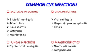
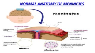
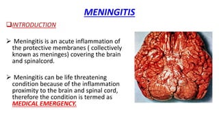
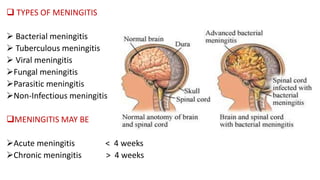
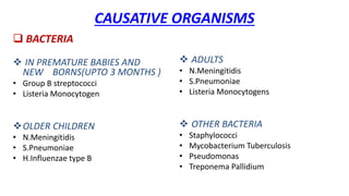
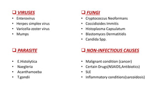
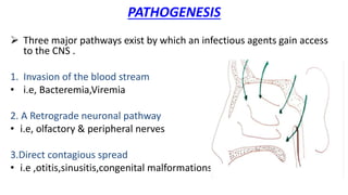
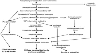
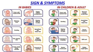
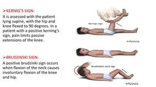
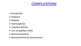
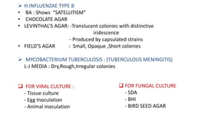
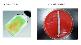
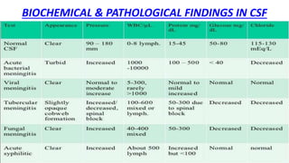
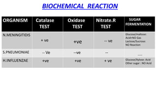
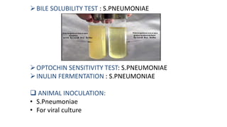
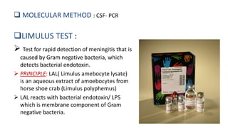
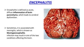
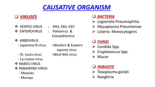
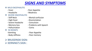
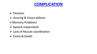
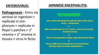
CNS INFECTIONS
Meningitis
Encephalitis
Brain Abscess
BY : VISHRANT AMIN & FRIENDS
COMMON CNS INFECTIONS
 BACTERIAL INFECTIONS
 Bacterial meningitis
 Tuberculosis
 Brain abscess
 Lysteriosis
 Neurosyphilis
 FUNGAL INFECTIONS
 Cryptococcal meningitis
 VIRAL INFECTIONS
 Viral meningitis
 Herpes simplex encephalitis
 Rabies
 PARASITIC INFECTIOS
 Neurocysticercosis
 Toxoplasmosis
CASE
• A seven year old boy presented to emergency department with high
grade fever on the previous day. He complained of headache, was
disoriented and had projectile vomiting. On examination, the neck
was found to be rigid and kerning’s sign was positive. A lumbar
puncture was carried out along with complete blood counts and
serum biochemistry. Cytology showed polymorphs at
1000/cumm,protein 250 mg/dl and glucose20 mg/dl. Blood glucose
level normal.
• On gram stain, gram negative cocci seen in pairs, most of them inside
polymorphs.
• The latex agglutination test was positive for N.Meningitidis antigen.
NORMAL ANATOMY OF MENINGIES
MENINGITIS
INTRODUCTION
 Meningitis is an acute inflammation of
the protective membranes ( collectively
known as meninges) covering the brain
and spinalcord.
 Meningitis can be life threatening
condition because of the inflammation
proximity to the brain and spinal cord,
therefore the condition is termed as
MEDICAL EMERGENCY.
 TYPES OF MENINGITIS
 Bacterial meningitis
 Tuberculous meningitis
 Viral meningitis
Fungal meningitis
Parasitic meningitis
Non-Infectious meningitis
MENINGITIS MAY BE
Acute meningitis < 4 weeks
Chronic meningitis > 4 weeks
CAUSATIVE ORGANISMS
 BACTERIA
 IN PREMATURE BABIES AND
NEW BORNS(UPTO 3 MONTHS )
• Group B streptococci
• Listeria Monocytogen
OLDER CHILDREN
• N.Meningitidis
• S.Pneumoniae
• H.Influenzae type B
 ADULTS
• N.Meningitidis
• S.Pneumoniae
• Listeria Monocytogens
 OTHER BACTERIA
• Staphylococci
• Mycobacterium Tuberculosis
• Pseudomonas
• Treponema Pallidium
 VIRUSES
• Enterovirus
• Herpes simplex virus
• Varicella-zoster virus
• Mumps
 PARASITE
• E.Histolytica
• Naegleria
• Acanthamoeba
• T.gondii
 FUNGI
• Cryptococcus Neoformans
• Coccidioides Immitis
• Histoplasma Capsulatum
• Blastomyces Dermatitidis
• Candida Spp.
 NON-INFECTIOUS CAUSES
• Malignant condition (cancer)
• Certain Drugs(NSAIDS,Antibiotics)
• SLE
• Inflammatory conditions(sarcoidosis)
PATHOGENESIS
 Three major pathways exist by which an infectious agents gain access
to the CNS .
1. Invasion of the blood stream
• i.e, Bacteremia,Viremia
2. A Retrograde neuronal pathway
• i.e, olfactory & peripheral nerves
3.Direct contagious spread
• i.e ,otitis,sinusitis,congenital malformations
SIGN & SYMPTOMS
IN BABIES IN CHILDREN & ADULT
KERNIG’S SIGN:
It is assessed with the patient
lying supine, with the hip and
knee flexed to 90 degrees. In a
patient with a positive kerning’s
sign, pain limits passive
extensions of the knee.
BRUDZINSKI SIGN:
A positive brudzinki sign occurs
when flexion of the neck causes
involuntary flexion of the knee
and hip.
COMPLICATIONS
 Encephalitis
 Deafness
 Epilepsy
 Hydrocephalus
 Cognitive Deficits
 Loss of pupillary reflex
 Abnormal posture
 Decreased level of consciousness
LABORATORY DIAGNOSIS
SPECIMEN
 CSF : By Lumbar puncture (L3-L4 level)
See for the turbidity
Divide into 4 parts
 For Microbiological Test
 For Pathological Test
 For Biochemical Test
 For Preservation
 BLOOD
 serology & culture
 URINE
 For antigen detection
• S.Pneumoniae
• C.Neoformans
MICROSCOPY
 STAINING
 GRAM’S STAIN:
• N.Meningitidis : Intracellular, gram negative, diplococci
• S.Pneumoniae : gram positive, diplococci(lanceolated shaped)
• H.Influenzae : gram negative, pleomorphic, coccobacilli
 Z-N STAIN:
• To demonstrate AFB along with LYMPHOCYTE (tuberculous meningitis)
 INDIAN INK: ( For demonstration of capsule of organisms)
• C.Neoformans
• S.Pneumoniae
 WET-MOUNT & GIEMSA STAIN : For Parasite
 QUELLANG REACTION:
• C.Neoformans
• S.Pneumoniae
CULTURE
 FOR BACTERIA:
 N.MENINGITIDIS:
(1)ON SOLID MEDIA:
- Blood agar Small,Translucent,Round,Convex,
- Chocolate agar Bluish-Grey colour,
Weak haemolysis
- Muller-Hinton Agar
- Thayer-Martin agar : selective media
(2) ON LIQUID MEDIA:
- Turbidity with little or no surface growth
 S.PNEUMONIAE:
- BA Small,Dome-Shaped,Glistening with
- CA an area of green discolouration ( α- haemolysis)
N.MENINGITIDIS ( BA )
• S.PNEUMONIAE ( CA ) • S.PNEUMONIAE(BA )
 H.INFLUENZAE TYPE B
• BA : Shows “SATELLITISM”
• CHOCOLATE AGAR
• LEVINTHAL’S AGAR: -Translucent colonies with distinctive
iridescence
- Produced by capsulated strains
• FIELD’S AGAR : Small, Opaque ,Short colonies
 MYCOBACTERIUM TUBERCULOSIS : (TUBERCULOUS MENINGITIS)
L-J MEDIA : Dry,Rough,Irregular colonies
 FOR VIRAL CULTURE :
- Tissue culture
- Egg Inoculation
- Animal inoculation
 FOR FUNGAL CULTURE
- SDA
- BHI
- BIRD SEED AGAR
• L-J MEDIUM • H.INFLUENZAE
BIOCHEMICAL & PATHOLOGICAL FINDINGS IN CSF
BIOCHEMICAL REACTION
ORGANISM Catalase
TEST
Oxidase
TEST
Nitrate.R
TEST
SUGAR
FERMENTATION
N.MENINGITIDIS
+ ve +ve -- ve
Glucose/maltose:
Acid+NO Gas
Lactose/Sucrose:
NO Reaction
S.PNEUMONIAE -- Ve --ve -- -----
H.INFLUENZAE +ve +ve + ve Glucose/Xylose: Acid
Other sugar : NO Acid
BILE SOLUBILITY TEST : S.PNEUMONIAE
OPTOCHIN SENSITIVITY TEST: S.PNEUMONIAE
INULIN FERMENTATION : S.PNEUMONIAE
 ANIMAL INOCULATION:
• S.Pneumoniae
• For viral culture
SEROLOGY
FOR N.MENINGITIDIS
(1)Slide Agglutination Test
(2)Latex Agglutination Test
(3)Haemagglutination Test
FOR S.PNEUMONIAE :
(1) Latex Agglutination Test
(2) Demonstration of SSS
(3) Immunochromatography
(4) CRP Test (biomarker)
(5) Procalcitonin Level (biomarker)
FOR H.INFLUENZAE Latex agglutination test
FOR L.MONOCYTOGENS
C.NEOFORMANS
 MOLECULAR METHOD : CSF- PCR
LIMULUS TEST :
 Test for rapid detection of meningitis that is
caused by Gram negative bacteria, which
detects bacterial endotoxin.
 PRINCIPLE: LAL( Limulus amebocyte lysate)
is an aqueous extract of amoebocytes from
horse shoe crab (Limulus polyphemus)
 LAL reacts with bacterial endotoxin/ LPS
which is membrane component of Gram
negative bacteria.
ENCEPHALITIS
 Encephalitis is defined as acute,
diffuse inflammation of brain
parenchyma, which leads to cerebral
dysfunction.
 Sometimes, it is associated with
meningitis, which is known as
Meningoencephalitis
infection may result in one of the two
conditions affecting the brain.
PRIMARY ENCEPHALITIS
 Occurs when a virus / other
infectious agent infects the
brain.
 May be reactivation after
previous illness
 SECONDARY ENCEPHALITIS
(POST INFECTIONAL ENCEPHALITIS)
 It is faulty immune system
reaction in response to a
infection elsewhere in body.
 Occurs 3 weeks after initial
infection.
CAUSATIVE ORGANISM
 VIRUSES
 HERPES VIRUS : HSV, EBV, VZV
 ENTEROVIRUS : Poliovirus &
Coxsackievirus
 ARBOVIRUS
- Japanese B virus - Western & Eastern
equine virus
- St. Louis virus - West Nile virus
- La crosse virus
 RABIES VIRUS
 PARAMYXO VIRUS
- Measles
- Mumps
 BACTERIA
 Legionella Pneumophilia
 Mycoplasma Pneumoniae
 Listeria Monocytogens
 FUNGI
 Candida Spp.
 Cryptococcus Spp.
 Mucor
 PARASITE
 Toxoplasma gondii
 Naegleria
SIGNS AND SYMPTOMS
 MILD ENCEPHALITIS
- Fever - Poor Appetite
- Headache - Weakness
 SEVERE ENCEPHALITIS
- Stiff Neck - Mental confusion
- High Fever - Disorientation
- Severe headache - Convulsion
- Memory loss - Problems with Speech
- Hallucination - Coma
 IN INFANTS
-Vomiting - Poor Appetite
- Body stiffness - Poor memory
 BRUDZINSKI SIGN:
 KERNING’S SIGN :
COMPLICATION
 Paralysis
 Hearing & Vision defects
Memory Problems
 Speech impairment
 Lack of Muscle coordination
 Coma & Death
PATHOGENESIS
 Transmission into human depend on causative organism :
• HSV & NAEGLERIA : Respiratory Tract
• ARBOVIRUS : Vector ( Mosquito & Tick )
• ACANTHAMOEBA : Eye & Nasal route
• RABIES : Dog bite
 Spread to CNS : ( 3 Routes )
1. Haematogenous Route
2. Neuronal Route
3. Contagious Route
 Damage to the Brain Parenchyma
 Causes Encephalitis
ENTEROVIRUS: JAPANESE ENCEPHALITIS:
Pathogenesis : Entry via
aerosol or ingestion->
replicate in oro-
pharynx-> replicate in
Peyer's patches-> 1°
viremia-> 2° viremia in
tissues-> virus in feces
LABORATORY DIAGNOSIS
SPECIMENS:
 CSF 4. THROAT SWAB
 BLOOD 5. NASOPHARYNGEAL SWAB
 URINE
MICROSCOPY:
 VIRAL:
 Herpes virus: Tzank smear Multinucleated giant cells and Ground glass
chromatin and giemsa stained smears and flurescent antibody techniqe
 Rabies virus: Negri bodies
 Measles: multinucleated giant cells in Giemsa stained smears
 BACTERIAL:
 Legionella pneumophilia: Legionella stains poorly with Gram stain, stains
positive with silver
 Mycoplasma pneumoniae: Giemsa stain
3. FUNGAL: Candida spp.: Gram stain +ve
Cryptococcus: India ink stain capsulated budding yeast cells
4.PARASITIC: Naegleria fowleri : wright or giemsa stain pink nuclei and blue cytoplasm
Toxoplasma gondii: Periodic acid Schiff Bradyzoites are PAS +ve
CULTURE:
 VIRAL: Limited utility as culture may be insensitive
 BACTERIAL: Listeria monocytogens: Listeria grows on media such as Mueller-Hinton agar.
Identification is enhanced if the primary cultures are done on agar containing sheep blood, because
the characteristic small zone of hemolysis can be observed around and under colonies.
Legionella pneumophilia: Cultured on charcoal yeast extract with iron & cysteine
 FUNGAL: Candida spp. & Cryptococcus: Sabouraud agar creamy white, smooth colony.
 PARASITIC: Naegleria fowleri: Non nutrient agar Plaques formation
SPINAL TAP: A spinal tap is done by inserting a needle into the spinal column.
Another name for a spinal tap is a lumbar puncture. The CSF fluid taken during a spinal
tap will also be tested to find the cause of encephalitis. It may show which type of virus
or germ is causing the inflammation.
ISOLATION OF VIRUS:
 HSV : Human Diploid Fibroblast – Typical CPE ( 24-48 hrs)
 RABIES : CPE is minimal so by Immunofluorescence
 ARBOVIRUS : Vero,BHK-21 & Mosquito cell line ( Immunofluorescence)
SEROLOGY:
 DEMONSTRATION OF IgM ANTIBODY
 IMMUNOFLUORESCENCE
 ELISA
 LATEX AGGLUTINATION TEST
MOLECULAR METHOD: PCR.
BRAIN ABSCESS
 Brain abscess (or cerebral abscess) is an abscess caused by inflammation and
collection of infected material, coming from local or remote infectious sources,
within the brain tissue.
A).LOCAL SOURCES B). REMOTE SOURCES
Ear infection, Dental abscess Lung, heart, kidney
Infection of Paranasal sinuses
Infection of the Mastoid air cells of the temporal bone,
Epidural abscess
 The infection may also be introduced through a skull fracture following a
headtrauma or surgical procedures.
 Brain abscess is usually associated with congenital heart disease in young
children.
 It may occur at any age but is most frequent in the third decade of life.
CAUSATIVE ORGANISMS
BACTERIA
• S.Aureus
• S.Intermidius
• Bacteroides
• Fusobacterium
• Enterobacteriaceae
• Pseudomonas spp
 LESS COMMON
• N.Meningitidis
• S.Pneumoniae
• H.Influenzae
 FUNGI
• Aspergillus
• Candida
• Cryptococcus
• Mucor
• Coccidioides
• Histoplasma capsulatum
• Blastomyces dermatitidis
PARASITE
• Toxoplasma gondii
• Entamoeba histolytica
• Trypanosoma cruzi
• Schistosoma
SIGN & SYMPTOMS
• Headache
• Drowsiness
• Fever
• Confusion
• Seizures
• Speech difficulties
• Hemiparesis
STAGES OF BRAIN ABSCESS
1. Early cerebritis
2. Late cerebritis
3. Early Capsule
4. Late Capsule
RADIOLOGY IS IMPORTANT FOR DIAGNOSIS
OF ALL CNS INFECTION
 CT-SCAN
 MRI
 CHEST X-RAY
CNS INFECTION

CNS INFECTION

  • 1.
  • 2.
    COMMON CNS INFECTIONS BACTERIAL INFECTIONS  Bacterial meningitis  Tuberculosis  Brain abscess  Lysteriosis  Neurosyphilis  FUNGAL INFECTIONS  Cryptococcal meningitis  VIRAL INFECTIONS  Viral meningitis  Herpes simplex encephalitis  Rabies  PARASITIC INFECTIOS  Neurocysticercosis  Toxoplasmosis
  • 3.
    CASE • A sevenyear old boy presented to emergency department with high grade fever on the previous day. He complained of headache, was disoriented and had projectile vomiting. On examination, the neck was found to be rigid and kerning’s sign was positive. A lumbar puncture was carried out along with complete blood counts and serum biochemistry. Cytology showed polymorphs at 1000/cumm,protein 250 mg/dl and glucose20 mg/dl. Blood glucose level normal. • On gram stain, gram negative cocci seen in pairs, most of them inside polymorphs. • The latex agglutination test was positive for N.Meningitidis antigen.
  • 4.
  • 5.
    MENINGITIS INTRODUCTION  Meningitis isan acute inflammation of the protective membranes ( collectively known as meninges) covering the brain and spinalcord.  Meningitis can be life threatening condition because of the inflammation proximity to the brain and spinal cord, therefore the condition is termed as MEDICAL EMERGENCY.
  • 6.
     TYPES OFMENINGITIS  Bacterial meningitis  Tuberculous meningitis  Viral meningitis Fungal meningitis Parasitic meningitis Non-Infectious meningitis MENINGITIS MAY BE Acute meningitis < 4 weeks Chronic meningitis > 4 weeks
  • 7.
    CAUSATIVE ORGANISMS  BACTERIA IN PREMATURE BABIES AND NEW BORNS(UPTO 3 MONTHS ) • Group B streptococci • Listeria Monocytogen OLDER CHILDREN • N.Meningitidis • S.Pneumoniae • H.Influenzae type B  ADULTS • N.Meningitidis • S.Pneumoniae • Listeria Monocytogens  OTHER BACTERIA • Staphylococci • Mycobacterium Tuberculosis • Pseudomonas • Treponema Pallidium
  • 8.
     VIRUSES • Enterovirus •Herpes simplex virus • Varicella-zoster virus • Mumps  PARASITE • E.Histolytica • Naegleria • Acanthamoeba • T.gondii  FUNGI • Cryptococcus Neoformans • Coccidioides Immitis • Histoplasma Capsulatum • Blastomyces Dermatitidis • Candida Spp.  NON-INFECTIOUS CAUSES • Malignant condition (cancer) • Certain Drugs(NSAIDS,Antibiotics) • SLE • Inflammatory conditions(sarcoidosis)
  • 9.
    PATHOGENESIS  Three majorpathways exist by which an infectious agents gain access to the CNS . 1. Invasion of the blood stream • i.e, Bacteremia,Viremia 2. A Retrograde neuronal pathway • i.e, olfactory & peripheral nerves 3.Direct contagious spread • i.e ,otitis,sinusitis,congenital malformations
  • 11.
    SIGN & SYMPTOMS INBABIES IN CHILDREN & ADULT
  • 12.
    KERNIG’S SIGN: It isassessed with the patient lying supine, with the hip and knee flexed to 90 degrees. In a patient with a positive kerning’s sign, pain limits passive extensions of the knee. BRUDZINSKI SIGN: A positive brudzinki sign occurs when flexion of the neck causes involuntary flexion of the knee and hip.
  • 13.
    COMPLICATIONS  Encephalitis  Deafness Epilepsy  Hydrocephalus  Cognitive Deficits  Loss of pupillary reflex  Abnormal posture  Decreased level of consciousness
  • 14.
    LABORATORY DIAGNOSIS SPECIMEN  CSF: By Lumbar puncture (L3-L4 level) See for the turbidity Divide into 4 parts  For Microbiological Test  For Pathological Test  For Biochemical Test  For Preservation  BLOOD  serology & culture  URINE  For antigen detection • S.Pneumoniae • C.Neoformans
  • 15.
    MICROSCOPY  STAINING  GRAM’SSTAIN: • N.Meningitidis : Intracellular, gram negative, diplococci • S.Pneumoniae : gram positive, diplococci(lanceolated shaped) • H.Influenzae : gram negative, pleomorphic, coccobacilli  Z-N STAIN: • To demonstrate AFB along with LYMPHOCYTE (tuberculous meningitis)  INDIAN INK: ( For demonstration of capsule of organisms) • C.Neoformans • S.Pneumoniae  WET-MOUNT & GIEMSA STAIN : For Parasite  QUELLANG REACTION: • C.Neoformans • S.Pneumoniae
  • 16.
    CULTURE  FOR BACTERIA: N.MENINGITIDIS: (1)ON SOLID MEDIA: - Blood agar Small,Translucent,Round,Convex, - Chocolate agar Bluish-Grey colour, Weak haemolysis - Muller-Hinton Agar - Thayer-Martin agar : selective media (2) ON LIQUID MEDIA: - Turbidity with little or no surface growth  S.PNEUMONIAE: - BA Small,Dome-Shaped,Glistening with - CA an area of green discolouration ( α- haemolysis)
  • 17.
    N.MENINGITIDIS ( BA) • S.PNEUMONIAE ( CA ) • S.PNEUMONIAE(BA )
  • 18.
     H.INFLUENZAE TYPEB • BA : Shows “SATELLITISM” • CHOCOLATE AGAR • LEVINTHAL’S AGAR: -Translucent colonies with distinctive iridescence - Produced by capsulated strains • FIELD’S AGAR : Small, Opaque ,Short colonies  MYCOBACTERIUM TUBERCULOSIS : (TUBERCULOUS MENINGITIS) L-J MEDIA : Dry,Rough,Irregular colonies  FOR VIRAL CULTURE : - Tissue culture - Egg Inoculation - Animal inoculation  FOR FUNGAL CULTURE - SDA - BHI - BIRD SEED AGAR
  • 19.
    • L-J MEDIUM• H.INFLUENZAE
  • 20.
  • 21.
    BIOCHEMICAL REACTION ORGANISM Catalase TEST Oxidase TEST Nitrate.R TEST SUGAR FERMENTATION N.MENINGITIDIS +ve +ve -- ve Glucose/maltose: Acid+NO Gas Lactose/Sucrose: NO Reaction S.PNEUMONIAE -- Ve --ve -- ----- H.INFLUENZAE +ve +ve + ve Glucose/Xylose: Acid Other sugar : NO Acid
  • 22.
    BILE SOLUBILITY TEST: S.PNEUMONIAE OPTOCHIN SENSITIVITY TEST: S.PNEUMONIAE INULIN FERMENTATION : S.PNEUMONIAE  ANIMAL INOCULATION: • S.Pneumoniae • For viral culture
  • 23.
    SEROLOGY FOR N.MENINGITIDIS (1)Slide AgglutinationTest (2)Latex Agglutination Test (3)Haemagglutination Test FOR S.PNEUMONIAE : (1) Latex Agglutination Test (2) Demonstration of SSS (3) Immunochromatography (4) CRP Test (biomarker) (5) Procalcitonin Level (biomarker) FOR H.INFLUENZAE Latex agglutination test FOR L.MONOCYTOGENS C.NEOFORMANS
  • 24.
     MOLECULAR METHOD: CSF- PCR LIMULUS TEST :  Test for rapid detection of meningitis that is caused by Gram negative bacteria, which detects bacterial endotoxin.  PRINCIPLE: LAL( Limulus amebocyte lysate) is an aqueous extract of amoebocytes from horse shoe crab (Limulus polyphemus)  LAL reacts with bacterial endotoxin/ LPS which is membrane component of Gram negative bacteria.
  • 25.
    ENCEPHALITIS  Encephalitis isdefined as acute, diffuse inflammation of brain parenchyma, which leads to cerebral dysfunction.  Sometimes, it is associated with meningitis, which is known as Meningoencephalitis infection may result in one of the two conditions affecting the brain.
  • 26.
    PRIMARY ENCEPHALITIS  Occurswhen a virus / other infectious agent infects the brain.  May be reactivation after previous illness  SECONDARY ENCEPHALITIS (POST INFECTIONAL ENCEPHALITIS)  It is faulty immune system reaction in response to a infection elsewhere in body.  Occurs 3 weeks after initial infection.
  • 27.
    CAUSATIVE ORGANISM  VIRUSES HERPES VIRUS : HSV, EBV, VZV  ENTEROVIRUS : Poliovirus & Coxsackievirus  ARBOVIRUS - Japanese B virus - Western & Eastern equine virus - St. Louis virus - West Nile virus - La crosse virus  RABIES VIRUS  PARAMYXO VIRUS - Measles - Mumps  BACTERIA  Legionella Pneumophilia  Mycoplasma Pneumoniae  Listeria Monocytogens  FUNGI  Candida Spp.  Cryptococcus Spp.  Mucor  PARASITE  Toxoplasma gondii  Naegleria
  • 28.
    SIGNS AND SYMPTOMS MILD ENCEPHALITIS - Fever - Poor Appetite - Headache - Weakness  SEVERE ENCEPHALITIS - Stiff Neck - Mental confusion - High Fever - Disorientation - Severe headache - Convulsion - Memory loss - Problems with Speech - Hallucination - Coma  IN INFANTS -Vomiting - Poor Appetite - Body stiffness - Poor memory  BRUDZINSKI SIGN:  KERNING’S SIGN :
  • 29.
    COMPLICATION  Paralysis  Hearing& Vision defects Memory Problems  Speech impairment  Lack of Muscle coordination  Coma & Death
  • 30.
    PATHOGENESIS  Transmission intohuman depend on causative organism : • HSV & NAEGLERIA : Respiratory Tract • ARBOVIRUS : Vector ( Mosquito & Tick ) • ACANTHAMOEBA : Eye & Nasal route • RABIES : Dog bite  Spread to CNS : ( 3 Routes ) 1. Haematogenous Route 2. Neuronal Route 3. Contagious Route  Damage to the Brain Parenchyma  Causes Encephalitis
  • 31.
    ENTEROVIRUS: JAPANESE ENCEPHALITIS: Pathogenesis: Entry via aerosol or ingestion-> replicate in oro- pharynx-> replicate in Peyer's patches-> 1° viremia-> 2° viremia in tissues-> virus in feces
  • 33.
    LABORATORY DIAGNOSIS SPECIMENS:  CSF4. THROAT SWAB  BLOOD 5. NASOPHARYNGEAL SWAB  URINE MICROSCOPY:  VIRAL:  Herpes virus: Tzank smear Multinucleated giant cells and Ground glass chromatin and giemsa stained smears and flurescent antibody techniqe  Rabies virus: Negri bodies  Measles: multinucleated giant cells in Giemsa stained smears  BACTERIAL:  Legionella pneumophilia: Legionella stains poorly with Gram stain, stains positive with silver  Mycoplasma pneumoniae: Giemsa stain
  • 34.
    3. FUNGAL: Candidaspp.: Gram stain +ve Cryptococcus: India ink stain capsulated budding yeast cells 4.PARASITIC: Naegleria fowleri : wright or giemsa stain pink nuclei and blue cytoplasm Toxoplasma gondii: Periodic acid Schiff Bradyzoites are PAS +ve CULTURE:  VIRAL: Limited utility as culture may be insensitive  BACTERIAL: Listeria monocytogens: Listeria grows on media such as Mueller-Hinton agar. Identification is enhanced if the primary cultures are done on agar containing sheep blood, because the characteristic small zone of hemolysis can be observed around and under colonies. Legionella pneumophilia: Cultured on charcoal yeast extract with iron & cysteine  FUNGAL: Candida spp. & Cryptococcus: Sabouraud agar creamy white, smooth colony.  PARASITIC: Naegleria fowleri: Non nutrient agar Plaques formation
  • 35.
    SPINAL TAP: Aspinal tap is done by inserting a needle into the spinal column. Another name for a spinal tap is a lumbar puncture. The CSF fluid taken during a spinal tap will also be tested to find the cause of encephalitis. It may show which type of virus or germ is causing the inflammation. ISOLATION OF VIRUS:  HSV : Human Diploid Fibroblast – Typical CPE ( 24-48 hrs)  RABIES : CPE is minimal so by Immunofluorescence  ARBOVIRUS : Vero,BHK-21 & Mosquito cell line ( Immunofluorescence) SEROLOGY:  DEMONSTRATION OF IgM ANTIBODY  IMMUNOFLUORESCENCE  ELISA  LATEX AGGLUTINATION TEST MOLECULAR METHOD: PCR.
  • 36.
    BRAIN ABSCESS  Brainabscess (or cerebral abscess) is an abscess caused by inflammation and collection of infected material, coming from local or remote infectious sources, within the brain tissue. A).LOCAL SOURCES B). REMOTE SOURCES Ear infection, Dental abscess Lung, heart, kidney Infection of Paranasal sinuses Infection of the Mastoid air cells of the temporal bone, Epidural abscess  The infection may also be introduced through a skull fracture following a headtrauma or surgical procedures.  Brain abscess is usually associated with congenital heart disease in young children.  It may occur at any age but is most frequent in the third decade of life.
  • 37.
    CAUSATIVE ORGANISMS BACTERIA • S.Aureus •S.Intermidius • Bacteroides • Fusobacterium • Enterobacteriaceae • Pseudomonas spp  LESS COMMON • N.Meningitidis • S.Pneumoniae • H.Influenzae  FUNGI • Aspergillus • Candida • Cryptococcus • Mucor • Coccidioides • Histoplasma capsulatum • Blastomyces dermatitidis PARASITE • Toxoplasma gondii • Entamoeba histolytica • Trypanosoma cruzi • Schistosoma
  • 38.
    SIGN & SYMPTOMS •Headache • Drowsiness • Fever • Confusion • Seizures • Speech difficulties • Hemiparesis
  • 39.
    STAGES OF BRAINABSCESS 1. Early cerebritis 2. Late cerebritis 3. Early Capsule 4. Late Capsule
  • 40.
    RADIOLOGY IS IMPORTANTFOR DIAGNOSIS OF ALL CNS INFECTION  CT-SCAN  MRI  CHEST X-RAY