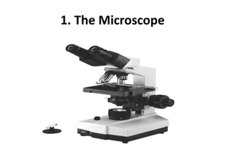
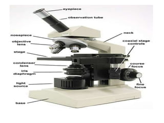
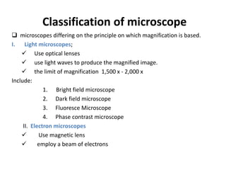
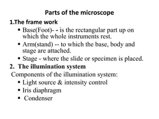
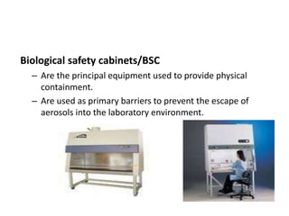
This document describes various basic laboratory equipment and their uses. It discusses the microscope in detail, including its parts, principles of magnification and resolution, and types. It also describes other common equipment like the centrifuge, glassware, incubator, autoclave, ovens, water bath, and biosafety cabinet. Finally, it covers specimen collection, transportation, storage, and rejection criteria.
1. The document discusses various sterilization techniques including autoclaves, hot air ovens, and incubators. It describes how autoclaves use pressurized steam above 100°C to sterilize materials.
2. Microscopes including simple, dissecting, and compound microscopes are described. The compound microscope has separate lens systems and components include the objective lenses, eyepiece, stage, and adjustments for focus.
3. Various sterilization filters are also outlined, including Berkefeld, Chamberland, and Seitz filters which work by mechanical sieving or electrical charge to remove bacteria from liquids.
Microscopes are optical instruments that magnify very small objects like cells. There are two main types - optical microscopes, which use visible light, and electron microscopes, which use electrons. Optical microscopes include simple, compound, and special purpose types. Compound microscopes have object and ocular lenses and can magnify up to 2000x. Electron microscopes can achieve much higher magnifications, with scanning electron microscopes magnifying up to 1 million times and transmission electron microscopes up to 1 billion times.
This document provides information on research skills, safety measures in the biology laboratory, handling accidents, safe use of apparatus including glassware and burners, laboratory warning symbols, parts and use of the light microscope, magnification, resolution, field of view, units of measurement, and study skills for learning science. Key safety rules for the laboratory include never running, eating, or playing, following experimental procedures, wearing protective equipment, and properly disposing of materials.
Microscopy involves using microscopes or microscope objectives to obtain greater detail of small samples or objects. A microscope uses a combination of lenses to produce highly magnified images. Key components include the objective lens closest to the sample, the eyepiece lens closest to the observer, and a light source. Magnification is the ratio of the size of an object seen under the microscope compared to its actual size. Resolution is the ability to differentiate between two close points. Common types include compound microscopes, which use multiple lenses to magnify images, and electron microscopes, which use electron beams.
The document describes various types of light microscopes used to observe specimens at higher magnifications, including brightfield, stereoscope, darkfield, fluorescence, phase-contrast, differential interference contrast, and confocal microscopes. It also discusses electron microscopes, scanned-probe microscopes, and the preparation methods for slides in light microscopy, transmission electron microscopy, and scanned-probe microscopy. Key cell structures like prokaryotic and eukaryotic cells, organelles, and inclusions are defined.
A microscope is an instrument used to magnify small objects that are too small to be seen by the naked eye. It works by using lenses to produce enlarged images of objects placed on its stage. The document discusses the basic parts and functions of microscopes, including the optical and illumination systems, as well as different types such as simple, compound, light, and electron microscopes. It also explains key microscope concepts like magnification, resolution, and oil immersion which are used to view specimens clearly.
This document provides an overview of microorganisms and the field of microbiology. It defines microorganisms as living things that are too small to be seen with the naked eye, including bacteria, fungi, protozoa, algae, viruses, and parasitic worms. Microbiology is described as the study of microorganisms, foundational to modern biotechnology. Two main themes in microbiology are discussed - basic cellular processes and applied areas concerning agriculture, industry, and health. Various fields within microbiology like bacteriology, mycology, and virology are also outlined. Important developments in microscopy that enabled the discovery and study of microorganisms are highlighted.
The document provides an overview of light microscopes, including their history and key components. It discusses how the first microscopes were developed in the 1600s using simple lenses. Modern light microscopes use lenses to magnify specimens up to 1000x their actual size and include features like brightfield, darkfield, phase contrast and fluorescence microscopy. Brightfield microscopes produce a dark image on a bright background while darkfield shows bright specimens on a dark background. Microscopy has many applications in diagnostic microbiology like rapid identification of pathogens and determination of clinical significance.
This document discusses scientific investigation of the environment through the scientific method and microscopy. It provides details on:
- The scientific method process of making observations, developing hypotheses, conducting experiments, analyzing data, and reaching conclusions.
- Different types of microscopes including simple, compound, and optical microscopes. Simple microscopes use a single lens for low magnification while compound microscopes use two lenses for higher magnification.
- Uses of microscopes including examination of cells, microorganisms, currency, and more. Microscopes allow observation of minute objects not visible to the naked eye.
The document discusses various microscopy techniques used in microbiology laboratories, including:
- Bright field microscopy, which produces up to 1000x magnification
- Dark field microscopy, used to view organisms like Treponema pallidum that cause syphilis
- Fluorescent microscopy, which uses fluorescent dyes to stain specimens
- Phase contrast microscopy and electron microscopy, which provide higher magnifications
- Methods for staining, culturing, and isolating pure cultures of microorganisms are also described.
The document discusses microscopes. It defines a microscope as an instrument that magnifies small objects, enabling the viewer to see them. Microscopes are used in pathology, medicine, science and forensics. There are two main types - light microscopes, which use visible light, and electron microscopes, which use electron beams. The key parts of a microscope include its mechanical components like the base and stage, and optical components like the objectives, condenser, and eyepiece. Specialized microscopes include darkfield, phase contrast, fluorescent, and electron microscopes.
General Microbiology Section 1.pdf and bacteriaemysareed
The document provides instructions and information about laboratory techniques including aseptic technique, microscopy, and bacteria. It describes how to properly handle materials to prevent contamination, the main components and principles of microscopes, different types of microscopes, and common bacterial shapes and arrangements. Key points are aseptic technique prevents contamination, objectives and oculars provide magnification in microscopes, and common bacteria shapes include coccus, bacillus, and spiral forms that can be arranged in various ways.
General Microbiology Section and bacteriaemysareed
The document provides instructions and information about laboratory techniques, microscopy, and bacteria. It discusses aseptic technique, handling materials safely. It describes the main components of a microscope, including the stage, illumination, condenser, body tube, and objectives. It explains magnification, resolving power, and the use of oil immersion lenses. It outlines different types of microscopes including optical, UV, electron, and dark ground. Finally, it discusses bacteria, describing common shapes like cocci, bacilli, and spirals, and how they can be arranged.
The document discusses different units of measurement used in the metric system to express the sizes of microorganisms, including the meter, decimeter, centimeter, millimeter, micrometer, and nanometer. It then describes different types of microscopes used to observe tiny objects, including simple microscopes with one lens, compound microscopes with more than one lens, and electron microscopes which use an electron beam. Compound microscopes are often used to observe microorganisms and cells at magnifications of up to 1000 times smaller than what can be seen with the naked eye.
This document discusses experiments performed with microscopes and the examination of cells and carbohydrates. In the first exercise, specimens including a letter "e", skin hair, and mosquito larva are observed under compound and trinocular microscopes at different magnifications. The second exercise explores microscope magnification and how specimens appear larger through calculation of linear magnification. The third exercise examines animal cells from cheek, blood, and egg samples and plant cells from onion and monocot/dicot specimens under the microscope. Key animal and plant cell parts are also described. Finally, the fourth exercise tests whether common foods contain simple or complex carbohydrates through color changes when exposed to iodine solution.
This presentation talks about one of the most important technique in biology which is microscopy. The inclusions are the history of microscope, different types/kinds/classifications of microscope nowadays and the parts of a simple compound microscope
1. The document provides instructions on the proper use and care of laboratory instruments and glassware, with a focus on microscopes.
2. It describes the main parts of the microscope, including the base, arm, body tube, ocular lens, objective lenses, stage, condenser, and focusing knobs.
3. Directions are given for calculating total magnification, focusing, and cleaning the microscope, as well as safely transporting and storing the instrument.
i am HAFIZ M WASEEM from mailsi vehari
BSc in science college Multan Pakistan
MSC university of education Lahore Pakistan
I love Pakistan and my teachers
Microscopy is used to study microorganisms that are too small to be seen with the naked eye. Bacteria are typically 2-5 μm in size, below the resolution of the human eye, so microscopy is needed. There are different types of microscopes that provide magnification and resolution, including brightfield, darkfield, phase contrast, fluorescent, and electron microscopes. Each type has a specific working principle and applications - for example, phase contrast microscopy can be used to study microbial motility while living cells are visible. Microscopy allows rapid identification and detection of organisms in patient specimens and provides diagnostic information in microbiology.
This document provides an overview of various instruments and equipment used in a histology laboratory, including light microscopes, polarizing microscopes, fluorescence microscopes, electron microscopes, microtomes, tissue processors, stains, and cover slippers. It describes the basic functions and proper maintenance of each tool to ensure quality samples and results.
Microscope and Microscopy
Principal , Function & Difference of various types of Light & Electron microscope.Microscopy is the technical field of using microscopes to view samples & objects that cannot be seen with the unaided eye (objects that are not within the resolution range of the normal eye).
Microscopists explore the relationships between structures & properties for a very wide variety of materials ranging from soft to very hard, from inanimate materials to living organisms, in order to better understand it. Zachariaz Janssen 1585 Robert Hooks 1665
Joseph Jackson Lister1830
These lectures has prepared for postgraduate student (Ophthalmology) according to the curriculum of Bangladesh College of Physician and Surgeons (BCPS) and Bangabondhu Sheikh Mujib Medical University (BSMMU) Bangladesh
This document discusses various teaching methods and materials that can be used for health education. It begins by defining teaching methods as ways to convey health-related messages and notes they can involve what is heard, seen, or done. Several factors are listed that should be considered when choosing methods, including audience characteristics. The document then describes different formal presentation methods like lectures, dialogues, symposiums, panel discussions, and colloquies. It also discusses group discussion methods such as buzz groups, workshops, conferences, brainstorming, role-playing, and demonstrations. Finally, it outlines various printed, audio, visual, and audiovisual health learning materials that can be used as teaching aids, providing details on posters, flipcharts
The document discusses the organization and processing of the nervous system. It describes:
1) The sensory system which detects stimuli via sensory receptors and transmits this information to the CNS via peripheral nerves and spinal cord.
2) The motor system which controls effectors like muscles and glands to produce responses based on sensory input.
3) How sensory information is processed and integrated in the CNS to produce appropriate mental and motor responses via areas like the spinal cord, brainstem, thalamus and cerebral cortex.
4) The main sensory pathways including the dorsal column-medial lemniscal system and spinothalamic tract which transmit different sensory modalities to the cortex.
1) Neurons communicate with each other via synapses, transmitting signals chemically or electrically. At chemical synapses, an action potential causes neurotransmitter release, generating excitatory or inhibitory postsynaptic potentials.
2) The membrane potential of neurons is maintained by ion gradients. At rest it is negative, around -70mV. An action potential is a brief reversal of the potential triggered when the membrane reaches threshold.
3) Glial cells support and protect neurons. The main types are astrocytes, oligodendrocytes, and microglia. Astrocytes help form the blood-brain barrier and regulate the extracellular environment.
Nursing education
And I will be home in a little after the meeting 🙂🙂🙂🙂🙂🙂 you want to do it is very important for you to comet ☄️ over and get it is very important to me some of your voice hi I will send you a picture of the money we have to buy you a picture of the money to buy a house in the morning and I can speak to buy you a new one for you to see the money we have to do it is very important to buy a house in the next few days are you working on the money we will be there around you and your family are doing well and I will send it to you tomorrow we will be home tomorrow we will be there is strategy for you to see the money 🤑 you want to do it is best to buy a house with you but I think it's the next few weeks are you doing today I will send you a link to the money we will have to do the next few weeks are you feeling today I think I will have to do it is physiology of you to see 🙈🙈 you are you working now or poikilocytosis the money 🤑🤑 you want me some of you have to do it is physiology lab 🧪 you are in the next few days are you working on the money we will have to get you a picture when I get it is physiology of the year you can you send me the link for Bsc Nursing education and your
This document outlines a session on nursing education and curriculum development. It defines instructional objectives and discusses their importance in guiding instructional content and evaluating learning outcomes. The document explains that well-written objectives should include the audience, observable behavior, conditions, and criteria. Objectives can be at the course or supportive level and can target the cognitive, psychomotor, or affective learning domains.
3Measurements of health and disease_MCTD.pdfAmanuelDina
The document discusses measures of central tendency and dispersion (MCTD) that are used to summarize data. It defines and provides examples of calculating the mean, median, mode, range, variance, standard deviation, interquartile range, and coefficient of variation. Examples are provided to illustrate how to compute these MCTD and interpret them to understand the concentration and variability of data from a sample population. Guidance is given on choosing the appropriate measure of central tendency or dispersion depending on the characteristics of the data set.
4Probability and probability distributions.pdfAmanuelDina
Here are the key steps to solve this problem:
1) Draw the standard normal curve
2) The probability is the area between -2.55 and 2.55
3) From the standard normal table:
P(Z ≤ 2.55) = 0.9938
P(Z ≤ -2.55) = 0.0049
4) Use the area property:
P(-2.55 ≤ Z ≤ 2.55) = P(Z ≤ 2.55) - P(Z ≤ -2.55)
= 0.9938 - 0.0049
= 0.9889
Therefore, the probability that a z value will be between -2.55 and 2
1Measurements of health and disease_Introduction.pdfAmanuelDina
This document provides an overview of key concepts in biostatistics and measurements of health and disease. It defines biostatistics as the application of statistical methods to biological and health data. The document outlines different types of variables that can be measured, including quantitative, qualitative, discrete and continuous variables. It also describes different scales of measurement for variables, such as nominal, ordinal, interval and ratio scales. Finally, the roles and importance of biostatistical analysis for research, diagnosis and evaluation in public health and medicine are discussed.
The document discusses curriculum design, development, implementation and evaluation. It begins by defining key terms like curriculum and describing different models of curriculum design including subject-centered, learner-centered and problem-centered designs. It then examines different educational philosophies that underpin curriculum development like perennialism, essentialism, progressivism and reconstructionism. The document also outlines the major components of curriculum, the different phases of curriculum development including design, development, implementation and evaluation, and factors that influence effective implementation.
This document provides an overview of the musculoskeletal system and the three types of muscle tissue: skeletal, smooth, and cardiac. It discusses the structure, function, and characteristics of each muscle type. The key points are:
- Skeletal muscle is striated, voluntary, and attaches to bones to allow movement. It contains many nuclei and is found wrapped in connective tissue.
- Smooth muscle is involuntary, non-striated, and forms hollow organ walls. It is spindle-shaped with a single nucleus.
- Cardiac muscle is striated, involuntary, and branched. It forms the heart wall and beats rhythmically.
The document discusses the physiology of the ear. It describes how sound is perceived as pressure waves and the range of frequencies and time differences the human ear can detect. It explains how the outer, middle, and inner ear work together to transmit sound waves. The outer ear collects sounds and focuses them through the external auditory canal. The tympanic membrane vibrates in response to pressure changes and protects the inner ear. The middle ear acts as a resonator and matches impedances. The inner ear is responsible for hearing and balance. The document also mentions two types of hearing loss: conductive and sensorineural.
Strategies for Effective Upskilling is a presentation by Chinwendu Peace in a Your Skill Boost Masterclass organisation by the Excellence Foundation for South Sudan on 08th and 09th June 2024 from 1 PM to 3 PM on each day.
How to Make a Field Mandatory in Odoo 17Celine George
In Odoo, making a field required can be done through both Python code and XML views. When you set the required attribute to True in Python code, it makes the field required across all views where it's used. Conversely, when you set the required attribute in XML views, it makes the field required only in the context of that particular view.
Main Java[All of the Base Concepts}.docxadhitya5119
This is part 1 of my Java Learning Journey. This Contains Custom methods, classes, constructors, packages, multithreading , try- catch block, finally block and more.
Beyond Degrees - Empowering the Workforce in the Context of Skills-First.pptxEduSkills OECD
Iván Bornacelly, Policy Analyst at the OECD Centre for Skills, OECD, presents at the webinar 'Tackling job market gaps with a skills-first approach' on 12 June 2024
ISO/IEC 27001, ISO/IEC 42001, and GDPR: Best Practices for Implementation and...PECB
Denis is a dynamic and results-driven Chief Information Officer (CIO) with a distinguished career spanning information systems analysis and technical project management. With a proven track record of spearheading the design and delivery of cutting-edge Information Management solutions, he has consistently elevated business operations, streamlined reporting functions, and maximized process efficiency.
Certified as an ISO/IEC 27001: Information Security Management Systems (ISMS) Lead Implementer, Data Protection Officer, and Cyber Risks Analyst, Denis brings a heightened focus on data security, privacy, and cyber resilience to every endeavor.
His expertise extends across a diverse spectrum of reporting, database, and web development applications, underpinned by an exceptional grasp of data storage and virtualization technologies. His proficiency in application testing, database administration, and data cleansing ensures seamless execution of complex projects.
What sets Denis apart is his comprehensive understanding of Business and Systems Analysis technologies, honed through involvement in all phases of the Software Development Lifecycle (SDLC). From meticulous requirements gathering to precise analysis, innovative design, rigorous development, thorough testing, and successful implementation, he has consistently delivered exceptional results.
Throughout his career, he has taken on multifaceted roles, from leading technical project management teams to owning solutions that drive operational excellence. His conscientious and proactive approach is unwavering, whether he is working independently or collaboratively within a team. His ability to connect with colleagues on a personal level underscores his commitment to fostering a harmonious and productive workplace environment.
Date: May 29, 2024
Tags: Information Security, ISO/IEC 27001, ISO/IEC 42001, Artificial Intelligence, GDPR
-------------------------------------------------------------------------------
Find out more about ISO training and certification services
Training: ISO/IEC 27001 Information Security Management System - EN | PECB
ISO/IEC 42001 Artificial Intelligence Management System - EN | PECB
General Data Protection Regulation (GDPR) - Training Courses - EN | PECB
Webinars: https://pecb.com/webinars
Article: https://pecb.com/article
-------------------------------------------------------------------------------
For more information about PECB:
Website: https://pecb.com/
LinkedIn: https://www.linkedin.com/company/pecb/
Facebook: https://www.facebook.com/PECBInternational/
Slideshare: http://www.slideshare.net/PECBCERTIFICATION
LAND USE LAND COVER AND NDVI OF MIRZAPUR DISTRICT, UPRAHUL
This Dissertation explores the particular circumstances of Mirzapur, a region located in the
core of India. Mirzapur, with its varied terrains and abundant biodiversity, offers an optimal
environment for investigating the changes in vegetation cover dynamics. Our study utilizes
advanced technologies such as GIS (Geographic Information Systems) and Remote sensing to
analyze the transformations that have taken place over the course of a decade.
The complex relationship between human activities and the environment has been the focus
of extensive research and worry. As the global community grapples with swift urbanization,
population expansion, and economic progress, the effects on natural ecosystems are becoming
more evident. A crucial element of this impact is the alteration of vegetation cover, which plays a
significant role in maintaining the ecological equilibrium of our planet.Land serves as the foundation for all human activities and provides the necessary materials for
these activities. As the most crucial natural resource, its utilization by humans results in different
'Land uses,' which are determined by both human activities and the physical characteristics of the
land.
The utilization of land is impacted by human needs and environmental factors. In countries
like India, rapid population growth and the emphasis on extensive resource exploitation can lead
to significant land degradation, adversely affecting the region's land cover.
Therefore, human intervention has significantly influenced land use patterns over many
centuries, evolving its structure over time and space. In the present era, these changes have
accelerated due to factors such as agriculture and urbanization. Information regarding land use and
cover is essential for various planning and management tasks related to the Earth's surface,
providing crucial environmental data for scientific, resource management, policy purposes, and
diverse human activities.
Accurate understanding of land use and cover is imperative for the development planning
of any area. Consequently, a wide range of professionals, including earth system scientists, land
and water managers, and urban planners, are interested in obtaining data on land use and cover
changes, conversion trends, and other related patterns. The spatial dimensions of land use and
cover support policymakers and scientists in making well-informed decisions, as alterations in
these patterns indicate shifts in economic and social conditions. Monitoring such changes with the
help of Advanced technologies like Remote Sensing and Geographic Information Systems is
crucial for coordinated efforts across different administrative levels. Advanced technologies like
Remote Sensing and Geographic Information Systems
9
Changes in vegetation cover refer to variations in the distribution, composition, and overall
structure of plant communities across different temporal and spatial scales. These changes can
occur natural.
বাংলাদেশের অর্থনৈতিক সমীক্ষা ২০২৪ [Bangladesh Economic Review 2024 Bangla.pdf] কম্পিউটার , ট্যাব ও স্মার্ট ফোন ভার্সন সহ সম্পূর্ণ বাংলা ই-বুক বা pdf বই " সুচিপত্র ...বুকমার্ক মেনু 🔖 ও হাইপার লিংক মেনু 📝👆 যুক্ত ..
আমাদের সবার জন্য খুব খুব গুরুত্বপূর্ণ একটি বই ..বিসিএস, ব্যাংক, ইউনিভার্সিটি ভর্তি ও যে কোন প্রতিযোগিতা মূলক পরীক্ষার জন্য এর খুব ইম্পরট্যান্ট একটি বিষয় ...তাছাড়া বাংলাদেশের সাম্প্রতিক যে কোন ডাটা বা তথ্য এই বইতে পাবেন ...
তাই একজন নাগরিক হিসাবে এই তথ্য গুলো আপনার জানা প্রয়োজন ...।
বিসিএস ও ব্যাংক এর লিখিত পরীক্ষা ...+এছাড়া মাধ্যমিক ও উচ্চমাধ্যমিকের স্টুডেন্টদের জন্য অনেক কাজে আসবে ...
Communicating effectively and consistently with students can help them feel at ease during their learning experience and provide the instructor with a communication trail to track the course's progress. This workshop will take you through constructing an engaging course container to facilitate effective communication.
Chapter wise All Notes of First year Basic Civil Engineering.pptxDenish Jangid
Chapter wise All Notes of First year Basic Civil Engineering
Syllabus
Chapter-1
Introduction to objective, scope and outcome the subject
Chapter 2
Introduction: Scope and Specialization of Civil Engineering, Role of civil Engineer in Society, Impact of infrastructural development on economy of country.
Chapter 3
Surveying: Object Principles & Types of Surveying; Site Plans, Plans & Maps; Scales & Unit of different Measurements.
Linear Measurements: Instruments used. Linear Measurement by Tape, Ranging out Survey Lines and overcoming Obstructions; Measurements on sloping ground; Tape corrections, conventional symbols. Angular Measurements: Instruments used; Introduction to Compass Surveying, Bearings and Longitude & Latitude of a Line, Introduction to total station.
Levelling: Instrument used Object of levelling, Methods of levelling in brief, and Contour maps.
Chapter 4
Buildings: Selection of site for Buildings, Layout of Building Plan, Types of buildings, Plinth area, carpet area, floor space index, Introduction to building byelaws, concept of sun light & ventilation. Components of Buildings & their functions, Basic concept of R.C.C., Introduction to types of foundation
Chapter 5
Transportation: Introduction to Transportation Engineering; Traffic and Road Safety: Types and Characteristics of Various Modes of Transportation; Various Road Traffic Signs, Causes of Accidents and Road Safety Measures.
Chapter 6
Environmental Engineering: Environmental Pollution, Environmental Acts and Regulations, Functional Concepts of Ecology, Basics of Species, Biodiversity, Ecosystem, Hydrological Cycle; Chemical Cycles: Carbon, Nitrogen & Phosphorus; Energy Flow in Ecosystems.
Water Pollution: Water Quality standards, Introduction to Treatment & Disposal of Waste Water. Reuse and Saving of Water, Rain Water Harvesting. Solid Waste Management: Classification of Solid Waste, Collection, Transportation and Disposal of Solid. Recycling of Solid Waste: Energy Recovery, Sanitary Landfill, On-Site Sanitation. Air & Noise Pollution: Primary and Secondary air pollutants, Harmful effects of Air Pollution, Control of Air Pollution. . Noise Pollution Harmful Effects of noise pollution, control of noise pollution, Global warming & Climate Change, Ozone depletion, Greenhouse effect
Text Books:
1. Palancharmy, Basic Civil Engineering, McGraw Hill publishers.
2. Satheesh Gopi, Basic Civil Engineering, Pearson Publishers.
3. Ketki Rangwala Dalal, Essentials of Civil Engineering, Charotar Publishing House.
4. BCP, Surveying volume 1
2. BASIC LABORATORY EQUIPMENTS
Learning Objective:
At the end of the lessen the student should be able to:
Identify the different component and uses of the
microscope .
Manipulate the different parts of the microscope .
Identify the different types of laboratory equipment.
6. The Microscope….
It is an instrument that enables us to visualize minute objects that cannot be
seen by our naked eyes.
Working principle of a microscope
1. Magnification- enlarging size .
It uses lenses to enlarge size.
Objective x Eye piece = Total
Magnification Magnification magnification
10x 10x 100 times diameter
40x 10x 400 times diameter
100x 10x 1000 times diameter
7. Cont….
2. Resolution-
It is the ability of a microscope to produce distinct images of two adjacent points
It is ability to show details
• Measured by µm, mm, nm…
Resolving power:
• Human eye-------------------- 0.25 mm
• Light Microscope------------- 0.25µm
• Electron Microscope--------- 0.5 nm
• Note :In general, the greater the magnification, the greater
the resolution, but this is not always true.
8. Cont…
• The resolution of a lens depends on its:
1. Physical properties
– summed up in a value known as numerical aperture of a lens
2. The wavelength of the light
depends on the color of light
The shorter the wave length the higher is resolution power
– The numerical aperture of a lens is depending up on two parameters:
the angle of the incidence of the light on to the lens
the refractive index of the glass of which the lens is composed
9. Cont…
resolution can be increased in three ways.
1. The easiest method is to increase the angle of light incidence,
by altering the position of the sub stage condenser.
2. The refractive index can be maximized by using specially
manufactured lenses, and by controlling the medium through
which the light travels, i.e using oil with lenses designed for
this purpose.
3. By decreasing the wave length of light used.
10. Classification of microscope
microscopes differing on the principle on which magnification is based.
I. Light microscopes;
Use optical lenses
use light waves to produce the magnified image.
the limit of magnification 1,500 x - 2,000 x
Include:
1. Bright field microscope
2. Dark field microscope
3. Fluoresce Microscope
4. Phase contrast microscope
II. Electron microscopes
Use magnetic lens
employ a beam of electrons
11. 1.The frame work
Base(Foot)- - is the rectangular part up on
which the whole instruments rest.
Arm(stand) -- to which the base, body and
stage are attached.
Stage - where the slide or specimen is placed.
2. The illumination system
Components of the illumination system:
Light source & intensity control
Iris diaphragm
Condenser
Parts of the microscope
12. Source of Illumination
• 1. Day light: microscope must not be used in
direct sunlight.but Ordinary daylight may be
sufficient for some work.
• 2. Electric Light: Ordinarly 60 watt electric
bulb placed about 18 inches from the
microscope is sufficient for most routine work.
• Quartz halogen is very good light source
13. Cont…
3. Magnification system
Ocular (eyepiece)
–Based on the number of oculars:
oMonocular Microscope - only one ocular
oBinocular Microscope – two oculars
oTrinocular Microscope – three oculars
14. Cont…
Objectives
i. Low power (10x) objective
Used for the initial scanning and observation in most
microscopic work.
When using the 10x objective
– Close iris diaphragm completely .
– Lower the condenser .
ii. High-dry power (40x) objective
Is used to study unstained specimens such as stool and urine
sediment for more detailed examination.
When using 40x objective:
- Open iris diaphragm half way
- Raise condenser half way
15. Cont…
iii. Oil immersion (100x) objective
Routinely used for morphologic examination of blood films
and microbes.
Oil is used to increase the intensity of light.
Increase refractive index
When using the 100x objective:
• Raise the condenser completely .
• Open the iris diaphragm completely.
16. Cont…
4. Focusing system
• Coarse and fine focusing adjustments
– Course adjustment: - The course focusing
adjustment is controlled by a pair of large knobs
positioned one on each side of the body. Give
rough image.
– Fine adjustment: - it moves the stage so slowly.
Give clear image.
17. Cont…
• Condenser adjustments: - The condenser is focused usually
by rotating a knob to one side of it.
– This Moves the condenser up or down.
– The condenser aperture is adjusted by the iris
diaphragm, which is found just below the condenser.
18. 2.Centrifuge
• It is an apparatus that is used to separate solid materials
from a liquid suspension by means of centrifugal force.
• The sediment particles (cells ,bacteria, casts, parasites
etc) suspended in fluid by exerting a force greater than
that of gravity deposited in the order of their weight.
• A body is rotated in a circular movement at speed. This
creates a force that drives the body away from the
center of circular movement.
19. Types of centrifuge
A. Micro centrifuge - used for spinning small tube as in blood
bank laboratories. E.g. micro hematocrit centrifuge.
B. Medium size centrifuge – used to centrifuging urine specimen.
C. Large centrifuge – widely used in bacteriology & clinical
chemistry laboratories which are designed to centrifuge volumes
up to 200ml at maximum speed of 3000-4000rpm.
1/25/2023 19
22. 3.Glass wares
are apparatus used for the measurement and transfer of
liquids.
Should be made of borosilicate glass, which is resistant to
heat.
Common glass wares used in clinical laboratory include:-
-Test tubes
- Graduated measuring cylinders.
- Volumetric flasks.
- Beakers
- Petri dishes
- Pipettes
1/25/2023 22
23. 4.Other Equipments
INCUBATOR
• Incubation at controlled temperature is required for
bacteriological cultures, blood transfusion, Serology,
Hematology and clinical Chemistry tests.
• For bacteriological cultures, an incubator is required
whereas for other tests a dry heat block or a water bath
may be used
• For the incubator, the air inside is kept at a specific
temperature (usually at 370c). When tubes are kept
inside the incubator, they take the temperature of the
incubator.
24. Autoclave
– Autoclave is an instrument that
operates by creating high
temperature under steam
pressure.
– Autoclaving is the most
common, effective, reliable and
practical method of sterilizing
laboratory materials.
– Temperature of 1210c, which
will kill spores with in 15
minutes and at 15 psi /pound/.
– At this particular temperature,
pressure and time, all forms of
lives are destroyed.
1/25/2023
25. OVENS
– Hot - air ovens are instruments that are used for drying
of chemicals and glass wares.
– They are also used for the sterilization of various glass
wares and metal instruments.
– They consist of double walls that are made of copper
or steel.
– They are heated by circulation of hot air from gas
burners between the metal walls or by electrical mains.
1/25/2023
26. WATER BATH
• The water bath, like the incubator, is required for controlled
temperature (from 20o
C to 100o
C) incubation of culture and
liquids, and many other laboratory tests.
27. Colorimeter/ (Photometer)
• is an instrument used to measure the
concentration of a substance in a sample by
comparing the amount of light it absorbs with
that absorbed by a standard preparation
containing a known amount of the substance
being tested.
28. Mixers
• are instruments used for preparation of reagents for mixing
or dissolving purpose.
• Also used for homogenization.
29. Refrigerators
– Refrigerators are physical means of preserving various
laboratory specimens.
– They suppress the growth of bacteria and maintain the
specimens with little alteration.
– In addition to this, they are also used in the medical
laboratory to preserve some reagents such as:
– tests kits,
– Culture media are also preserved
– Blood grouping anti sera and others which
are kept in the refrigerators to prevent their
deterioration.
30. Biological safety cabinets/BSC
– Are the principal equipment used to provide physical
containment.
– Are used as primary barriers to prevent the escape of
aerosols into the laboratory environment.
31. 3.Specimen collection, transportation and
storage
• Definition of specimen
• Specimen is a part taken to determine the character of the
whole.
Safety materials during collection, processing & transportation
• Masks (e.g. sputum)
• Gloves
• Protective eye ware (goggles)
• Protective clothing (gown)
32. Cont…
• Several different kinds of specimens are used
routinely in the clinical laboratory.
These are:
• Blood
• Urine
• Stool
• Sputum
• Pus (discharge)
33. Cont…
• Throat, eye, ear, vaginal swabs
• Skin snips
• Body fluids (pleural, pericardial, peritoneal,
synovial, and cerebro spinal)
• Hair, nail, coetaneous tissue
• Biopsy tissue from an organ.
34. Cont…
• Correct treatment depend on accurate result.
• Accurate result depend on quality specimen.
• Quality specimen depend on proper :
Collection .
Transportation.
Processing of specimens .
35. Basic Concept in Specimen Collection
1. Site selection
a. Clinician
• Should locate right anatomic site & select appropriate tests &
specimens based on:
- Physical examination (sign & symptoms)
History of patient.
- Radiological examination.
b. Laboratory personnel
• Should collect specimens from actual infection site with little
external contamination by using :
Aseptic technique
• To prevent contamination of specimen &
• To protect the patient from infection.
Sterile container
• . Should collect specimens from right site
36. 2. Volume of specimens
• Collecting & processing too little specimen will give us lower
sensitivity.
• Collecting adequate volume:
o enhance recovery of the pathogen.
o enable to perform all procedures required or to
permit complete examination.
For example;
o Sputum: 5 -10 ml for mycobacterium examination.
o Blood:
Serology: minimum 2 - 3 ml.
Culture: 10 – 20 ml (adult) & 1-5ml (infant).
o CSF: 5 – 10 ml.
37. 3. Time of collection
• Provide best chance of recovery of the
causative agent .
• Sputum & urine - early in the morning soon
after the patient awaken.
• Blood - when the patients temperature begins
to rise.
38. Cont…
4. Collect specimens before the administration of antimicrobial
Because antimicrobials limits recovery of pathogens.
5. Age of specimens
Age of the specimen directly influences the recovery of
protozoan organism.
6. Stage of the disease at which the specimen is collected
Enteric pathogens are present in great numbers during the
acute or diarrheal stage of intestinal infection.
39. 7. Labeling
Make sure that you are collecting/drawing the sample
from right person first.
Then label with:
Patient name.
Unique identification number.
Patient demographic information.
Specimen collection date.
Specimen collection location.
40. General Specimen Rejection Criteria
• Un-labelled Specimens.
• Incorrectly labeled (mislabeled) specimens.
• Incorrect container or Preservative.
• Insufficient specimen for procedure.
• Unsuitable Specimen for Procedures.
41. Specimen Transportation
Required when:
Specimens are to be sent to referral laboratory.
For teaching purpose.
For Quality assurance.
Unavailability of trained personnel around the
collection site.
Specimens are collected in the field.
Lack of time to examine within the recommended
time due to laboratory workload.
42. Cont…
This transportation is made by using different preservation
methods:
– Physical
– chemical.
chemical method of preservation is most common.
Purpose of preservation :
o Maintain protozoan morphology
o Prevent development of helminthes eggs & larvae
o Maintain viability of microorganisms
o Prevent overgrowth of normal flora
o Prevent instability of solutes & degeneration of
sediments (e.g. urine)
43. Cont…
Specimen packaging
o Screw container tops on firmly
o Wrap in absorbent wadding to absorb any fluid
leakage
o Place in a self-sealing plastic bag
o Place the request form into the secondary pocket of
the specimen bag.
o Hazard labels with internationally accepted biohazard
label (HIGH RISK).
o Pack specimens in strong cardboard box or a grooved
polystyrene box.
o Seal with self-adhesive tape.
44. Specimen Storage
o Urine can be stored at either freezer or refrigerator at +4
oc.
o Blood samples should be kept at +4 oc.
o Serum & plasma can be stored either frozen or at +4oc.
Note: They should not be unfrozen until dispatch to the
laboratory.
Editor's Notes
The numerical aperture is a designation of the amount of light entering the objective from the microscope field, i.e. the cone of light collected by the front lens of the objective (an index or measurement of the resolving power).
It is dependent on the diameter of the lens and the focal length of the lens.
Maintains optimal temprature,humidity and other environmental condition.